Skeletal Physiology What do bones do for us

Skeletal Physiology What do bones do for us? ?

Bone Parts

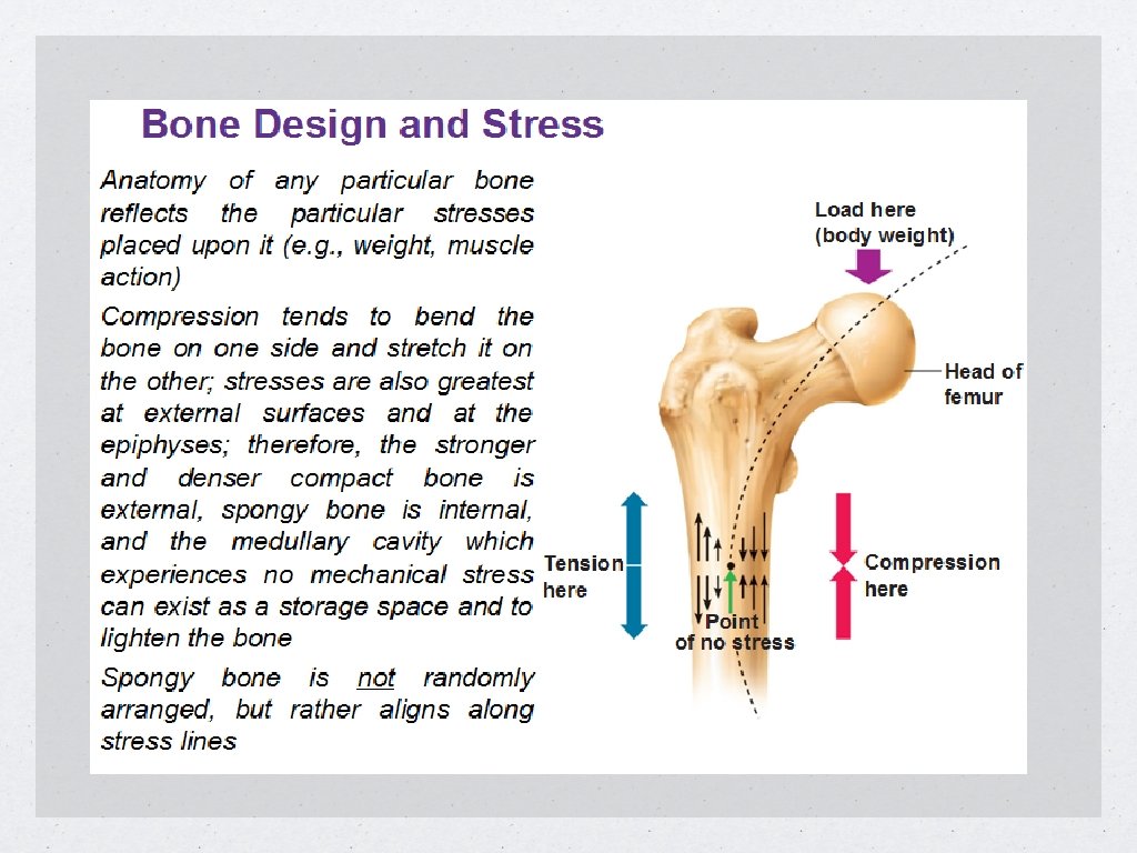
Bone Parts Diaphysis- shaft of the bone Epiphysis- Ends of the bone (mainly spongy bone) Metaphysis- Border of the diaphysis and epiphysis Marrow Cavity- central cavity space of the bone for storage and circulation. Many think the bones are rather dull but to be true it changes constantly based upon the stresses we place on it.
. 2")
Bone Histology Osteocytes- mature bone cells contained within a the bone matrix (Lamellae). 2 Functions Maintain the protein and mineral content of the surrounding matrix. Rebuild the matrix. Participate in the repair of damaged bone. Canaliculi- connect each lamellae with nutrients.

Bone Histology continued Osteoblasts- new cells that develop into osteocytes. Help maintain the matrix and mineral concentrations. Osteoclasts- remove bone matrix that is replaced by osteoblasts. huge cells containing 50+nuclei. When osteoblasts work faster than osteoclasts, bone get stronger When osteoclasts work faster than osteoblasts, bones get weaker

diaphysis shaft metaphysis narrow zone between epiphysis and diaphysis epiphysis end of bone; consists of spongey bone (also, cancellous, trabecular) cortex thin covering of compact bone (over spongy bone) matrix -very dense -contains osteocytes within pockets called lacunae -calcium phosphate - 2/3 of weight of bone; interacts with calcium hydroxide to form crystals of hydroxyapatite canaliculi narrow passageways thru matrix; exchange of nutrients periosteum covers outer surfaces of bones; fibrous outer layer, cellular inner layer -isolates bone from surrounding tissues -provides a route for circulatory and nervous supply -participates in bone growth and repair

calcium phosphate two-thirds of weight of bone; interacts with calcium hydroxide to form crystals of hydroxyapatite collagen fibers stronger than steel when pulled; easily tolerate twisting and bending; weak at compression osteocyte mature bone cells; each occupies a lacuna; cannot divide; repair damaged bone; maintain protein and mineral content of matrix lamellae layers of matrix osteoblast produce new bone matrix; osteoclast remove and recycle bone matrix; fifty or more nuclei

Bone Histology

Bone Parts
")
Bone Parts Compact Bone- Organized as osteons which are circular patterns of bone (osteocytes) around a central canal called a Haversian canal. Very strong because the osteons run parallel with the bone Haversian canal has blood vessels that give nutrients to the osteocytes. pull or push these osteons and it is strong Hold one end and push side to side, it is weak.

Bone Parts Spongy bone- not arranged as osteons. Matrix is arranged as trabeculae, which are struts or plates that branch out as an open matrix. Red marrow is contained within the openings of the matrix. Make blood cells (red blood cells and white blood cells) Located where bones are not heavily stressed or where stresses come from many directions Much lighter than compact bone which allows muscles to move them. Yellow marrow is stored within (Adipose cells)

Bone Parts Periosteum- superficial layer that wraps bone isolates the bone from surrounding tisue provides a route for circulatory and nervous supply actively participates in bone growth and repair The fibers of the periosteum are interwoven with those of the ligaments and tendons that connect to the bone.

Bone Parts Endosteum- lining of the spongy matrix and marrow cavities, it is where deposits from osteoblasts and removals from osteoclasts can take place during bone formation, repair and remodeling.

Ossification

Ossification is the process of replacing tissues with bone, this process occurs because of calcification which is the process of depositing calcium. All bones start as hyaline cartilage Chondrocytes are cartilage cells and during the process, they die and are replaced bone by osteoblasts. As bone is created, cavities are also created to allow for circulation of nutrients and waste. Epiphyseal plates control the rate and secondary ossification continues in the epiphysis. Same action as in the shaft

Ossification terms Endochondral ossification - hyaline cartilage replace by bone Primary ossification center- point at which bone development begins Secondary ossification center-osteoblasts and capillaries develop within the cavities of the ossification process Articular cartilage- thin cap of cartilage that is left at the ends of bones for protection at a joint Epiphyseal cartilage- separates epiphysis from diaphysis

Ossification continued Epiphyseal cartilage continues to grow= bone elongation At puberty, sex hormones and growth hormones make this process occur dramatically. Osteoblasts produce cells faster than chondrocytes thus making cartilage disappear. This rate can be monitored at the epiphyseal line (growth plate). They monitor the width of the cartilage with successive x-rays
, mandible, and clavicle) Mesenchymal cells")
Intramembranous Ossification that creates dermal bones (flat bones (skull), mandible, and clavicle) Mesenchymal cells cluster together and differentiate into osteoblasts within the dermis.

Bone Repair

Bone Repair terms Fracture- the crack or break of a bone due to extreme loads, sudden impacts, or stresses from an unusual direction. Fracture hematoma- a large blood clot that leaves a fibrous meshwork in the damaged area. External callus- enlarged collar of cartilage and bone that forms at the level of the fracture. Internal callus- organized within the marrow of the fracture that creates new bone to attach the two ends.

Types of fractures

Other fracture types

Osteoporosis • Due to the lack of bone mass from the reduction of sex hormones that maintain normal rates of bone deposition. • Accelerates in women after menopause due to the lack of estrogen production. Men less common due to the fact that androgens continue to be produced through older age. • Cancer can also be a factor because that activate osteoclast-activating factors which make bones replace mineral faster than they can be created.

Joint Classification • Synarthrosis- non moving joints found within the skull, teeth and jaw, and epiphyseal cartilage. • Amphiarthrosis- semi-moveable joints found within the ribs, vertebrae, pubic bones and pelvis. • Diarthrosis- moveable joints known as synovial joints that have various ranges of motion based upon structure. Elbow, Knee, wrist, ankle, neck, back fingers, toes, hip, and shoulder.

Aspects of a synovial joint • Synovial fluid- viscous fluid like molasses that provides 3 functions. • Lubrication from compression- it reduces friction • Nutrient Distribution circulates nutrients and picks up waste every time the joint is in motion • Shock Absorption cushions joints when subjected to compression. Distributes shock evenly within the joint.

Other synovial joint structures • Cartilage/Fat Pads- meniscus is a pad of cartilage that cushions opposing bones. • Fat pads- fill in the spaces of a joint as it changes in shape due to movement. • Ligaments- Intrinsic ligaments are localized thickening of the joint capsule, while extrinsic ligaments are on the outside to give support to the joint. A sprain is the stretching of the ligaments where small tears can occur. Usually the bone will break before ligaments tear because healing is much quicker.

More synovial structures • Tendons- although not really part of articulation, tendons can limit the movement of a joint and help in it’s support. • Bursae- fluid filled pockets that hold synovial fluid. They cushion and reduce shock to the joint.

Joint dislocation • Luxation- extreme stresses may cause for articulating surfaces to become displaced. Damage to the articular cartilage and joint capsule results and due to nerves that monitor the location, the event is very painful. • Subluxation- partial dislocation. People who are double jointed have weaker joint stabilization and are more at risk for dislocations. Of course the larger range of motion becomes a focus for conversation.

Dynamic motions • Linear- a gliding motion where the shaft of the bone from the joint allows forward, backwards, and side to side. • Angular- tip of the bone stays at the point of origin but the shaft can move and change angles relative to the surface. • Circumduction- tip of the bone is stationary to the point of origin, but the shaft when held under a 90 degree angle and move in a 360 degree motion. • Rotation- Angle of the tip and shaft are unchanged and the bone can spin around its longitudinal axis.

Dynamic motions

Flexion Refers to movement where the angle between two bones decreases. Flexion is commonly known as bending. Extension Refers to movement where the angle between two bones increases. Extension is otherwise known as straightening Hyperextension greater than normal extension Abduction (physiology) moving of a body part away from the central axis of the body Adduction MOVEMENT TOWARD THE MIDLINE OF THE BODY Circumduction Proximal end of limb remains stationary while distal end moves in a circle. Occur commonly at ball and socket, condyloid, and saddle joints. Pronation palm down

Supination rotation of the hands and forearms so that the palms face upward Medial rotation rotates towards the middle of the body Rotation Movement of bone around longitudinal axis without lateral or medial displacement. Common movement of ball and socket as well as atlas around dens of axis Lateral rotation Movement around an axis away from the mid-line Dorsiflexion toes raised, heels down Plantar flexion bending of the sole of the foot by curling the toes toward the ground Lateral Flexion Vertebral column bends to the side. Most pronounced in cervical and thoracic regions. Opposition Movement of the thumb to touch the fingertips

Depression Structure moves in inferior direction Elevation structure moves in a superior direction Protraction Top: Moving body part anteriorly in the horizontal plane Retraction bottom: Moving body part posteriorly in the horizontal plane Inversion sole of foot turned inward Eversion Sole of foot turns outward

Synovial Joint Types Hinge Joint elbow and knee are this kind of synovial joints Pivot Joint a synovial joints that permit rotation like your head Gliding Joints what synovial joints are between ankle and wrist bones; between vertebrae; end of collarbones and permit the least amount of movement Saddle Joint synovial joints that are modified ellipsoidal; one side of concave and the other bone is convex (like a rider sitting in a saddle) found in joint between metacarpal bone of thumb and the carpal bone allowing the opposition of the thumb Ball and socket joint synovial joint where round head of one bone rests in a socket of another bone; allows wide range of motion, most moveable of all body joints -very loosely joined

Ellipsoidal joint- oval articulation resting on a depression in the opposing surface.

- Slides: 37