Skeletal Muscle Voluntary Striated Multinucleated Long cylindrical cells

")

 Draw a cross-sectional view of a muscle.")


 fascicle b)")
 Sarcolemma: cell membrane Sarcoplasma: cytoplasm Sarcoplasmic reticulum (SR): ~Endoplasmic")


: Thin filaments (actin) Z-line H-Zone Thick filament (myosin) I-band A-band")


 NMJ: site where axon and muscle fiber communicate motor neuron- stimulates")
 action potential sweeps across the sarcolemma,")


- Slides: 32
Skeletal Muscle Voluntary Striated Multinucleated Long, cylindrical cells Non-branching Attached to bones Rapid & vigorous contraction, tire easily, but exert great force GENERAL FUNCTIONS: Movement Posture maintenance Joint stability Heat generation Smooth Muscle Cardiac Muscle Involuntary Non-striated Single nucleus Spindle shaped cells Non-branching Internal organs Involuntary Striated Single nucleus Cylindrical cells Branching, Intercalated discs Heart Contractions are slow & sustained Contracts at a rhythmic, steady rate set by “pacemaker” Similarities for all muscle types: All muscle cells are elongated (muscle fibers) Sarcolemma = cell membrane of muscle cell Sarcoplasm = cytoplasm of muscle cell Take the Moodle Quiz over this before moving on! QUIZ: Skeletal, Cardiac or Smooth Muscle Tissue?
Connective Tissue Coverings of the Skeletal Muscle 1. ENDOMYSIUM surrounds each muscle fiber (cell) 2. PERIMYSIUM surrounds groups muscle fibers called FASCICLES 3. EPIMYSIUM surrounds the entire muscle 4. DEEP FASCIA outermost layer that surrounds the entire muscle to form the tendon (connects muscle to bone) or aponeurosis (connects muscle to muscle). Fascicle surrounds fascicle
Skeletal Muscle Tissue Organization Connective Tissue Wrappings of Skeletal Muscle Practice CLICK HERE! http: //media. pearsoncmg. com/bc/bc_marieb_ehap_9/activities/chapter 6/Act 6 A. html
Structure of a Skeletal Muscle (ie. Biceps) Draw a cross-sectional view of a muscle. Include the following labels w/definitions: Fascicle, muscle fiber, muscle, epimysium, perimysium, endomysium, thin filament, thick filament
Fascicle: bundle of muscle cells surrounded by perimysium Fascia: outermost covering very thick CT becomes tendon Muscle fiber: ~a muscle cell ~made of myofibrils (made of protein filaments) surrounded by endomysium Epimysium: connective tissue surrounding muscle Perimysium: -ct that surrounds fascicles -contains blood vessels and Endomysium: nerves ct that surrounds each muscle fiber Protein filaments ~Thick filament = myos ~Thin filament = actin
PROJECT: Build a model of a muscle. Include the following labels: fascicle muscle fiber epimysium perimysium endomysium thin filament (actin) thick filament (myosin) fascia blood vessels/nerves IDEAS: I have built a model using saran wrap, yarn, straws, pipe cleaner, foil, etc. BE CREATIVE!
QUIZ: Draw a model of a muscle. Include the following labels: a) fascicle b) muscle fiber c) epimysium d) perimysium e) endomysium f) thin filament (actin) g) thick filament (myosin) h) fascia
Skeletal Muscle Cells (MUSCLE FIBERS) Sarcolemma: cell membrane Sarcoplasma: cytoplasm Sarcoplasmic reticulum (SR): ~Endoplasmic reticulum ~Channels that surround each myofibril and run parallel to them. ~High concentration of calcium ions needed for contractions Transverse tubules (T-tubules): Channels allow for ion movement between SR and sarcoplasm (into muscle cell). Myoglobin -a protein similar to hemoglobin that stores oxygen until needed by the mitochondria
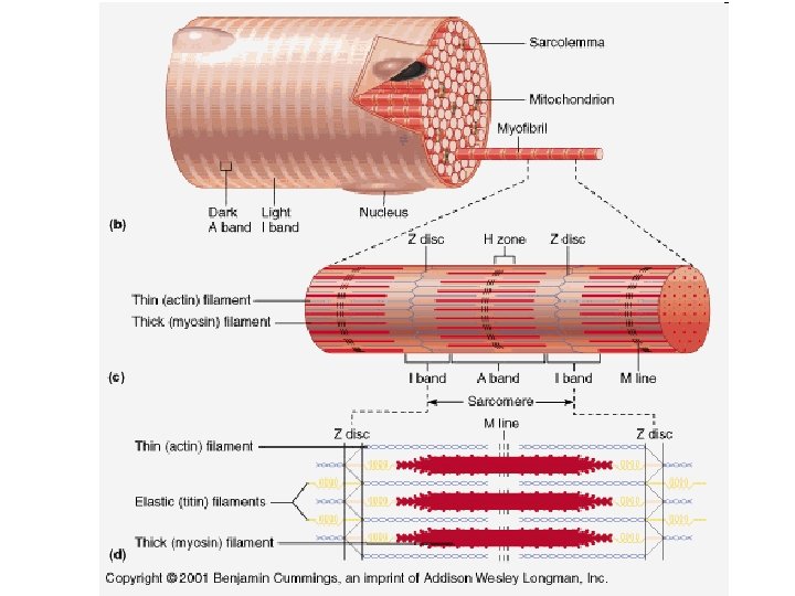
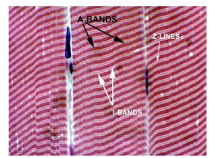
Functional Unit of Muscle: SARCOMERE Myofibrils -- long, thin, cylindrical rods, usually 1 -2 µm in diameter, that run within and parallel to the long axis of the muscle fiber. Made of myofilaments. Myofilaments -- comprised of thick and thin filaments. The thick are comprised of myosin and the thin are comprised of actin, troponin, and tropomyosin. Sarcomere -- the basic contractile unit of the muscle. Has Z-lines on either end along with A -band two 1/2 I-bands. Z-line connects the sarcomeres together. During contraction, the H-zone and I-bands decrease in size (HI…bye…get it? ). All the remaining areas do not change in length.
Draw a diagram of a sarcomere at rest. Label the sarcomere, M-line, Z-line, A-band, and I-band, and H-zone, act Draw arrows showing the direction of movement. A sarcomere at rest: Explain what changes occur to a sarcomere during full contraction:
Myofibril Anatomy (a sarcomere): Thin filaments (actin) Z-line H-Zone Thick filament (myosin) I-band A-band M-line
A diagrammatic view of contraction at the sarcomere level Draw a diagram of a sarcomere at rest and once fully contracted. Label the sarcomere, M-line, Z-line, A-band, and I-band. Draw arrows showing the direction of movement. A sarcomere at rest: A sarcomere during full contraction:
PROJECT: Build a model of a sarcomere. Make sure it can contract/relax. Label the following areas/parts: actin (thin filaments) myosin (thick filaments) Z-line H-zone I-band A-band M-line IDEAS: You may look at a model that I have from students. is not perfect, but it may get you thinking! Research the internet…there are some examples! It
Functional Characteristics of Muscle EXCITABILITY ability to receive and respond to stimuli EXTENSIBILITY ability to be stretched or extended CONTRACTILITY ability to shorten forcibly when stimulated ELASTICITY ability to go back to original shape and length after contraction or extension
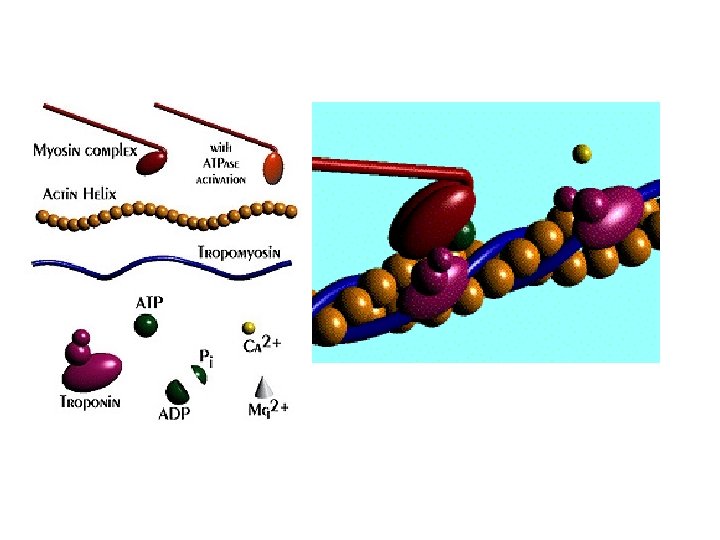
More about actin and myosin… Muscle contraction animation http: //entochem. tamu. edu/Muscle. Struc. Contractswf/index. html Myosin heads Cover binding sites where myosin heads will attach for a contraction
1 - Calcium released from sarcoplasmic reticulum 2 - Myosin head energized via myosin-ATPase activity which converts the bound ATP to ADP + Pi 3 - Calcium binds to troponin 4 - Tropomyosin translocates to uncover the cross-bridge binding sites 5 - The energized myosin heads approach the binding sites 6 - The first myosin head binds to actin 7 - The bound myosin head releases ADP + Pi, flips and the muscle shortens 8 - The second myosin head binds to actin 9 - The first myosin head binds ATP to allow the actin and myosin to unbind 10 - The second myosin head releases its ADP + Pi, flips & the muscle shortens further 11 - The second myosin head binds to ATP to allow the actin and myosin to unbind 12 - The second myosin head unbinds from the actin, flips back and is ready for the next cycle 13 - The cross-bridge cycle is terminated by the loss of calcium from the troponin 14 - Tropomyosin translocates to cover the cross-bridge binding sites 15 - The calcium returns to the sarcoplasmic reticulum, the muscle relaxes & returns to the resting state
1 - Calcium released from sarcoplasmic reticulum 2 - Myosin head energized via myosin-ATPase activity which converts the bound ATP to ADP + Pi 3 - Calcium binds to troponin 4 - Tropomyosin translocates to uncover the cross-bridge binding sites 5 - The energized myosin heads approach the binding sites 6 - The first myosin head binds to actin 7 - The bound myosin head releases ADP + Pi, flips and the muscle shortens 8 - The second myosin head binds to actin 9 - The first myosin head binds ATP to allow the actin and myosin to unbind 10 - The second myosin head releases its ADP + Pi, flips & the muscle shortens further 11 - The second myosin head binds to ATP to allow the actin and myosin to unbind 12 - The second myosin head unbinds from the actin, flips back and is ready for the next cycle 13 - The cross-bridge cycle is terminated by the loss of calcium from the troponin 14 - Tropomyosin translocates to cover the cross-bridge binding sites 15 - The calcium returns to the sarcoplasmic reticulum, the muscle relaxes & returns to the resting state
Motor Unit • single motor neuron and many skeletal muscle • all muscle fibers controlled by motor neuron small muscles that require fine control have many motor units, with few fibers per unit (2 -3 possible) large muscles that do not require fine control typically have fewer motor units, with many fibers per unit To get larger force production, more motor units are recruited, more individual fibers contract
Recruitment of Motor Units • recruitment - increase in the number of motor units activated • whole muscle composed of many motor units • as intensity of stimulation increases, recruitment of motor units continues until all motor units are activated • smaller motor units recruited first • larger motor units recruited later • produces smooth movements • muscle tone – continuous state of partial
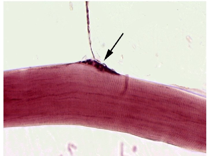
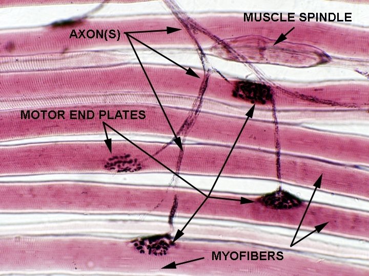
Neuromuscular Junction (NMJ) NMJ: site where axon and muscle fiber communicate motor neuron- stimulates muscle fiber to contract motor end plate- nuclei and mitochondria abundant; sarcolemma folded; directly under NMJ synaptic cleft- small space between neuron and muscle fiber synaptic vesicles- store neurotransmitters (chemical in
Muscle Fiber Twitch Stimulus applied Latent Period 1) action potential sweeps across the sarcolemma, T-tubules, opens Ca+2 gates in SR 2) Ca+2 ions released by SR (no tension yet), travel to troponin, pulling tropomyosin off binding sites on the actin molecules Contraction Period 3) cross bridges form myosin heads are interacting with active sites on the actin A single twitch will NOT create a contraction. Need a sustained contraction requires repeated stimulations (action potentials) Relaxation Period 4) Ca ions returned to SR 5) cross-bridges detach 6) sarcomere relaxes Refractory Period “rest” phase
Summation ~process by which individual twitches combine ~produces sustained contractions ~can lead to tetanic contractions (tetanus) (no relaxation period)
Types of Contractions Most muscular movements involve both isotonic and isometric contractions. ISOTONIC– muscle moves muscle contracts and changes length (attachment moves) ISOMETRIC – tensing a muscle contracts but does not change length (attachment does not move)
Fast and Slow Twitch Muscle Fibers Muscle fibers vary in contraction speed (slow or fast twitch) Slow-twitch fibers (red fibers)—long distance runners • resistant to fatigue, endurance muscles • tons of mitochondria (ATP) • most myoglobin • rich blood supply • AEROBIC Fast-twitch fibers (white fibers less myoglobin)-sprinters • contract rapidly, fatigue easily (lactic acid build up) • poorer blood supply • fewer mitochondria, but extensive SR (Ca store) • ANAEROBIC
Energy Sources for Contraction *The energy used to power the interaction between actin and myosin is ATP. *ATP must be regenerated constantly if contraction is to continue. Three ways to regenerate ATP: 1) Anaerobic cellular respiration Glycolysis Occurs in cytoplasm Makes pyruvic acid + 2 ATP 2) Aerobic cellular respiration Citric acid cycle and electron transport chain Oxygen required, occurs in mitochondria Makes carbon dioxide, water and 34 ATP 3) Coupled reaction with CP (creatine phosphate*) CP + ADP creatine + ATP *Muscle stores creatine phosphate
Muscle Fatigue • inability to contract • commonly caused from • decreased blood flow • ion imbalances • deficit of ATP • accumulation of lactic acid (soreness) (pyruvic acid lactic acid) • cramp – sustained, involuntary contraction Oxygen Debt The amount of oxygen needed by the liver to convert lactic acid to • glucose oxygen not available to muscle • glycolysis continues • pyruvic acid converted to lactic acid • liver converts lactic acid to glucose (requires oxygen…muscle doesn’t get O 2 (debt!))