Skeletal Muscle Cell Contraction Sliding Filament Mechanism The


 is stored in")
 occurs due")

 – The ATP(bound to myosin) breaks")
 – The myosin cross-bridges again attach")
 – When the Ach is no longer")
- Slides: 9
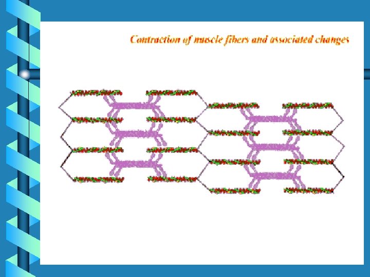
Skeletal Muscle Cell Contraction
Sliding Filament Mechanism • The sliding movement of filaments that cause muscle contraction. • Thin filaments slide inward due to actin an myosin connections
Sliding Filament Mechanism • Fiber at Rest – At rest, Ca+(calcium) is stored in the sarcoplamic reticulum. – At rest, ATP is bound to myosin – At rest; actin, toponin and tropomyosin are all present on thin filaments.
Sliding Filament Mechanism • Role of stimulus: – Action potential (on sarcolemma) occurs due to ACh binding at its receptor sites. – This action potential then travels across the muscle cell membrane down the T-tubules and across the sarcoplasmic reticulum (SR). – This initiates the diffusion of Ca+ out of the SR and into the sarcoplasma.
Sliding Filament Mechanism • Muscle Contraction: – The diffused Ca+ reaches the thin filaments and binds to the troponin. – The troponin and tropomyosin then move and expose the actin binding sites. – Myosin cross-bridges then attach to the exposed actin binding sites.
Sliding Filament Mechanism • Muscle Contraction (Cont. ) – The ATP(bound to myosin) breaks down to ADP, releasing energy that is used to move the cross-bridges inward. – New ATP attaches to myosin causing the cross-bridges to break and allowing it back to its original position.
Sliding Filament Mechanism • Muscle Contraction (Cont. ) – The myosin cross-bridges again attach to actin, ATP breaks down and moves the cross-bridges inward. – The cycle continues until the ACh is no longer present on the motor end plate.
Sliding Filament Mechanism • Muscle Contraction (End) – When the Ach is no longer present the action potential is stopped and the Ca+ is actively pumped back into the SR, the troponin & tropomyosin cover up the binding sites and the filaments return to the original position. • ATP is quickly regenerated by the mitochondria and reattached to the myosin.