Skeletal and smooth muscle cells are elongated muscle
")
")


 Blood vessel Fascicle (bundle of muscle fibers) Epimysium (wraps entire muscle)")


 exercise - (biking, jogging) - results in")


 •")


 Bare zone Thin filament")


- Slides: 46
• Skeletal and smooth muscle cells are elongated (muscle cell = muscle fiber) • Contraction of muscles is due to the shortening of microfilaments called myofibrils – Prefixes myo and mys refer to “muscle” – Prefix sarco refers to “flesh”
Sarcolemmaplasma membrane Myofibril Dark band Light band Nucleus Segment of a muscle fiber (cell)
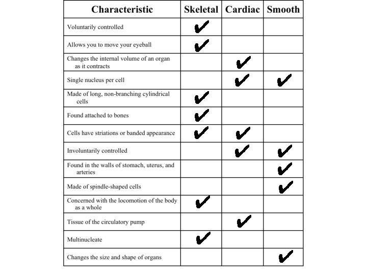
Skeletal Muscle Characteristics • • Attached by tendons to bones Multinucleate Striated—have visible banding Voluntary—subject to conscious control
Cardiac Muscle Characteristics • • • Branching cells Joined at intercalated discs Found only in the walls of the heart Striated w/single nucleus Involuntary
Smooth Muscle Characteristics • • • Lacks striations Spindle-shaped cells Single nucleus Involuntary—no conscious control Found mainly in the walls of hollow organs
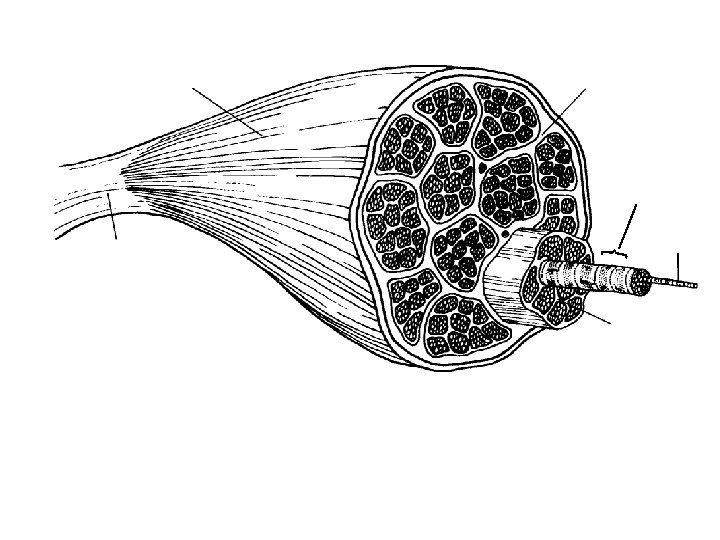
Muscle fiber (cell) Blood vessel Fascicle (bundle of muscle fibers) Epimysium (wraps entire muscle) Perimysium Endomysium (between fibers) Tendon Bone Figure 6. 1
Connective Tissue Wrappings of Skeletal Muscle • Cells are surrounded and bundled by connective tissue – Endomysium—encloses a single muscle fiber – Perimysium—wraps around a fascicle (bundle) of muscle fibers – Epimysium—covers the entire skeletal muscle
Skeletal Muscle Functions • • Produce movement Maintain posture Stabilize joints Generate heat
Contraction of Skeletal Muscle • Graded responses can be produced by changing: – The frequency of muscle stimulation – The number of muscle cells being stimulated at one time
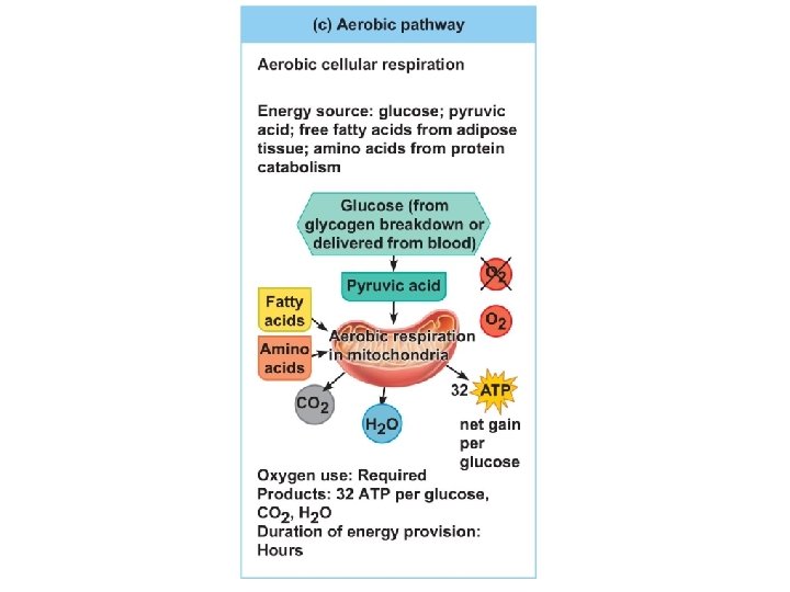
Energy for Muscle Contraction • Aerobic respiration – Uses O 2 to break down glucose (in mitochondria – Releases energy (about 32 ATP) – Slower reaction – CO 2 and H 2 O are produced
Energy for Muscle Contraction • Anaerobic respiration – Without O 2, occurs in cytosol – One glucose produces 2 ATP – Lactic acid is produced which causes muscle fatigue – Fast reaction but uses lots of glucose
Figure 6. 10 c
Muscle Fatigue and Oxygen Deficit • Muscle fatigue - muscle is unable to contract even when stimulated. – Caused by oxygen debt or deficit – Increasing lactic acid and lack of ATP causes the muscle to contract less
Types of Muscle Contractions • Isotonic contractions – The muscle contracts and movement occurs • Isometric contractions – Pushing against an unmovable object. – Muscles do not contract building tension in the muscles.
Effect of Exercise on Muscles Aerobic (endurance) exercise - (biking, jogging) - results in stronger, more flexible muscles with greater resistance to fatigue - More efficient body metabolism - Improves digestion, coordination Resistance (isometric) exercise - (weight lifting) - increases muscle size and strength - by increasing the size of the muscle fibers,
Figure 6. 11 a-b
Muscles and Body Movements • Muscles are attached to at least two points – Origin • Attachment to a immoveable bone – Insertion • Attachment to an movable bone
Immovable bone Muscle contracting Origin Relaxed muscle Movable bone Tendon Insertion Figure 6. 12
Naming Skeletal Muscles • By direction of muscle fibers – Example: Rectus (straight) • By relative size of the muscle – Example: Maximus (largest) • By location of the muscle – Example: Temporalis (temporal bone) • By number of origins – Example: Triceps (three heads)
Naming Skeletal Muscles • By location of the muscle’s origin and insertion – Example: Sterno (on the sternum) • By shape of the muscle – Example: Deltoid (triangular) • By action of the muscle – Example: Flexor and extensor (flexes or extends a bone)
Types of Body Movements • Flexion – Decreases the angle of the joint and brings two bones closer together • Extension – Increases angle between two bones – Extension beyond 180° is hyperextension
Figure 6. 13 a
Figure 6. 13 b
Types of Body Movements • Rotation – Movement of a bone around its longitudinal axis
Figure 6. 13 c
Types of Body Movements • Abduction – Movement of a limb away from the midline • Adduction – Movement of a limb toward the midline
Figure 6. 13 d
Types of Body Movements • Circumduction – Common in ball-and-socket joints
Figure 6. 13 d
Special Movements • Dorsiflexion – Lifting the foot so that the superior surface approaches the shin (toward the dorsum) • Plantar flexion – Depressing the foot (pointing the toes) – “Planting” the foot toward the sole
Figure 6. 13 e
Special Movements • Inversion – Turn sole of foot medially • Eversion – Turn sole of foot laterally
Figure 6. 13 f
Special Movements • Supination – Forearm rotates laterally so palm faces anteriorly – Radius and ulna are parallel • Pronation – Forearm rotates medially so palm faces posteriorly – Radius and ulna cross each other like an X
Figure 6. 13 g
Special Movements • Opposition – Move thumb to touch the tips of other fingers on the same hand
Figure 6. 13 h
Muscle Tone • Some fibers are contracted even in a relaxed muscle • Different fibers contract at different times to provide muscle tone and to be constantly ready
Microscopic Anatomy of Skeletal Muscle • Sarcomere—contractile unit of a muscle fiber • Organization of the sarcomere – Myofilaments • Thick filaments = myosin filaments • Thin filaments = actin filaments
Thick filament Myofilament structure (within one sarcomere) Bare zone Thin filament
Stimulation and Contraction of Single Skeletal Muscle Cells • Excitability (also called responsiveness or irritability)—ability to receive and respond to a stimulus • Contractility—ability to shorten when an adequate stimulus is received • Extensibility—ability of muscle cells to be stretched • Elasticity—ability to recoil and resume resting length after stretching
Axon terminals at neuromuscular junctions Spinal cord Motor unit 1 unit 2 Nerve Motor neuron cell bodies Muscle (a) Axon of motor neuron Muscle fibers Figure 6. 4 a
Synaptic vesicle containing ACh 1 Action potential reaches axon terminal of motor neuron. Axon terminal of motor neuron Mitochondrion 2 Calcium (Ca 2+) channels open and Ca 2+ enters the axon terminal. 3 Ca 2+ entry causes some synaptic vesicles to release their contents (acetylcholine, a neurotransmitter) by exocytosis. Ca 2+ Synaptic cleft Ca 2+ ACh receptor Sarcolemma Fusing synaptic vesicle Sarcoplasm of muscle fiber Folds of sarcolemma 4 Acetylcholine diffuses across the synaptic cleft and binds to receptors in the sarcolemma. Figure 6. 5, step 4