SINUS AND FISTULA Dr Varun S Dept of


 (or)")
a pipe (or)a tube.")

following surgery : eg. , intestinal fistulas (faecal, biliary, pancreatic) (B)following")


 fistula Presence of a foreign body. e. g.")
 endothelisation of the track. e. g. , AVF Malignancy. Dense fibrosis")


 actinomycosis. 3. Opening:")

Temperature and tenderness: b)Discharge: after application of pressure over the surrounding area. c)")

fistula. • Water")


 Non cryptoglandular (10%cases) TB Diabetes mellitus Crohn’s disease Carcinoma rectum")

 • Inter sphincteric (45%) •")


 • Pain (which increases until temporary relief")


 Fistulectomy Seton techniques Fibrin glue Anal fistula plug Advancement flap")


 and also for posterior horse-shoe shaped fistulas. • Excision")


- Slides: 70
SINUS AND FISTULA Dr. Varun. S Dept. of Surgery SKHMC
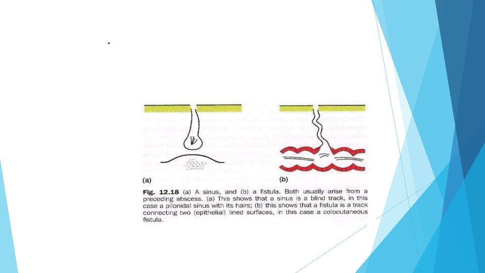
DEFINITION SINUS: Blind track lined by granulation tissue leading from epithelial surface down into the tissues. Latin: Hollow (or) a bay
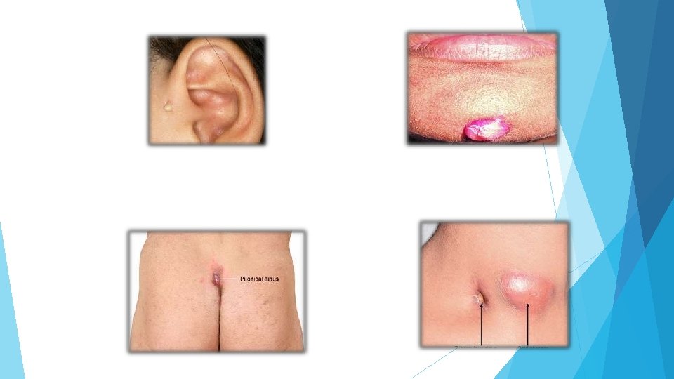
CAUSES CONGENITAL ACQUIRED Preauricular sinus TBsinus Pilonidal sinus Median mental sinus Actinomycosis
FISTULA: ABNORMAL communication between lumen of one viscus and lumen of another (INTERNALFISTULA) (or) between lumen of one hollow viscus to the exterior (EXTERNAL FISTULA) (or) between any two vessels
Latin : flute (or)a pipe (or)a tube.
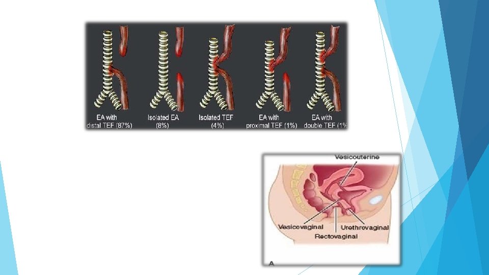
CAUSES CONGENITAL ACQUIRED Branchial fistula I. Traumatic Tracheo-esophageal II. Inflammatory Umbilical III. Malignancy Congenital AV fistula IV. Iatrogenic Thyroglossal fistula
ACQUIRED I. TRAUMATIC: (A)following surgery : eg. , intestinal fistulas (faecal, biliary, pancreatic) (B)following instrumental delivery (or) difficult labour e. g. , vesicovaginal, rectovaginal, ureterovaginal fistula
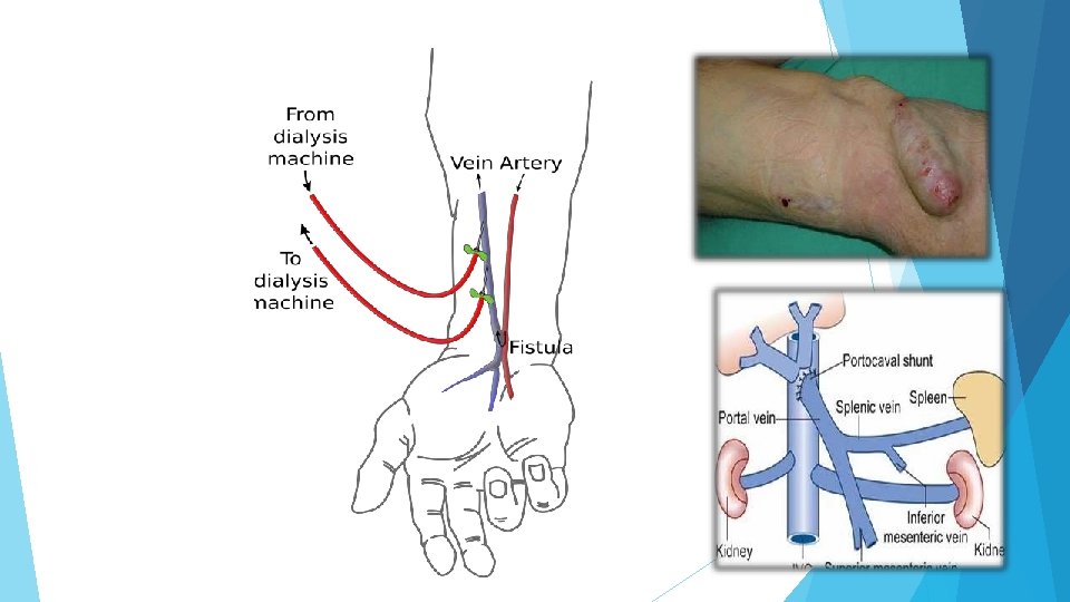
II. INFLAMMATORY: Intestinal actinomycosis, TB III. MALIGNANCY: when growth of one organ penetrates into the nearby organ. e. g. , Rectovesical fistula in carcinoma rectum IV. IATROGENIC: Cimino fistula- AVF for hemodialysis ECK fistula- to treat esophageal varices in portal HTN
FISTULA EXTERNAL INTERNAL Orocutaneous Tracheo-esophageal Colovesical Enterocutaneous Rectovesical Appendicular AVF Thyroglossal Cholecystoduodenal Branchial
Causes for persistence of sinus (or) fistula Presence of a foreign body. e. g. , suture material Presence of necrotic tissue underneath. e. g. , sequestrum Insufficient (or) non-dependent drainage. e. g. , TBsinus Distal obstruction. e. g. , faecal (or) biliary fistula Persistent drainage like urine/faeces/CSF Lack of rest [contd. ]
Epithelialisation (or) endothelisation of the track. e. g. , AVF Malignancy. Dense fibrosis Irradiation Malnutrition Specific causes. e. g. , TB, actinomycosis Ischemia Drugs. e. g. , steroids Interference by the patient
PATHOPHYSIOLOGY CONGENITAL: Arise from remnants of embryonic ducts that persist instead of being obliterated and disappearing completely during embryonic development. e. g. , pre-auricular sinus, branchial fistula, TOF, congenital AVF.
ACQUIRED : Usually secondary to presence of foreign body, necrotic tissue in affected area (or) microbial infection (or) following inadequate drainage of abscess. e. g. , perianal abscess when bursts spontaneously into skin forming a sinus and when bursts into both skin and anal canal forming a fistula.
CLINICAL FEATURES Usually asymptomatic but when infected manifest as- • Recurrent/ persistent discharge. • Pain. • Constitutional symptoms if any deep seated origin.
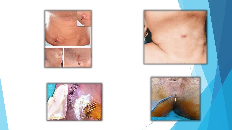
CLINICAL EXAMINATION INSPECTION: 1. Location: usually gives diagnosis in most of the cases. SINUS: pre-auricular- root of helix of ear. median mental- symphysis menti. TB- neck. FISTULA: branchial- sternomastoid ant border. parotid- parotid region thyroglossal- midline of neck below hyoid.
2. Number: usually single but multiple seen in HIV patients (or) actinomycosis. 3. Opening: a) sprouting with granulation tissue-foreign body. b) flushing with skin- TB 4. Surrounding area: erythematous- inflammatory bluish- TB excoriated- faecal pigmented- chronic sinus/fistulae.
5. Discharge: White thin caseous, cheesy like- TBsinus Faecal- faecal fistula Yellow sulphur granules- actinomycosis Bony granules- osteomyelitis Yellow purulent- staph. infections Thin mucous like- brachial fistula Saliva- parotid fistula
Palpation: a)Temperature and tenderness: b)Discharge: after application of pressure over the surrounding area. c) Induration: present in chronic fistulae/sinus as in actinomycosis, OM TBSinus induration absent. d) Fixity: e) Palpation at deeper plane: lymph nodes- TB Thickening of bone underneath- OM
INVESTIGATIONS CBP- Hb, TLC, DLC, ESR. Discharge for C/S , AFB, cytology, Gram staining. X-RAY of the part to rule out OM, foreign body. X-RAY KUBand USG abdomen in cases of lumbar fistula to rule out staghorn calculi. MRI BIOPSYfrom edge of sinus CT Sinusogram
FISTULOGRAPHY/ SINUSOGRAPHY: • For knowing the exact extent/origin of sinus (or)fistula. • Water soluble or ultrafluid lipoidal iodine dye is used. • Lipoidal iodine is poppy seed oil containing 40% iodine.
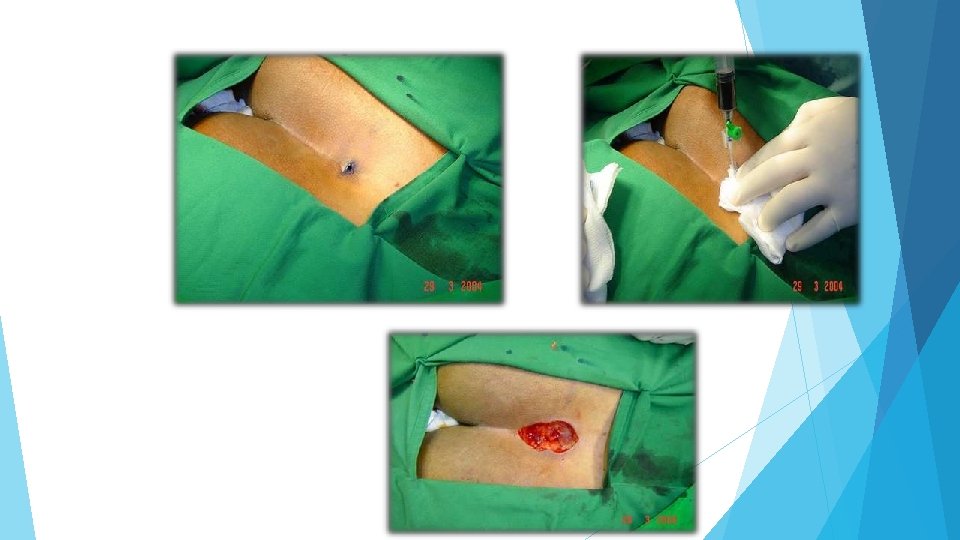
TREATMENT BASIC PRINCIPLES: Antibiotics Adequate rest Adequate excision Adequate drainage.
After excision specimen SHOULD be sent for HPE. Treating the cause. e. g. , ATTfor TBsinus. removal of any foreign body. sequestrectomy for OM.
TUBERCULAR SINUS OF NECK Causative organism: mostly M. tuberculosis but also M. bovis Site and mode of infection: a) lymph nodes in anterior triangle from tonsils. b) lymph nodes in posterior triangle from adenoids. c) supraclavicular nodes from apex of the lung.
Clinical stages:
Stage of lymphadentis: non-tender, discrete, mobile, firm lymph nodes Stage of peridenitis: due to involvement of capsule. non-tender, MATTED, mobile together, firm pathognomic of TB
Stage of cold abscess: due to caseating necrosis. non-tender, cystic, fluctuant swelling not adherent to overlying skin. Sternocleidomastoid contraction testpresent deep to deep fascia trans illumination negative
TREATMENT: Zig-zag aspiration by wide bore needle in nondependent area to avoid a persistent sinus. Instillation of 1 g streptomycin +/-INHin solution with closure of wound without placing a drain. ATT NOTE: I&Dnot done-persistent TBsinus.
Stage of collar stud abscess: cold abscess ruptures through deep fascia forming an another swelling in sub-cutaneous plane. Fluctuant, adherent to skin. Treated like a cold abscess.
Collar stud abscess
Stage of sinus: collar stud abscess bursts out leading to a persistent discharging sinus. Can be multiple, wide opening, undermined edges, non-mobile. Bluish discoloration around the edges. NO INDURATION.
• INVESTIGATIO NS Hematocrit, ESR , S. albumin , S. globulin • FNAC of lymph nodes and smear for AFB and C/S • Open node biopsy of lymph nodes. • Edge biopsy of sinus- granuloma. • mantoux test • Chest X ray • Sputum for AFB
Sometimes, USG neck to detect cold abscess. Hypoechoeic lesions with internal echoes S/O debris within. Guided aspiration of cold abscess.
ATT TREATME NT Excision of sinus tract with excision of diseased lymph nodes.
FISTULA-IN-ANO Chronic abnormal communication usually lined to some degree by granulation tissue, which runs outwards from anorectal lumen (internal opening) to skin of perineum or the buttocks (external opening)
AETIOPATHOGENE SIS Cryptoglandular (90%cases) Non cryptoglandular (10%cases) TB Diabetes mellitus Crohn’s disease Carcinoma rectum Trauma Lymphogranuloma venereum Radiotherapy Immunocompromised patients (HIVetc. , )
CRYPTOGLANDULAR HYPOTHESIS
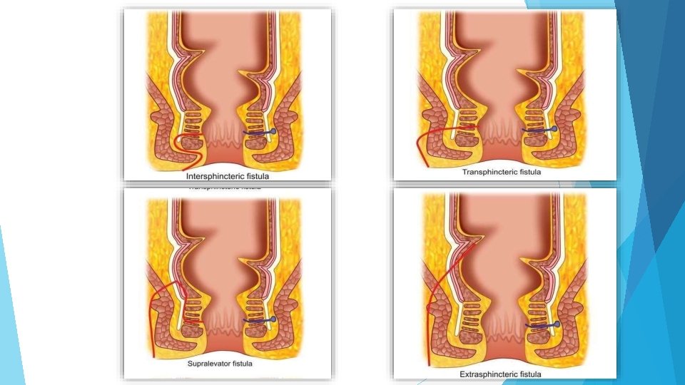
CLASSIFICATION PARK’SCLASSIFICATION: (relation of primary tract to external sphincter) • Inter sphincteric (45%) • Trans sphincteric (40%) • Supra sphincteric • Extra sphincteric
STANDARD CLASSIFICATION Sub cutaneous Sub mucous Low anal High anal Pelvi rectal
Can be low level fistula- open into anal canal below the internal ring. high level fistula- at/ above the internal ring. Can be Simple- without any extensions Complex- with extensions Can be single multiple- TB, ulcerative colitis, crohn’s, HIV, LGV
CLINICAL PRESENTATION • Intermittent discharge (sero-purulent/ bloody) • Pain (which increases until temporary relief occurs when pus discharges) • Pruritus ani • Previous h/o anal gland infection
CLINICAL ASSESMENT HISTORY: full medical history incl. obstetric, anal, gastrointestinal, surgical, continence DRE: area of induration, fibrous tract and internal opening may be felt (“button-hole” defect in Ca rectum) PROCTOSIGMOIDOSCOPY: To evaluate rectal mucosa for any underlying disease process.
GOODSALL’S RULE • Ifexternal opening in anterior half of anus, fistula usually runs directly into anal canal. • Ifexternal opening in posterior half of anus, fistula usually curves midline of the anal canal posteriorly.
IMAGING Fistulography Endoanal ultrasound MRI
Fistulography: Reveals primary and secondary tracts. Useful if extra sphincteric fistula suspected.
END 0 ANAL ULTRASOUND • Determines sphincter integrity. • Complexity of fistula. horse-shoe fistula
MRI “GOLD STANDARD” for fistula-in-ano imaging. high variety supra sphincteric fistula. horse-shoe fistula.
MRI Abscesses and extensions contralateral disease
PRINCIPLES OF TREATMENT Control sepsis EUA Laying open abscesses and secondary tracts Adequate drainage –seton insertion Define anatomy • Openings and tracts Internal and External Single –v- multiple Extensions /Horseshoe • Relation to sphincter complex High –v- Low Exclude co-existent disease
SURGICAL MANAGEMENT Fistulotomy(Thelaying opentechnique) Fistulectomy Seton techniques Fibrin glue Anal fistula plug Advancement flap LIFTprocedure. sphincter preserving techniques.
FISTULOTOMY Ininter-sphincteric and low trans-sphincteric fistulas. Identification of tract with probe followed by division of all structures between external and internal openings. Secondary tracts laid open. +/- marsupialization.
Advantages least chance of recurrence relatively easy procedure minor degree of incontinence. Risks results in large and deep wounds that might take months to heal.
FISTULECTOMY • All chronic (low) and also for posterior horse-shoe shaped fistulas. • Excision of entire fibrous tissue and tract and wound kept open. • Sphincter repair +/-advancement flap. • High anal fistulas +/-colostomy.
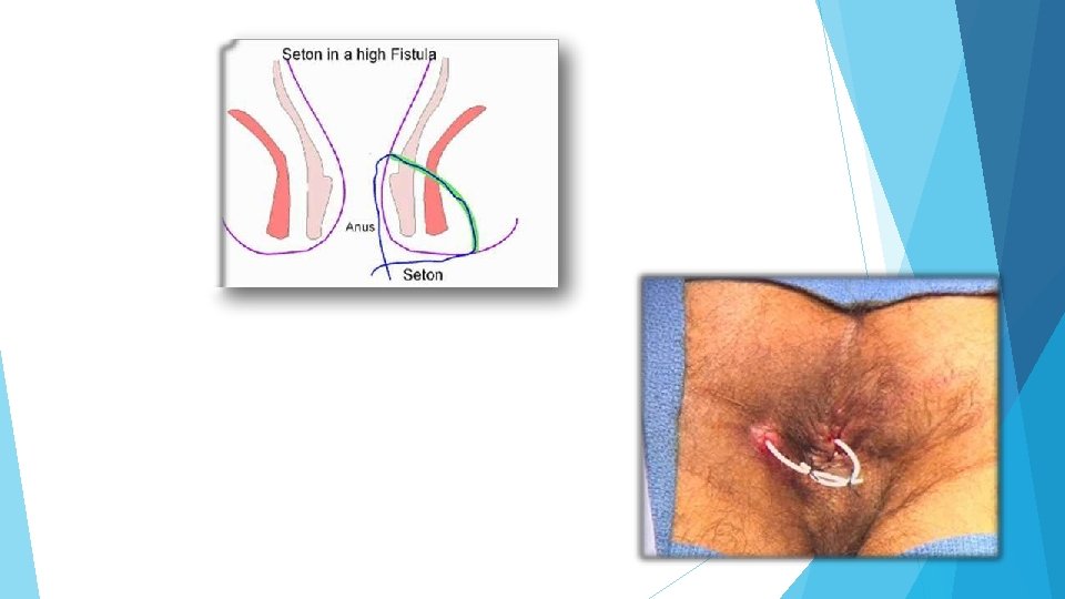
SETON SUTURE PLACEMENT • Preferable surgical option for high variety. • Setons are usually made from rubber slings • 2 types of seton suture can be placed • Draining Seton Facilitates draining of sepsis Left loose and allows fistula to heal by fibrosis • Cutting Seto Slowly "cheese-wires" though the sphincter muscle Allows fibrosis to take place behind as it gradually cuts through
FIBRIN GLUE Multi component system containing mainly human plasma fibrinogen and thrombin. Injected into fistula track which hardens in few minutes and fills the track.
ANAL FISTULA PLUG The Anal fistula plug is a minimally invasive and sphincter-preserving alternative to traditional fistula surgery. The plug is a conical device and is placed by drawing it through the fistula tract and suturing it in place. the plug, once implanted, incorporates naturally over time into the human tissue (human cells and tissues will 'grow' into the plug), thus facilitating the closure of the fistula.
FISTULA PLUG
FISTULA PLUG:
ADVANCEMENT FLAPS Endorectal Fistula tract probed Flap raised • Mucosa +Int. Sphincter Internal opening excised/closed Flap advanced & sutured
ADVANCEMENT FLAP Anodermal Fistula tract probed Flap raised • Anodermal Flap advanced & sutures External defect closed
LIFT PROCEDURE Ligation of Inter sphincteric Fistula Tract Trans sphincteric fistula Draining seton – 6 weeks Tract prepared with fistula brush Debrides De-epithelializes
FOLLOW UP As with most anorectal disorders, follow-up care includes: Perianal baths, analgesics for pain, stool bulking agents, and good perianal hygiene