Single slice CT generations 11 Electron Beam CT
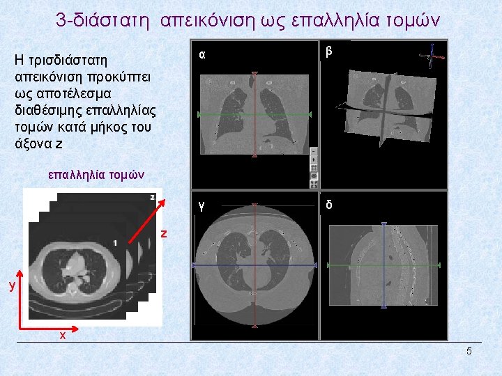

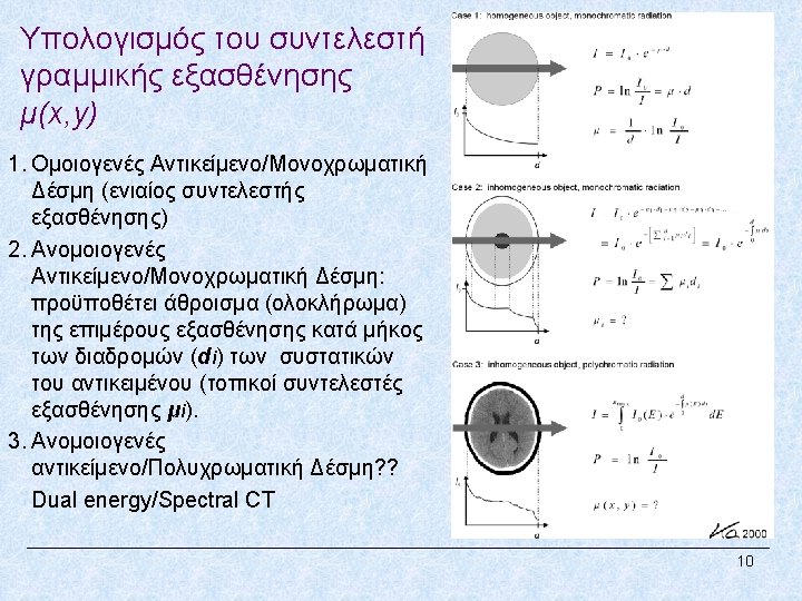

Single slice CT generations 11
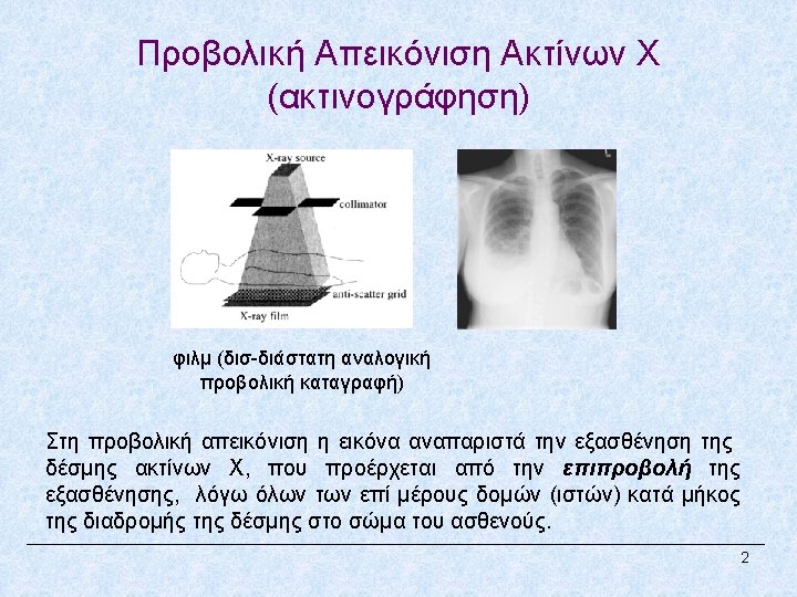
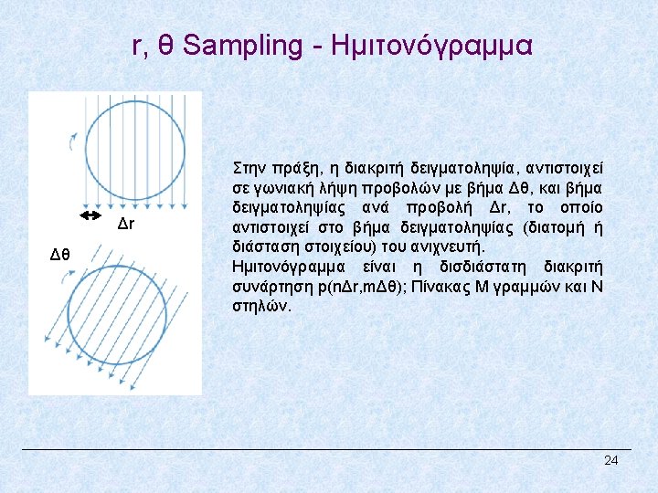
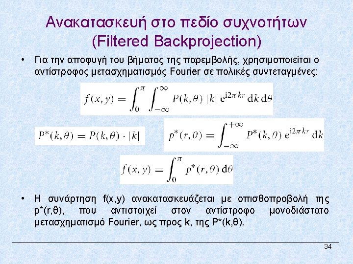
 An electron beam is swept across a semicircular")
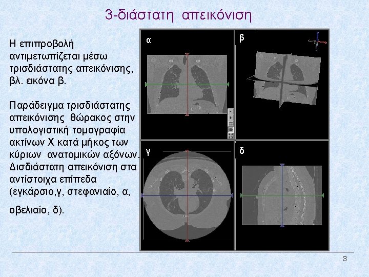
Electron Beam CT (5 th generation) An electron beam is swept across a semicircular anode, ring which encloses the patient. It permits single scans in the range of 30 to 100 ms without mechanical motion. 12

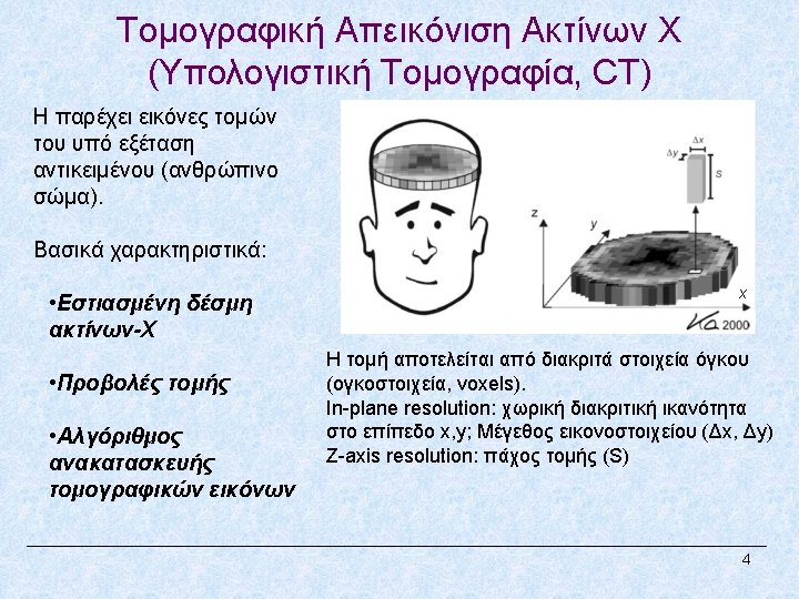
Single Detector vs. Multi-detector CT Single slice CT D: beam collimation/pitch d: detector row collimation/pitch 4 slice MDCT d: detector row collimation N: #detector rows 13

Sequential vs. Hellical Data Acquisition CT Sequence-axial mode: Circular motion of the tube for a specific z axis location. To increase the coverage of the z-axis the table has to be linearly moved at a new position for a new acquisition (step and shoot) y z x Hellical mode: Comination of the circular motion of the tube in the x, y plane with linear motion of the patient table at the same time, resulting in inceased coverage with important time savings (continuous acquisition mode) 14

Concepts of Hellical-MDCT Pitch, p, of a helical scan: the ratio of the table translating distance per helical gantry rotation (s) to the thickness of the x-ray beam. Single slice CT MDCT 15
.")
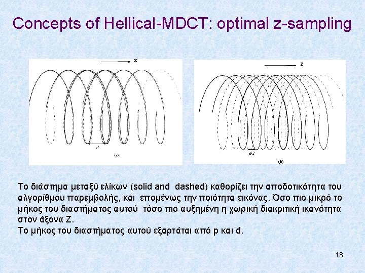
Concepts of Hellical-MDCT: Z-axis resolution Helical z sampling pattern: single slice CT (solid line). Helical z sampling pattern: single slice CT, exploiting 180 ° projection measurements periodicity (dashed line). Z-axis resolution: s and s/2 respectively. Helical z sampling pattern: 4 -slice MDCT (solid lines). Helical z sampling pattern: 4 -slice CT, exploiting 180 ° projection measurements periodicity (dashed lines). β: rotation angle, z: sampling positions 16

Concepts of Hellical-MDCT: Z-axis resolution Gantry angle β 0 when table location is at z 0 d: the gap of the solid helix, s=pd for the solid helix and s/2=p/2 x d, p: pitch n: detector index (1, …, N: #detector rows) 17
 Uniform size of detector elements. Non-uniform size of")
Uniform vs. Variable detector size (sampling) Uniform size of detector elements. Non-uniform size of detector elements, enables efficient slice thicknesses, by combining detector signals. 19

Multidetrector row CT: “slice race” 20

CT image reconstruction 21

 f(x, y)= 1 2 3 4 αρχική 0 0 0 0 8")
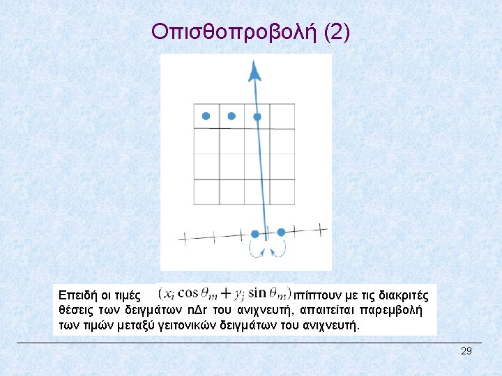
Οπισθοπροβολή (Παράδειγμα) f(x, y)= 1 2 3 4 αρχική 0 0 0 0 8 14 16 17 back-project (4, 6) at 00 4 6 13 16 19 22 back-project (1, 5, 4) at 450 αρχικοποίηση 5 11 9 10 back-project (3, 7) at 900 back-project (3, 5, 2) at 1350 τελική 30
 f(x, y)= 1 2 3 4 αρχική 0 0 0 0")
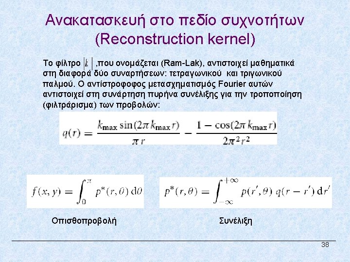
Φιλτραρισμένη οπισθοπροβολή (Παράδειγμα) f(x, y)= 1 2 3 4 αρχική 0 0 0 0 -2 17/2 23/2 7 αρχικοποίηση -3/2 9 6 3/2 back-project (-3/2, -1/2, 11/2, -7/2) at 900 back-project ( -2, 1, 4, -3) at 0 0 back-project -5/2, 5, -5/2) 1350 ( at 1 4 3 6 9 12 back-project -5/2, 5, -5/2) 450 τελική 41 ( at
 scale is a linear transformation of the")
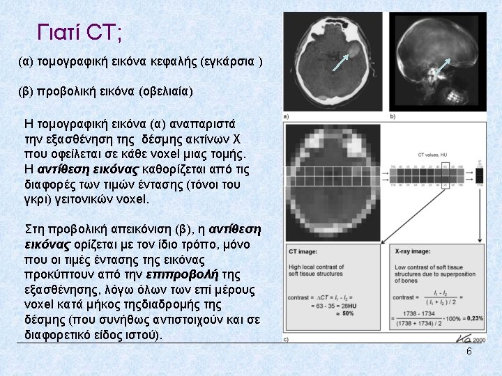
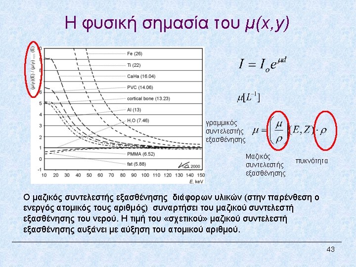
Η κλίμακα Hounsfιeld The Hounsfield Unit (HU) scale is a linear transformation of the linear attenuation coefficient measurement into one in which the radiodensity of distilled water (at standard pressure and temperature) is defined as zero HU, while the radiodensity of air at STP is defined as -1000 HU. For a material X with linear attenuation coefficient μX, the HU value is therefore given by: Where μwater and μair are the linear attenuation coefficients of water and air, respectively. 44

Προ-επεξεργασία: Αλγόριθμοι τμηματοποίησης πνευμονικών πεδίων και αγγειακού δέντρου P. Korfiatis, C. Kalogeropoulou, A. Karahaliou, A. Kazantzi, S. Skiadopoulos, L. Costaridou, “Texture classification-based segmentation of lung affected by interstitial pneumonia in high-resolution CT”, Medical Physics; 2008 35(12): 5290 -5302. P. Korfiatis, C. Kalogeropoulou, A. Karahaliou, A. Kazantzi, L. Costaridou, “Vessel tree segmentation in presence of interstitial lung disease in MDCT”, IEEE Transactions on Information Technology in Biomedicine; 2011 15(2): 214 -220. 48
")
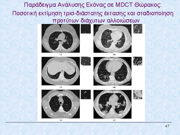
Ταξινόμηση του πνευμονικού παρεγχύματος σε φυσιολογικό και 2 πρώτυπα παθολογίας (Interstitial Lung Disease, ILD) P. Korfiatis, A. Karahaliou, C. Kalogeropoulou, A. Kazantzi, L. Costaridou, “Texture Based Identification and Characterization of Interstitial Pneumonia Patterns in Lung Multidetector CT”, IEEE Trans. Inform Tech. Biomed. ; 2010 14(3): 675680. Kazantzi L, Costaridou L, Skiadopoulos S, Korfiatis P, Karahaliou A Daoussis D, Andonopoulos A, Kalogeropoulou C. Automated 3 D Ιnterstitial Lung Disease Εxtent Quantification: Performance Evaluation and Correlation to PFTs J Digit Imaging 2014 DOI 10. 1007/s 10278013 -9670 -z 49
, 1999;")
Βιβλιογραφία 1. Hui Hu. Multi-slice helical CT: Scan and Reconstruction. Med. Phys. 26(1), 1999; 5 -18. 2. Willi A. Kallender. Chapter 1: Principles of Computed Tomography. Publicis MDC Verlag, 2000, Munich, Germany. 3. David J. Goodenough, Chapter 8: Tomographic Imaging, In Handbook of Medical Imaging Vol. 1. Physics and Psychophysics, Society of Photo-Optical Instrumentation Engineers, SPIE, Press, 2000, Bellingham, Washington, USA. 4. Mahadevappa Mahesh, The AAPM/RSNA Physics Tutorial for Residents: Search for Isotropic Resolution in CT from Conventional through Multiple-Row Detector. Radio. Graphics 2002; 22: 949– 962 5. Dianna D. Cody, Mahadevappa Mahesh, AAPM/RSNA Physics Tutorial for Residents: Technologic Advances in Multidetector CT with Focus on Cardiac Imaging. Radio. Graphics 2007; 27: 1829– 1837. 6. Flohr et al, Multi–Detector Row CT Systems and Image-Reconstruction Techniques. Radiology 2005; 235: 756– 773. 7. Paul Suetens. Chapter 3: X-ray Computed Tomography. Fundamentals of Medical Imaging. Cambridge University Press, second ed. 2009, New York, USA. 8. Willi A Kalender, X-ray computed tomography (Review). Phys. Med. Biol. 51 (2006) R 29–R 43. 9. Philippe Cattin, Computed Tomography: Principles of Medical Imaging, 2013. 50
- Slides: 50