Single bipolar nuclei Cytologic Findings Cellularity low Nucleus


Single bipolar nuclei


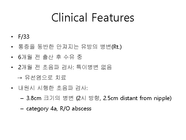
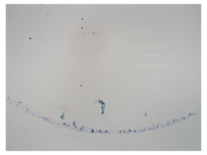
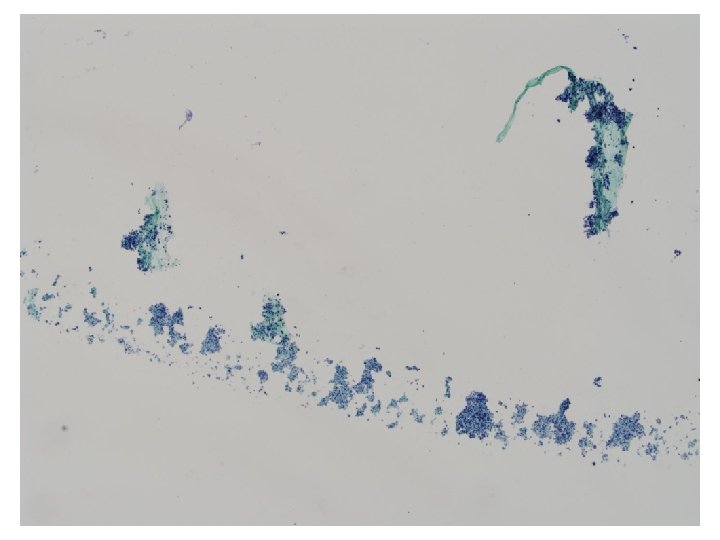
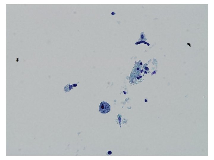
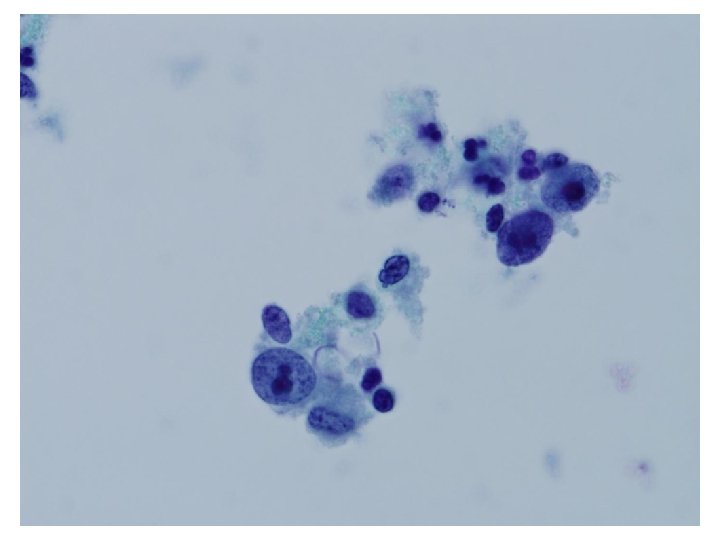
Cytologic Findings • • • • Cellularity ; low Nucleus ; large, no hyperchromasia, smooth nuclear membrane Nuclear chromatin ; fine Inflammatory cell infiltrates; neutrophils and lymphocytes Presence of bipolar cells (myoepithelial cell) Arrangement ; loose clusters Background ; presence of degenerated cell debris Cytoplasm ; rich, granular, fragile Presence of atypical bare nuclei Cell shape ; polygonal, ill-defined Cell size ; large Nucleoli ; prominent macronucleoli A few mitoses

Differential Diagnosis Benign Malignant • Abscess • Apocrine Ca • Lactating adenoma • Medullary Ca • Apocrine adenosis • Inavsive ductal Ca

Pregnancy-related Changes • Cellular aspirates • Single or dispersed cells • Lipid rich foamy or granular background • Large size of cells and nuclei • Abundant vacuolated or wispy cytoplasm • Round and uniform nuclei • Granular or vesicular, but evenly distributed chromatin • Single prominent nucleoli • Bare nuclei are common

Breast Cancer in Pregnancy and Lactation • The histological spectrum is not significantly different from breast carcinoma unrelated to pregnancy. (ref; Rosen’s breast pathology 3 rd edition, p. 721)

Conventional Breast Adenocarcinoma • High cellularity • Syncytial tissue fragments and loosely cohesive groups • Branching and anastomosing or acinar, nests, cords • Marked variation of cell size and shape • Nucleus – – – high N/C ratio smooth to irregular nuclear membrane fine to coarse, granular chromatin Single/multiple, micro/macronucleoli Frequent mitoses in poorly differentiated tumors (Ref; color atlas of differential diagnosis in exfoliative and aspiration cytopathology 2 nd edition p. 698)

Medullary Carcinoma • • • Moderate to highly cellular Loose cohesive groups & singly dispersed cells Moderate to highly pleomorphic cells Dense lymphomononuclear cells in background Nuclear irregularity & prominent nucleoli Malignant bare nuclei

Apocrine Carcinoma • Highly Cellular • Abnormal apocrine cells in loose cohesive clusters • Pleomorphic nuclei with prominent nucleoli • Intracytoplasmic lumen with secretion • Irregularity in nuclear outline • Foam cells, mitotic figures, background mucin, and necrosis were rare

Apocrine Adenosis • • • Frequently cellular Small & compact cell clusters Prominent & large nucleoli Variation of nuclear size Less hyperchromatic nuclear features Naked nuclei in the background < Watanabe K. et al. Diagn Cytopathol 2007; 35: 296 >

Lactating adenoma • Celluar smear • Dispersed and poorly cohesive clusters • Fragile, frayed granular to foamy to vacuolated cytoplasm • Mildly enlarged, well-dispersed hyperchromatic nuclei with prominent nucleoli • Numerous stripped epithelial nuclei and few to no bipolar naked nuclei • Dirty background of cytoplasmic fragments and secretory materials

Abscess • Neutrophils and foamy macrophages with evidence of cytophagocytosis • Abundant cell debris in background • Atypical epithelial cells –Nuclear enlargement –Prominent nucleoli –N/C ratio is normal

Differential Diagnosis Apocrine carcinoma Apocrine adenosis Lactating adenoma Abscess KCP-746 Cellularity high varible high variable Low Cytoplasm Rich, secretion Granular, vacuolated rich granular Nucleus hyperchomati c, nuclear membrane irregularity Large, less hyperchomatic, smooth nuclear membrane Large, smooth nuclear membrane large, smooth nuclear membrane Chromatin fine to coarse fine Nucleoli prominent Prominent Mitosis rare A few Bipolar bare absent nuclei present rare present Inflammation absent present absent
 • Breast, Right, FNA : Atypia, favor abscess Note : The")
Diagnosis (KCP 746) • Breast, Right, FNA : Atypia, favor abscess Note : The cytologic findings disclose the possibility of abscess. But malignant tumor can not be completely excluded. The core needle biopsy is recommended.

- Slides: 24