Signal transduction by Dr Heba M Kareem Cell

Signal transduction by Dr / Heba M. Kareem

Cell to Cell Communication Can occur in both eukaryotes and prokaryotes, mostly through chemical signals to trigger specific reactions in the cell May cause growth, the production of specific molecules, and other actions (like muscle contraction)
 Synthesis and release of signaling molecules")
Signal transduction - general steps • • 1) Synthesis and release of signaling molecules 2) Transport of signal to target cell 3) Detection of signal by a specific receptor protein 4) Intracellular events, often mediated by second messengers, that change metabolism or gene expression • 5)Termination of the signal and response Reception: Chemical signal binds to the receptor molecule (on cell surface or inside cell) Transduction: Reaction or series of reactions (cascade) leading to the final response Response: The specific cellular response

Requirement of Biosignaling ØThe transduction system must meet four specific criteria.

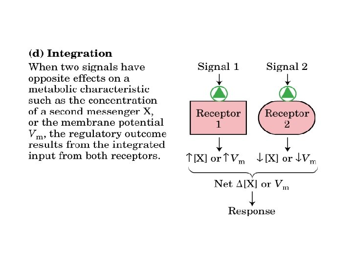
often short-lived & low concentration feedback control

, that forms a complex")
Ligands A Ligand is a substance (usually a small molecule), that forms a complex with a biomolecule by intermolecular forces (such as ionic bonds, hydrogen bonds and van der Waals forces) to serve a biological purpose. The tendency or strength of binding is called affinity and the association is usually reversible. A ligand that can bind to a receptor, alter the function of the receptor and trigger a physiological response is called an agonist for that receptor. Ligands that bind to a receptor but fail to activate the physiological response are receptor antagonists.

Receptors are protein molecules, embedded in either the plasma membrane or the cytoplasm or nucleus of a cell, to which one or more specific kinds of signalling molecules may attach. Each type of receptor recognizes and binds only certain ligand shapes (in analogy to a lock and key where the lock represents the receptor and the key, its ligand). Classification of receptors based on their location 1 - Intracellular receptors: found in the cytoplasm or inside nucleus and function in the nucleus as transcription factors to alter the rate of transcription of particular genes. Ligands: Steroid hormones and thyroid hormones (hydrophobic)

2 - Plasma-membrane receptors: Cell surface receptors are specialized integral membrane proteins that take part in communication between the cell and the outside world. The binding of extracellular molecules to the receptors initiates a chemical change on the intracellular side of the membrane. eg: Catecholamine hormones are hydrophilic and thus interact with cell surface receptors and transmit their signals through second messengers that are generated intracellularly Four main types of extracellular receptors 1 -Ion-channel receptors 2 -G protein-linked receptors 3 -Receptors associated with tyrosine kinases 4 -Receptors with intrinsic enzymatic activity
 which binds the hormone.")
Structure of cell surface receptors: 1 - Extracellular domain (N-terminal) which binds the hormone. 2 - Transmembrane domain: One or more than one membrane spanning region that are usually α-helices. 3 - Intracellular domain (C-terminal) which initiate the intracellular signaling cascade (Signal transduction). • Mechanisms of signal transduction that follow the binding of signaling molecules to plasma membrane receptors include: 1 - Phosphorylation of receptors at tyrosine residues (receptor tyrosine kinase activity), 2 - Conformational changes in signal transducer proteins (e. g. , proteins with SH 2 domains, heterotrimeric G proteins) 3 - Increases in the levels of intracellular second messengers Examples: c. AMP, inositol trisphosphate (IP 3), and diacylglycerol (DAG).
 - G proteins bind GTP (guanosine triphosphate), they control and")
G-protein Coupled Receptors (GPCRs) - G proteins bind GTP (guanosine triphosphate), they control and amplify intracellular signaling pathways Exist in two states 1) bound GTP: active 2) bound GDP: inactive • The extracellular domain connects to the intracellular domain through seven transmembrane spans • The intracellular domain is coupled to a heterotrimeric G-protein • The heterotrimeric G-protein is composed of 3 subunits: G , and G • When the G subunit is bound to GDP it is “OFF”; when it is bound to GTP it is “ON” • The conformational change relayed to the intracellular domain causes the G subunit to release GDP and bind to GTP thereby activating both the G and G /G subunits
 binds GTP, & can hydrolyze it to")
The subunit of a G-protein (G ) binds GTP, & can hydrolyze it to GDP + Pi. Adenylate Cyclase (AC) is a transmembrane protein, with cytosolic domains forming the catalytic site.
")
Signal Amplification (Cascade)
 • Protein kinases are enzymes that add a phosphate group")
Receptor Tyrosine Kinases (RTKs) • Protein kinases are enzymes that add a phosphate group from ATP onto a substrate protein; this reaction is called phosphorylation. RTKs are the high-affinity cell surface receptors for many polypeptide growth factors, and hormones This reversibility contributes to the dynamic nature of cells RTKs need to form dimers in the plasma membrane; the dimer is stabilized by ligands binding to the receptor. The interaction between the cytoplasmic domains stimulates the autophosphorylation of tyrosines within the domains of the RTKs, causing conformational changes.
")
Receptor Tyrosine Kinase steps involved: 1 -Ligand Reception 2 -Receptor Dimerization 3 -Catalysis (Phosphorylation) 4 -Subsequent Protein Activation 5 -Further Transduction 6 -Response

Ion-Channel Receptor are a group of transmembrane ion channels that are opened or closed in response to the binding of a chemical messenger A ligand-gated ion channel, upon binding with a ligand, changes conformation to open a channel in the cell membrane through which ions relaying signals can pass Ca 2+ acts as a second messenger initiating signal transduction cascades and altering the physiology of the responding cell. Reversibility is assured by pumping ions back out again (using separate protein)

Intracellular receptors They are receptors located inside the cell rather than on its cell membrane. The typical ligands for nuclear receptors are usually intracellular second messengers like inositol trisphosphate (IP 3), and lipophilic hormones like the steroid hormones testosterone and progesterone and derivatives of vitamins A and D To initiate signal transduction, the ligand must pass through the plasma membrane. On binding with the receptor, the ligands pass through the nuclear membrane into the nucleus, enabling gene transcription and protein production.

Second messengers Small molecules or ions that occur in the cytoplasm of a cell, and they are generated in response to a hormone binding to a cell-surface receptor, and activates various kinases that regulate the activities of other enzymes But in addition to their job as relay molecules, second messengers serve to greatly amplify the strength of the signal. There are three major classes of second messengers: 1. cyclic nucleotides (e. g. c. AMP) 2. inositol trisphosphate (IP 3) and diacylglycerol (DAG) 3. calcium ions (Ca 2+ ) The effect of c. AMP on cellular metabolism • Increased [c. AMP] increases glycogen breakdown • Increased [c. AMP] increases phosphorylase activity (via a complex cascade) • Phosphorylase (or glycogen phosphorylase) is a key enzyme involved in glycogen breakdown
 and diacylglycerol (DAG) 1 -IP 3 opens channels to release")
Inositol trisphosphate (IP 3) and diacylglycerol (DAG) 1 -IP 3 opens channels to release calcium ions from intracellular stores - IP 3 is able to increase [Ca 2+] by associating with the IP 3 channel or IP 3 receptor; - At least three molecules of IP 3 must bind to sites on the cytosolic side of the membrane protein to open the channel and release Ca 2+. - Increase [Ca 2+] activates protein kinase C (PKC) - IP 3 is a short-lived messenger (less than a few seconds) 2 -Diacylglycerol activates protein kinase C (PKC) - PKC phosphorylates Ser or Thr residues of specific target proteins, changing their catalytic activities; PKC is a family of protein kinase enzymes that are involved in controlling the function of other proteins through the phosphorylation of hydroxyl groups of serine and threonine amino acid residues on these proteins.

Ca 2+ as a second messenger
- Slides: 23