Signal Transduction BL 4010 12 07 06 Outline

Signal Transduction BL 4010 12. 07. 06

Outline • Extracellular signals • Signal-Transducing Receptors – single membrane spanning receptors – 7 TMS receptors • Intracellular Second Messengers – c. AMP – Calcium – G-proteins • Enzyme cascades

Additional resources • http: //www. signaling-gateway. org/ • http: //web. indstate. edu/thcme/mwking/signaltransduction. html • http: //stke. sciencemag. org/

Extracellular Signals • Light • Small molecules – hormones – toxins – metabolites • Large molecules – oligosaccharides – proteins

Transcriptional activation

Interferon

The phosphorelay system

Phosphorelay system
 Endocrine signals are directed")
Page 658 Figure 19 -1 a Classification of hormones. (a) Endocrine signals are directed at distant cells through the intermediacy of the bloodstream.
 Paracrine signals are directed")
Page 658 Figure 19 -1 b Classification of hormones. (b) Paracrine signals are directed at nearby cells.
 Autocrine signals are directed")
Page 658 Figure 19 -1 c Classification of hormones. (c) Autocrine signals are directed at the cell that produced them.

Classes of Hormones
 A hyperbolic")
Page 660 Figure 19 -3 a Binding of ligand to receptor. (a) A hyperbolic plot.
 A Scatchard")
Page 660 Figure 19 -3 b Binding of ligand to receptor. (b) A Scatchard plot.

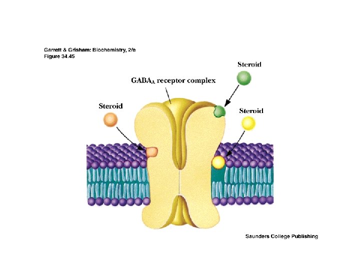
Classes of Hormones - Steroid Hormones • Derived from cholesterol- (e. g. Glucocorticoids, vitamin D, sex hormones) – regulate metabolism, salt/water balances, inflammation, sexual function. May bind to PM receptor or enter nucleus directly • May either act at nucleus or at plasma membrane • Steroids are hydrophobic and cannot diffuse freely to nucleus • Receptor proteins carry steroids to the nucleus • Steroid receptor proteins are all apparently members of a gene superfamily and have evolved from a common ancestral precursor

Classes of Hormones - Steroid Hormones
")
Classes of Hormones - Nonsteroid hormones • Amino Acid Derived Hormones (e. g. epinephrine) bind to PM receptors – regulate smooth muscle , blood pressure, cardiac rate, lipolysis, glycogenolysis

Effects of epinephrine Receptor Alpha 1 Alpha 2 Beta 1 Beta 2 • • Molecule Epinephrine, Norepinphrine Epinephrine Effect Increased Ca 2+ Decreased c. AMP Increased rate and force of contraction of heart muscle: predominantly an effect of epinephrine on beta receptors. Constriction of blood vessels: increased blood pressure. Dilation of bronchioles: assists in pulmonary ventilation. Stimulation of lipolysis in fat cells: provides fatty acids for energy production and conserves dwindling blood glucose. Increased metabolic rate Inhibition of certain "non-essential" processes: e. g. inhibition of gastrointestinal secretion and motor activity. Dilation of the pupils: particularly important in situations where you are surrounded by velociraptors under conditions of low ambient light
 - bind")
Classes of Hormones - Nonsteroid hormones • Peptide Hormones (e. g. insulin) - bind to PM receptors – regulate many processes in all tissues - including release of other hormones – All secreted polypeptide hormones are synthesized with a signal sequence (which directs them to secretory granules) – Usually synthesized as inactive preprohormones ("pre-pro" implies at least two precessing steps) – Proteolytic processing produces the prohormone and the hormone

Insulin is a peptide hormone

Secretion of insulin

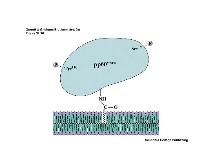
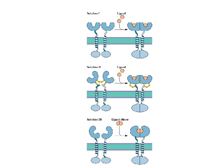
Single TMS Receptors • What is a receptor? • • Three main classes Extracellular domain to interact with hormone Single transmembrane segment Intracellular domain with enzyme activity Activity is usually tyrosine kinase or guanylyl cyclase Each of these has a "nonreceptor" counterpart src gene kinase - pp 60 v-src was first known Two posttranslational modifications
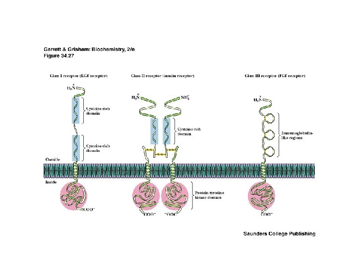

Receptor Tyrosine Kinases Membrane-associated allosteric enzymes • How do single-TMS receptors transmit the signal from outside to inside? ? • Oligomeric association is the key! • Extracellular ligand binding

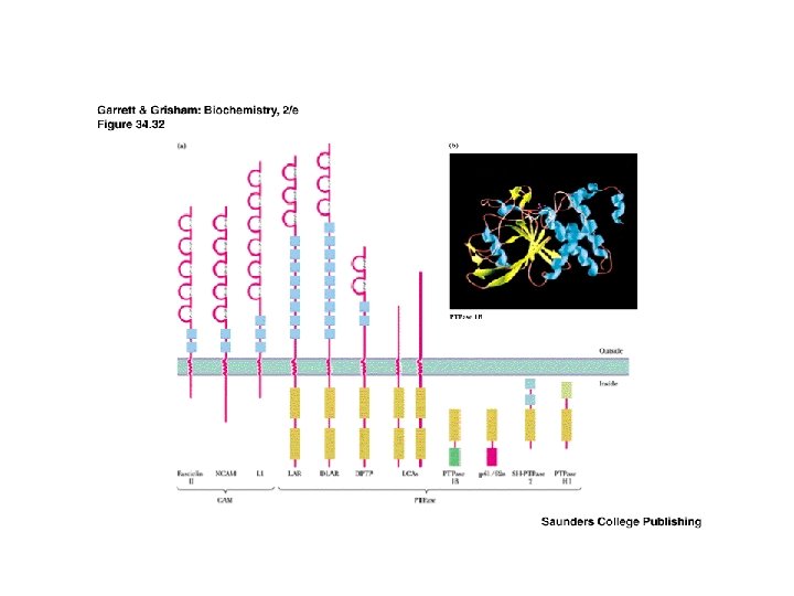
Protein-Tyrosine Phosphatases • • The enzymes that dephosphorylate Tyr Some PTPases are integral membrane proteins But there also lots of soluble PTPases Cytoplasmic PTPases have N-term. catalytic domains and Cterminal regulatory domains Membrane PTPases all have cytoplasmic catalytic domain, single transmembrane segment and an extracellular recognition site

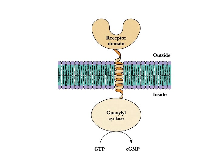
Guanylyl Cyclases • • Soluble or Membrane-Bound Membrane-bound GCs are the other group of singletransmembrane-segment receptors (besides RTKs) Peptide hormones activate the membrane-forms Note speract and resact, from mammalian ova Activation may involve oligomerization of receptors, as for RTKs

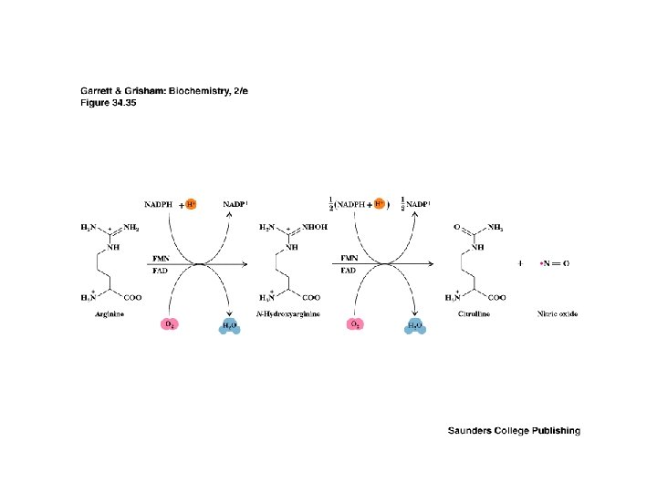
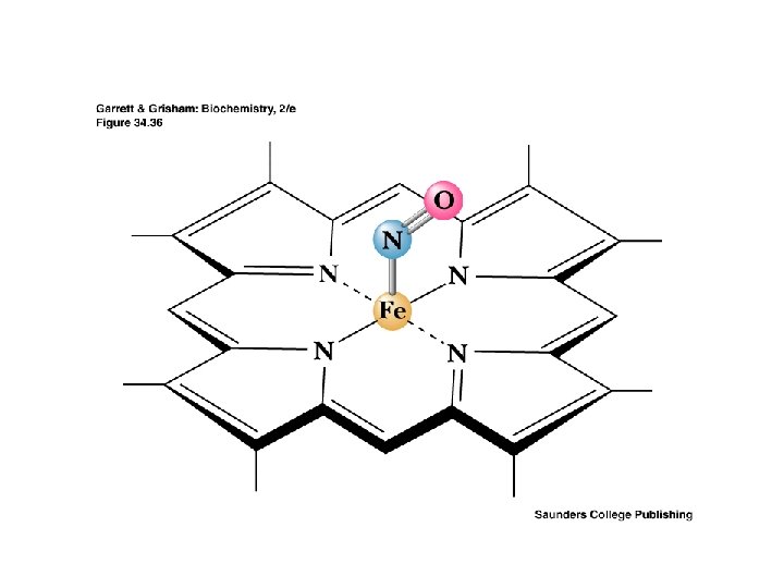
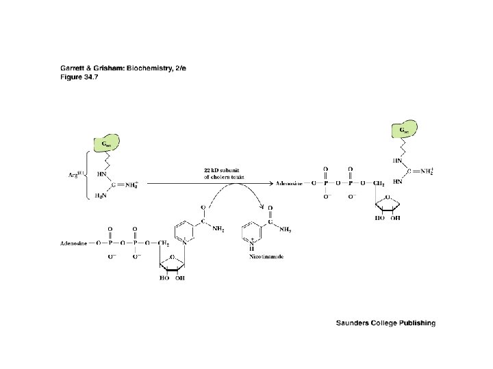
Soluble Guanylyl Cyclases • • • Receptors for Nitric Oxide NO is a reactive, free-radical that acts either as a neurotransmitter or as a second messenger NO relaxes vascular smooth muscle (and is thus involved in stimulation of penile erection) NO also stimulates macrophages to kill tumor cells and bacteria NO binds to heme of GC, stimulating GC activity 50 -fold Read about NO synthesis and also see box on Alfred Nobel
 – extracellular site for")
Types of Receptors • 7 -TMS receptors (G protein receptors) – extracellular site for hormone (ligand) – intracellular site for GTP-binding protein • Single-transmembrane segment receptors – extracellular site for hormone (ligand) – intracellular catalytic domain - e. g. kinase or guanylyl cyclase • Oligomeric ion channels

Second Messengers • • Many and there may be more! The hormone is the "first messenger" The second messenger - Ca 2+, c. AMP or other - is released when the hormone binds to its (extracellular) receptor The second messenger then activates (or inhibits) processes in the cytoplasm or nucleus Degradation and/or clearance of the second messenger is also (obviously) important

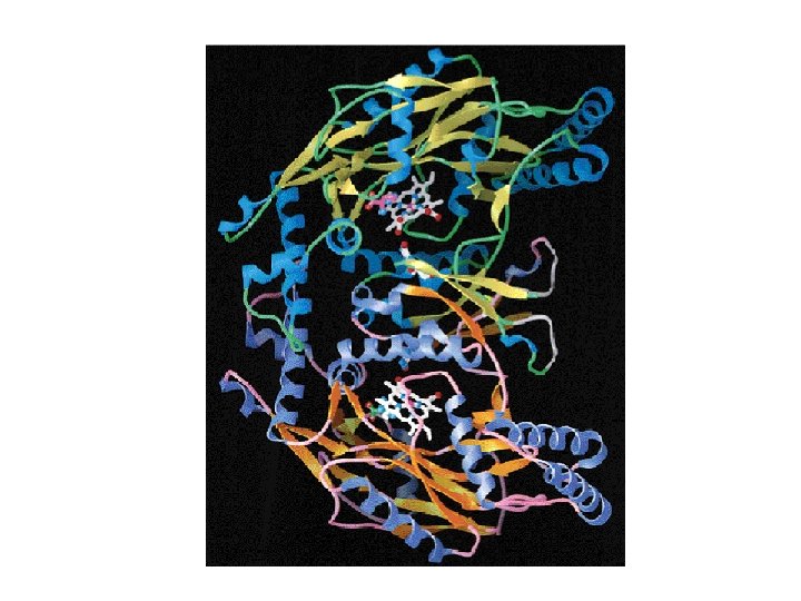
Steroid Receptor Proteins • Hydrophobic domain near C-terminus that interacts with steroid itself • Central, hydrophilic domain that binds to DNA • Central DNA-binding domains are homologous to one another, with 9 conserved Cys residues • Three pairs of Cys residues are in Cys-X-X-Cys sequences - as in Zinc-finger domains • Steroid-receptor complex may bind to DNA or to transcription factors • Thyroid hormone receptor proteins are similar

Steroid Receptor Proteins

Steroid Receptor Proteins

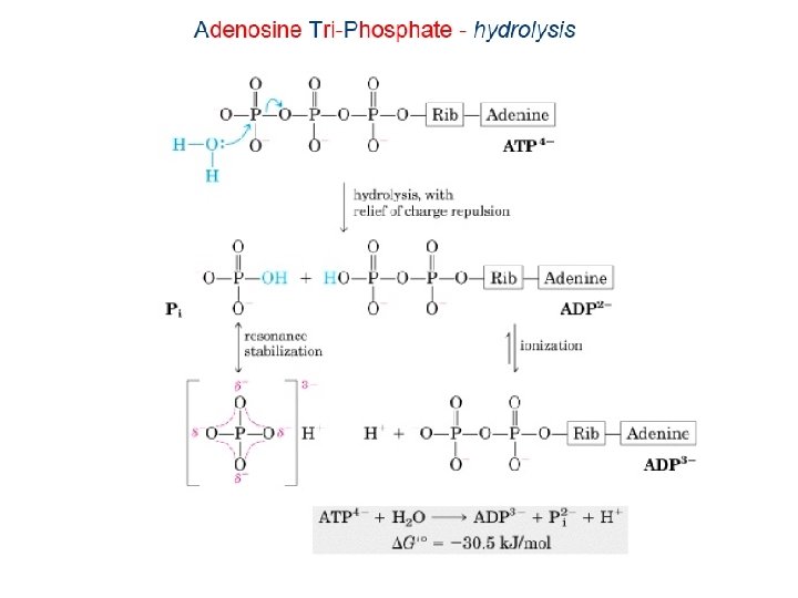
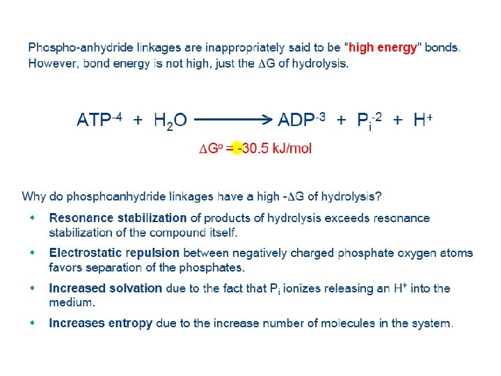
c. AMP and Glycogen Phosphorylase Earl Sutherland discovers the first second messenger • In the early 1960 s, Earl Sutherland showed that the stimulation of glycogen phosphorylase by epinephrine involved cyclic adenosine-3', 5'-monophosphate • He called c. AMP a "second messenger" • c. AMP is synthesized by adenylyl cyclase and degraded by phosphodiesterase


How are the hormone receptor and AC coupled? • Purified AC and purified receptor, when recombined, are not coupled. • Rodbell showed that GTP is required for hormonal activation of AC • In 1977, Elliott Ross and Alfred Gilman at Univ. of Virginia discovered a GTP-binding protein which restored hormone stimulation to AC • Hormone stimulates receptor, which activates GTP-binding protein, which activates AC

Heterotrimeric G Proteins A model for their activity • Binding of hormone, etc. , to receptor protein in the membrane triggers dissociation of GDP and binding of GTP to -subunit of G protein • G -GTP complex dissociates from G and migrates to effector sites, activating or inhibiting • But it is now clear that G also functions as a signalling device

Page 674 Figure 19 -13 Activation/deactivation cycle for hormonally stimulated AC.
 A partial list Potassium channel proteins Phospholipase A 2")
Signalling Roles for G( ) A partial list Potassium channel proteins Phospholipase A 2 Yeast mating protein kinase Ste 20 Adenylyl cyclase Phospholipase C Calcium channels • • Receptor kinases

Stimulatory and Inhibitory G G proteins may either stimulate or inhibit an effector. • In the case of adenylyl cyclase, the stimulatory G protein is known as Gs and the inhibitory G protein is known as Gi • Gi may act either by the Gi subunit binding to AC or by the Gi complexing all the Gi and preventing it from binding to AC
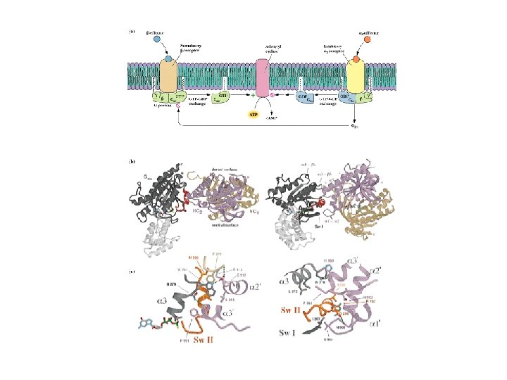

Page 676 Figure 19 -16 Mechanism of receptormediated activation/ inhibition of AC.

The ras Gene and p 21 ras • • An oncogene and its product a gene first found in rat sarcoma virus Normal cellular ras protein activates cellular processes when GTP is bound and is inactive when GTP has been hydrolyzed to GDP Mutant (oncogenic) forms of ras have severely impaired GTPase activity, so remain active for long periods, stimulating excessive growth and metabolic activity - causing tumors to form

G-protein coupled receptors • • • Receptors that interact with G proteins Seven putative alpha-helical transmembrane segments Extracellular domain interacts with hormone Intracellular domain interacts with G proteins Adrenergic receptors are typical Note desensitization by phosphorylationby protein kinase A
- Slides: 55