SHOULDER COMPLEX DR PURVI PATEL Objectives Student at

SHOULDER COMPLEX DR PURVI PATEL

Objectives Student at the end of session should be able to: � Enumerate joints of shoulder complex � Describe the osteokinematics and arthrokinematics � Explain scapulohumeral rhythm � Explain muscle function at the shoulder joint

� Shoulder complex consists of clavicle, scapula, and humerus � These three bones are joined by Sternoclavicular (SC) joint, Acromioclavicular (AC) joint, & Glenohumeral (GH) joint � Articulation between scapula & thorax is called as the scapulothoracic (ST) joint

scapulothoracic joint � ST “joint” is formed by articulation of scapula with thorax is not a true anatomic joint because it has none of usual joint characteristics (union by fibrous, cartilaginous, or synovial tissues) � It

Resting Position of Scapula � Normally, scapula rests at a position on posterior thorax approximately 2 inches from midline, between 2 nd through 7 th ribs � Scapula also is internally rotated 300 to 450 from coronal plane, is tipped anteriorly approximately 100 to 200 from vertical, & is upwardly rotated 100 to 200 from vertical

Motions of scapula from this resting position include 3 rotations: � upward/downward rotation, internal/external rotation, & anterior/posterior tipping � Translatory motions of scapula are elevation/depression & protraction/retraction

Motions of Scapula Upward/Downward Rotation � Plays a significant role in increasing range of elevation of arm � Motion: upward & downward rotation is upward & downward movement of glenoid fossa, respectively � Range: thorax 600 of upward rotation of scapula on
 or downward (caudally) along rib cage from")
Elevation/Depression � Motion: scapula moves upward (cephalad) or downward (caudally) along rib cage from its resting position Protraction/Retraction � Motion: Protraction & retraction of scapula on thorax are described as motion of scapula away from or towards vertebral column, respectively

Internal/External Rotation � IR of scapula on thorax results in prominence of vertebral border of scapula as a result of loss of contact with thorax � Often referred to clinically as SCAPULAR “WINGING” Anterior/Posterior Tipping � Anterior tipping occurs at AC joint & results in prominence of inferior angle of scapula
 Joint")
Sternoclavicular (SC) Joint

joint is a plane synovial joint with 3 rotatory & 3 translatory degrees of freedom � SC � It has synovial capsule, joint disk & 3 major ligaments � SC articulation consists of 2 saddle-shaped surfaces, which makes it incongruent � So, it has a fibrocartilage disk that increases congruence between joint surfaces

Sternoclavicular Motions � 3 rotatory degrees of freedom at SC joint are elevation/depression, protraction/retraction, & anterior/posterior rotation of clavicle � These motions should be visualized by referencing movement of lateral end of clavicle � Clavicular anterior/posterior rotation are long axis rolling motions of entire clavicle
 axis � MOTION: With elevation,")
Elevation & Depression of Clavicle � AXIS: Anteroposterior (A-P) axis � MOTION: With elevation, lateral clavicle rotates upward, & with depression, lateral clavicle rotates downward

Elevation & Depression of Clavicle
 Axis � MOTION: With protraction,")
Protraction & Retraction of Clavicle � AXIS: Vertical (superoinferior) Axis � MOTION: With protraction, lateral clavicle rotates anteriorly, & with retraction, lateral clavicle rotates posteriorly

Protraction & Retraction of Clavicle

Anterior & Posterior Rotation of Clavicle � Anterior/posterior occurs as a spin rotation of clavicle � MOTION: clavicle rotates posteriorly from neutral, bringing inferior surface of clavicle to face anteriorly � Also known as backward or upward rotation

Sternoclavicular ligaments �sternoclavicular �costoclavicular �interclavicular ligament

� Sternoclavicular ligament functions primarily to check anterior & posterior translatory movement of medial end of clavicle � Costo-clavicular ligament is found between clavicle & 1 st rib. It checks elevation of lateral end of clavicle � Interclavicular ligament resists excessive depression of distal clavicle & superior glide of medial end of clavicle

Acromioclavicular Joint is a plane synovial joint with 3 degrees of freedom � It has a joint capsule & 2 major ligaments � Primary function of AC joint is to allow scapula additional range of rotation on thorax

Acromioclavicular Joint ligaments � Acromioclavicular & coracoclavicular ligaments are the 2 ligaments � Coracoclavicular ligament, is divided into a lateral portion, trapezoid ligament & medial portion, conoid ligament � Both portions of coracoclavicular ligament limit upward rotation of scapula at AC joint

Acromioclavicular Motions � Primary rotatory motions that take place at AC joint are � internal/external rotation � anterior/posterior tipping or tilting � upward/downward rotation

Internal & External Rotation � AXIS: vertical axis � MOTION: Internal & external rotation at AC joint brings glenoid fossa of scapula anteromedially & posterolaterally, respectively

Anterior & Posterior Tipping � AXIS: oblique “coronal” axis � MOTION: Anterior tipping will result in acromion tipping forward & inferior angle tipping backward � Posterior tipping will rotate acromion backward & inferior angle forward

Upward/Downward Rotation � AXIS: oblique “A-P” axis � MOTION: Upward rotation tilts glenoid fossa upward & downward rotation is opposite motion

GLENOHUMERAL JOINT � GH joint is a ball-and-socket synovial joint with 3 degrees of freedom. It has a capsule, several ligaments and bursae � Articulation is composed of large head of humerus & smaller glenoid fossa � Humeral head faces medially, superiorly, and posteriorly with regard to its shaft & condyles � An axis through humeral head & neck in relation to a longitudinal axis through shaft of humerus forms an angle of 1300 to 1500 in frontal plane. It is known as angle of inclination of humerus

� In transverse plane, axis through humeral head & neck and axis through humeral condyles forms an angle which is usually 300 posteriorly � This angle is known as angle of torsion. The normal posterior position of humeral head with regard to humeral condyles may be termed posterior torsion, or retrotorsion, of humerus

� GH joint is surrounded by a large, loose capsule that is taut superiorly and slack inferiorly in resting position � When humerus is abducted & laterally rotated the capsule tightens, making abduction & lateral rotation the close-packed position for GH joint � Capsule is reinforced by the superior, middle, and inferior GH ligaments, as well as by coracohumeral ligament

Functions of ligaments of glenohumeral joint � Superior GH ligament contributes most to anterior & inferior stability when arm is at side (00 abduction) � Middle GH ligament contributes primarily to anterior stability with arm is at the side & up to 450 of abduction � With abduction beyond 450 or with combined abduction and rotation, IGHLC prevents inferior humeral head translations
 arch is formed by coracoid process, acromion,")
THE CORACOACROMIAL ARCH � Coracoacromial (or suprahumeral) arch is formed by coracoid process, acromion, & coracoacromial ligament that connects the two bones

BURSAE �Because of confined nature and closeness of structures in subacromial space, several bursae are present �Presence of bursae indicate possibility of frictional forces between structures �Most important are subacromial & subdeltoid bursae. These bursae separate supraspinatus tendon & head of humerus from acromion, coracoid process, coracoacromial ligament, & deltoid muscle

Glenohumeral Motions OSTEOKINEMATICS � GH joint has 3 degrees of freedom: flexion/extension, abduction/adduction, and medial/lateral rotation � Flexion & extension occur about a coronal axis & sagittal plane � Medial & lateral rotation occur about a vertical axis & horizontal plane � Abduction/adduction of GH joint occur around a sagittal axis & frontal plane
")
� Elevation in the plane of scapula (300 to 450 anterior to frontal plane) is called scapular abduction, or scaption

ARTHROKINEMATICS � According to convex-concave theory of joint motion with motions of humerus (physiological motions) convex head rolls in same direction & slides in opposite direction in glenoid fossa � Eg: With flexion, humeral head slides posteriorly & with abduction head slides inferiorly

Static Stabilization of the Glenohumeral Joint in the Dependent Arm With arm is hanging at the side, gravity acts on humerus parallel to shaft in a downward direction 1. But passive tension in structures of rotator interval capsule (superior capsule, superior GH ligament, and coracohumeral ligament) results in compressing the humeral head into the lower portion of the glenoid fossa 2. Capsule has an airtight seal, which produces negative intra-articular pressure. This pressure creates a vacuum that resists inferior humeral translation because of gravity
 Degree of glenoid inclination influences stability of GH joint. If there is a")
3) Degree of glenoid inclination influences stability of GH joint. If there is a slight upward tilt of glenoid fossa either anatomically in structure of scapula or through scapular upward rotation, tilt of the fossa will produce a partial bony block against humeral inferior translation When passive forces are inadequate for static stabilization, (eg. in heavily loaded arm, activity of supraspinatus is recruited

Dynamic Stabilization of the Glenohumeral Joint Deltoid and Glenohumeral Stabilization � The deltoid muscle is a prime mover (along with the supraspinatus) for GH abduction � The anterior deltoid is also considered the prime mover in GH flexion

� Large superiorly directed force of deltoid, if unopposed, would cause humeral head to impact coracoacromial arch before much abduction had occurred. � Inferior pull of gravity cannot offset the Fx component of deltoid, that is, deltoid cannot independently abduct (elevate) arm. Another force is required.

Action line of deltoid

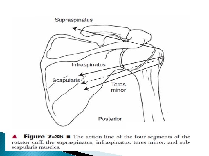
Rotator Cuff and Glenohumeral Stabilization � Supraspinatus, infraspinatus, teres minor, & subscapularis muscles make up rotator or musculotendinous cuff � Also known as SITS muscles

Action line of SITS MUSCLES

Long Head of the Biceps Brachii and Glenohumeral Stabilization � Biceps muscle is capable of contributing to the force of flexion & can, if humerus is laterally rotated, contribute to the force of abduction and anterior stabilization

Muscles of elevation Deltoid � Deltoid is an effective primary mover for flexion & abduction & anterior deltoid is the prime mover for flexion

Supraspinatus � Supraspinatus humerus � When is considered an abductor of deltoid is paralyzed, supraspinatus alone can bring arm through most if not all of the GH range, but motion will be weaker

� Secondary functions of supraspinatus are to compress GH joint & to assist in maintaining stability of dependent arm � With isolated & complete paralysis of supraspinatus, some loss of abduction force is evident, but most of its functions can be performed by remaining muscles

Infraspinatus, Teres Minor, & Subscapularis � Infraspinatus & Teres minor are external rotators of GH joint & subscapularis is an internal rotator � Lateral rotation is required during abduction to clear the greater tuberosity from under the coracoacromial arch. The medial rotatory function of the subscapularis muscle decreases with abduction.

Muscles of Depression Latissimus Dorsi Function �When UE is free to move in space, it may produce adduction, extension, or medial rotation of humerus �When hand &/or forearm is fixed in weight-bearing, latissimus dorsi muscle will pull its inferior attachment on pelvis towards its superior attachment on scapula & humerus

This results in lifting the body up as in a seated pushup. During crutch walking, a contraction of latissimus dorsi will unweight feet as trunk rises beneath the fixed scapula, allowing legs to swing forward through crutches

� Teres major muscle, like latissimus dorsi, adducts, medially rotates, and extends humerus Rhomboid Muscle Function � Rhomboid major and minor muscles are active in elevation of arm, especially in abduction � These muscles serve function as stabilizing synergists to muscles that upwardly rotate scapula

Upward Rotators of Scapula � Upward rotation of scapula is produced by a balance between trapezius & serratus anterior muscles through their attachments on clavicle & scapula � Upper portion of trapezius contributes directly to initial elevation of clavicle

� Serratus anterior contributes to combined clavicular & scapular motion � It plays a primary role in stabilizing scapula to thorax � Paralysis of serratus anterior muscle is classically characterized by scapular “winging”

Scapulothoracic Stability of scapula on thorax is provided by muscles that attach to both thorax & scapula & maintain contact between these surfaces by pulling or compressing scapula to thorax � Stability

� Ultimate functions of scapular motion are to orient glenoid fossa for ◦ optimal contact with moving arm ◦ to add range to elevation of arm ◦ to provide a stable base for controlled motions between humeral head & glenoid fossa

Integrated Function of Shoulder Complex/ Scapulohumeral rhythm Scapulothoracic & Glenohumeral Contributions contributes to elevation (flexion & abduction) of humerus by upwardly rotating glenoid fossa 500 to 600 from its resting position � Scapula

� GH joint contributes 1000 to 1200 of flexion & 900 to 1200 of abduction

� Combination of scapular & humeral movement results in a maximum range of elevation of 1500 to 1800

� Overall ratio of 20 of GH to 10 of ST motion during arm elevation & simultaneous GH & ST motion is called as “SCAPULOHUMERAL RHYTHM” � Ratio has variable consideration among individuals but is commonly accepted to be 2: 1 (20 of GH motion to 10 of scapular rotation) overall motion

� During setting phase (00 to 300 � During mid-range of humeral motion, abduction, 00 to 600 flexion), motion is primarily at GH joint, whereas, scapula seeks a stable position scapula has greater motion, approaching a 1: 1 ratio with humerus � Later in range, GH joint again dominates motion

PATHOMECHANICS OF SHOULDER JOINT Coracoacromial Arch � It protects structures beneath it from direct trauma from above � Such trauma can occur through even carrying a heavy bag over shoulder � contact of humeral head with undersurface of arch can simultaneously cause painful impingement or mechanical abrasion of structures lying in subacromial space

� Supraspinatus � When is particularly vulnerable subacromial space is narrowed (can be due to anatomic factors such as changes in shape of acromion inferiorly, changes in slope of acromion, acromial bone spurs, AC joint osteophytes), impingement of rotator cuff tendons & subacromial bursa during elevation of arm increases

� Subacromial bursa separates supraspinatus tendon & head of humerus from acromion, coracoid process, coracoacromial ligament, & deltoid muscle � It permits smooth gliding between humerus & supraspinatus tendon � Interruption or failure of this gliding mechanism is a common cause of pain & limitation of GH motion

MULTIPLE CHOICE QUESTIONS

1. a. b. c. d. Which of the following muscle helps effectively in crutch walking Serratus anterior Deltiod Latissimus dorsi Rhomboidus 2. Glenohumeral joint contributes 1000 to 1200 of flexion & 900 to 1200 of abduction a. True b. False

3. Axis through humeral head & neck in relation to longitudinal axis through shaft of humerus forms an angle which is known as a. Angle of torsion b. Angle of inclination 4. Which of the following provide static stability to shoulder joint a. Glenoidal inclination b. Corecohumeral ligament c. Rotator interval capsule d. All the above

5. SC joint is a plane synovial joint a. True b. False
- Slides: 66