Shoulder and Knee Exam Workshop Wade M Rankin


- Slides: 72

Shoulder and Knee Exam Workshop Wade M Rankin, DO, CAQSM Kelly Evans-Rankin, MD, CAQSM UK College of Medicine Department of Family and Community Medicine

• Thank you KAFP for allowing us to present at today’s workshop!

Disclosure We have no financial or other disclosures that would influence this presentation

Educational Need/Practice Gap = The number of unnecessary referrals to Orthopedics for common joint aches and pains. Need = To increase the comfort level of diagnosing and treating common musculoskeletal issues within the primary care setting.

Objectives • Discuss special tests and utility in both knee and shoulder exam • Demonstrate both the shoulder and knee exam • Provide opportunity for hands-on small group participation of exam techniques

Expected Outcome - Increase the comfort level of primary care physicians in diagnosing and treating common musculoskeletal issues.

The Shoulder Exam:

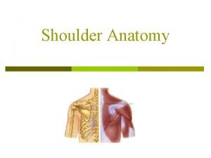
Anatomy • 30 muscles • 3 bones – Scapula, humerus, clavicle • 4 joints/articulations – Sternoclavicular joint, acromioclavicular joint, glenohumeral joint, scapulothoracic joint

Musculature • Abductors – Deltoid (ant/medial), supraspinatus • Adductors – Pecs, deltoid(rear), lats • Flexors – Deltoid • Extensors • External rotators – Supraspinatus, infraspinatus, teres minor • Internal rotators – Subscapularis, pectoralis minor

Rotator Cuff • 4 Muscles – – – Supraspinatus Infraspinatus teres minor Subscapularis “SIt. S”

Rotator Cuff • Supraspinatus – origin from superior scapula (above spine) and inserts on greater tubercle of humerus – Active in a. BDuction and external rotation – Tested with empty can test, resisted external rotation, Neer (ear) and Hawkin’s test – #1 rotator cuff muscle to be injured/inflamed

Rotator Cuff • Infraspinatus – Origin on Scapula (under spine) and inserts on greater tubercle of humerus – Externally rotates the humerus – Hard to isolate in testing – Can be injured along with supraspinatus, not usually isolated injury

Rotator Cuff • Teres Minor – Origin on the lateral border of the scapula, inserts on the greater tubercle of the humerus – Externally rotates the humerus – Hard to isolate in testing – Usually not injured in isolation

Rotator Cuff • Subscapularis – Origin on the anterior surface of scapula and inserts on the lesser tubercle of the humerus – Depresses the humeral head and internally rotates – Tested with the lift off test and can be tested with pushing on patients stomach (harder to isolate), Bear Hug test – Usually torn with fall on outstretched arm or with forced extension

Inspection • Look for symmetry between affected and unaffected side • Any atrophy to infraspinatus or supraspinatus? • One side higher than other? • Any scapular winging?

ROM Testing • Active Motion Testing – Check for Scapulothoracic Motion testing • Stand behind patient as he/she forward flexes • Is the motion fluid/smooth, any protraction/retraction of the scapula? – Active ROM • Full forward flexion, extension, abduction • Fluid or jerky?

ROM Testing • Passive ROM testing – You move the patient’s arm into the different positions – Are there anatomical limitations to movement? – Any crepitus or pops/cracks? – Fluid movement?

Palpation • Area of greatest tenderness- ask patient to point with one finger • Palpate over bicipital groove • Acromioclavicular joint • Subacromial scpace • Scapulothoracic musculature (trapezius, rhomboids, levator scapulae)

Special Tests: Glenohumeral Instability – Sulcus test – Apprehension/Relocation test

Special Tests: Glenohumeral Instability • Sulcus test • Pt holds his/her arm down by the side • Examiner grabs arm above elbow and gives a gentle downward force • With instability-the humerus will translate inferiorly and will become visible • Dyssymmetry important • Sens-90%, Spec-85%

Special Tests: Glenohumeral Instability • Apprehension/Relocation Test – Apprehension Test • Pt lays supine on table and examiner a. BDucts humerus to 90 degrees and ER the arm • Any pain or apprehension or unwillingness to complete the test is positive • Sens-69%, Spec-50% – Relocation Test • If pt has positive apprehension test, if the pain resolves when the examiner places a downward force on the humerus, the test is positive • Sens-68%, Spec-100% if used in combo with apprehension test

Special Tests: Acromioclavicular (AC) Joint – Cross arm a. DDuction test

Special Tests: Acromioclavicular (AC) Joint • Cross Arm ADDuction test (Apley Scarf Test) • Pt flexes to 90 degrees • ADDucts humerus • Test is positive if pain over AC joint with this maneuver • No Sens/Spec data

Special Tests: Rotator Cuff • Supraspinatus (RTC) Strength Testing – Empty can test (supraspinatus) – ER testing (supraspinatus, infraspinatus, and teres minor)

Special Tests: Rotator Cuff • Supraspinatus Strength- “empty can” test – Pt flexes to 135 degrees and IR (thumbs down) – Pt resists downward force applied by examiner – Smooth giving way of strength is positive test – Sens-18. 7%, Spec-100% for detecting tears

Special Tests: Rotator Cuff – IR/ER testing • Pt humerus a. DDucted with elbow at 90 degrees • Pt resists examiner with testing of ER and IR • Tests the infraspinatus, teres minor, supraspinatus, subscapularis (IR)

Special Tests: Rotator Cuff • Subscapularis Testing – Lift Off Test – Belly Press – Bear Hug Test

• Lift Off Test Special Tests: Rotator Cuff – Test subscapularis • Pt IR and places hand by back pocket • Sens-50%, Spec-95. 4% for detecting tears • Belly Press – Test subscapularis • Pt press palm of ipsilateral hand into belly • Examiner assess strength of IR

Special Tests: Rotator Cuff • Bear Hug test – Tests the subscapularis • Pt places palm of ipsilateral hand on the contralateral shoulder • Pt then resists anterior translation of the palm • Weakness is a positive test

Special Tests: Biceps Tendon • Biceps Tendon Tests – Speed’s Test – Yergason’s Test

• Special Tests: Biceps Tendon Speed’s Test – Test for long head of biceps tendon – Pt flexes to 90 degrees with palm/thumb up – Pt resists downward force applied by examiner to palm of patient – Positive test is pain in the bicipital groove – Sens-68. 5%, Spec-55. 5%

Special Tests: Biceps Tendon • Yergason’s Test – Test for long head of biceps tendon – Elbow flexed to 90 degrees and pronated – Examiner then resists the pt’s active supination – Pain over the bicipital groove is a positive test – Sens-37%, Spec-86. 1%

Special Tests: Impingement/RTC Tendonitis • Tests for Impingement/RTC Tendinitis – Neer’s Test (ear) – Hawkin’s Test

Special Tests: Impingement/RTC Tendonitis • Neer’s Test (ear) • Pt’s humerus is flexed to 180 degrees and a. DDucted by the ear • Test is positive when pain is elicited • Sens-75%, Spec-47. 5%

Special Tests: Impingement/RTC Tendonitis • Hawkin’s Test • Pt’s humerus is elevated to 90 degrees and forcibly internally rotated • Test is positive if pain elicited • Sens-91. 7%, Spec-44. 3% • Neer + Hawkin’s yields Sens-70. 8%, Spec-50. 8%

Special Tests: Labrum • Tests for labrum – SLAP or O’Brien’s Test

Special Tests: Labrum • SLAP test aka Obrien’s test – Test for labral pathology • Pt flexed to 90 degrees, adducted 15 degrees and internally rotated (thumb down) • Pt resists the downward force on the palm applied by the examiner • Positive test is pain/click with maneuver relieved when patient turns palm up • Sens-100%, Spec-98. 5%

Questions about the shoulder exam?

The Knee Exam:

Knee Anatomy • Bones – Femur, tibia, patella, fibula • Ligaments – – Anterior cruciate ligament Posterior cruciate ligament Medial collateral ligament Lateral collateral ligament • Cartilage/Meniscus – Medial/Lateral meniscus

Knee Anatomy • Tendons – Quadriceps Tendon – Patellar Tendon – Iliotibial Band

Inspection • Expose both knees completely • Observe walking-limp or antalgic? • Any muscle wasting? • Any varus or valgus deformity? • Any scars present? • Any redness or swelling? • Any rashes?

Palpation • Have patient point with one finger the area of maximal tenderness • Important to compartmentalize where the pain is coming from (ant/post/lateral/medial) for differential diagnosis

Palpation • Palpate pertinent anatomy – Joint lines-meniscal injuries – Med/lateral patella- PFS – Lateral femoral condyle- IT Band syndrome or LCL injury – Medial femoral condyle- Pes anserine bursitis or MCL injury – Posterior knee- Baker’s cyst – Patellar tendon- patellar tendinitis – Tibial tuberosity- Osgood Schlatter

ROM Testing • • Flexion- normal 130 degrees Extension- normal zero degrees Internal Rotation External Rotation

Special Tests: Patella • Patellar pathology – Patellar apprehension test – Patellar grind test

• Special Tests: Patellar apprehension test – Examiner puts lateral and medial glide to patella – Pt will have pain or feel as if his/her patella will sublux or dislocate

• Special Tests: Patellar grind test – Patient puts AP compression to patella and medially/laterally translates patella – Crepitus or grinding with translation is a positive test – Can also be performed by having the examiner translate the patella inferiorly and the pt contracts the quadriceps which makes the patella move in the trochlear groove- if crepitus/grinding-positive test – Test for patellar pathology including subluxation or chondromalacia patellae

Special Tests • Ligamentous Pathology – Lachman’s test – Reverse Lachman’s test – Ant/Post Drawer test – Pivot shift test – PCL sag test – Varus/valgus test – Figure Four test

• Special Tests: ACL Lachman’s Test – Test for ACL/PCL integrity – Examiner grasps the femur with one hand the tibia with the other (knee should be in 30 degrees of flexion) and moves the joint in the AP direction – Positive test is an endpoint that is not firm or increased motion in either ant/post direction compared to unaffected side

Special Tests: ACL • Reverse Lachman’s test – Hard to do Lachman’s test if pt with big legs or examiner with small hands – Have pt lie prone with knee in 30 degrees of flexion (resting distal tibia on examiner thigh) – Examiner then translates the thigh anteriorly, if no firm endpoint positive test for ACL injury

• Special Tests ACL/PCL Anterior/Posterior Drawer test – Test integrity of ACL/PCL – Pt flexes knee to 90 degree with foot flat on the table – Examiner sits on pt’s foot after externally rotating tibia about 15 degrees – Examiner then translates anteriorly and posteriorly after asking the pt to relax hamstring complex – Positive test is a non firm endpoint or increased translation when compared to the opposite side

Special Tests ACL • Pivot Shift Test – Test for ACL integrity – Examiner grasps the leg with both hands, one hand on foot/ankle with slight internal rotation, the other providing valgus force with knee in about 20 degrees of flexion – The knee is then extended and the test is positive if a subluxation or clunk of the tibia is noted

• PCL sag test Special Tests: PCL – Test integrity of the PCL – Pt lays supine with hips and knees both in 90 degrees of flexion – Examiner looks perpendicular to proximal tibia – Positive test if tibia “sags” when compared to unaffected side – If pt extends knee against resistance, PCL deficient knee will have superior translation of proximal tibia and be in same position as other knee

• Special Tests: MCL/LCL Varus/Valgus test – Test the integrity of the MCL/LCL – Test should be performed at both zero degrees and 30 degrees – Examiner puts one hand on distal tibia and the other hand on the knee – Varus test, 1 hand on medial knee, the other on the distal tibia- varus pressure applied – Valgus- reverse hands – Positive test is laxity with no firm endpoint

Special Tests: LCL • Figure Four Test – Test for LCL Sprain – Pt supine with heel of affected leg on the knee of the contralateral leg – Examiner palpates the LCL – Pos test if painful with maneuver or TTP over the LCL

Special Tests: Meniscus • Meniscal Pathology – Thessaly Test – Mc. Murray’s Test – Apley’s Grind Test

Special Tests: Meniscus

• Special Tests: Meniscus Mc. Murray’s Test – Test for meniscal pathology – Pt supine with knee/hip flexed to 90 degrees – Examiner ER lower leg and flexes/extends the knee while palpating medial/lateral joint line – Pos test is a click palpated in the joint line

Special Tests: Meniscus • Apley’s Grind Test – Test for meniscal pathology – Pt prone with knee in 90 degrees of flexion – Examiner applies force down through the heel while IR/ER – Positive test is pain with the maneuver

Strength Testing • Quadriceps testing • Hamstrings testing • Hip a. BDuctors/a. DDuctors

Questions about the knee exam? • LET’S PRACTICE…

Thank you very much for your time!

• Following Slides are for Information to review and put everything together • Advise to review and work through the test to arrive at your diagnosis.

Outcomes • Medial knee pain – MMT – Pes Anserine Bursitis – MCL sprain – OA

Outcomes • Medial Knee Pain – MMT- +Mc. Murray’s, +Apley, TTP medial joint, Neg Valgus test – Pes anserine bursitis- -Mc. Murray’s, -AGT, TTP over bursa, +Valgus test – MCL sprain- +Valgus test, TTP over insertion/origin of MCL – OA- +/-Mc. Murray’s test, +/-AGT, ? medial joint line tenderness, +/- Valgus test

Outcomes • Lateral knee pain – LCL sprain – LMT – Iliotibial Band Friction Syndrome

Outcomes • Lateral knee pain – LCL- + Varus test, + Figure Four test, TTP over LCL – LMT- +Mc. Murray’s, +AGT, TTP over lateral joint line – ITB Syndrome- TTP over lateral femoral condyle, +Ober’s test, otherwise normal

Outcomes • Anterior knee pain – Patellofemoral syndrome – Patellar tendinitis – Patellar subluxation/dislocation – Osgood Schlatter

Outcomes • PFS- +patellar grind, TTP around patella, weak hip a. BDuctors, increased Q angle • Patellar tendinitis- TTP over patellar tendon, jumper • Patellar sublux/D/L- +patellar apprehension, +patellar grind, TTP over patella • Osgood-Schlatter- TTP over tibial tuberosity, history

Outcomes • Posterior Knee Pain – PCL injury – Baker’s cyst – Can have post pain with post horn meniscal tear (uncommon-but keep in mind)

Outcomes • PCL tear- +Sag test, +Lachman’s, +Post Drawer test • Baker’s cyst- palpable fullness, lump
 Miserable malalignment syndrome
 Escala rankin
 Modified rankin scale
 James rankin barrister
 Rankin escala
 Rankine
 Rankin school district 98
 Florence rankin aa
 Gail rankin
 Rankin skore
 Thang điểm modified rankin scale
 Seth rankin
 Rankine
 Elizabeth wade nhs
 Kris kappel
 Thomas wade carter
 The roe wade
 Kiliani fischer synthesis
 Roe vs wade background
 A wade boykin
 Objectives of search and rescue team
 Sir william wade
 Wade lipscomb
 Felicia wade
 Creighton disability services
 Wade degottardi
 Wade trappe
 Sir william wade
 Wade alexander phipps
 Anoraks almanac
 Prtifo
 Craig v boren summary
 Wade henning
 Lashley wade theory
 Roe v wade summary
 Professor derick wade
 A spill at parsenn bowl: knee injury and recovery
 Knee flexors
 Knee anatomy chapter 16 worksheet 1
 Chapter 16 worksheet the knee and related structures
 Gastrocnemius origin and insertion
 Jacket restraints for pediatrics
 Coca valga
 Knee replacement
 Locking and unlocking of knee joint
 Prime mover of knee flexion
 Types of drowning
 Shoulder flexion agonist and antagonist
 Night i met einstein
 Beam knee in ship
 Knee joint capsule
 Primary function of the muscular system
 Youtube.com
 Patella tilt angle
 Difference between axial and pendular suspension
 Hip range of motion
 What knee do you genuflect on
 Principles of positioning in nursing
 Recumbent position
 Knee extension nerve
 Soft firm hard end feel
 Beam knee in ship
 Housemaids knee
 Knee flexion muscles
 Biomekanik knee joint
 Plc
Plc Biomekanik knee joint
 Knee walker rentals atlanta
 Plantar fascia anatomy
 What is a bone island
 Muscle around knee
 Knee screw home mechanism
 Joint resting position
