Shoulder Anatomy and Arthroscopy Mohsen MardaniKivi M D


 II. Labral Avulsion (55%)")

 *Lateral")

")

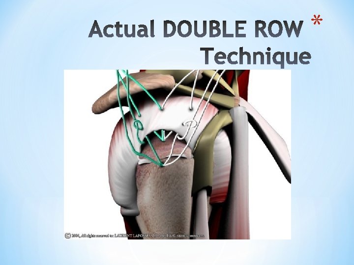
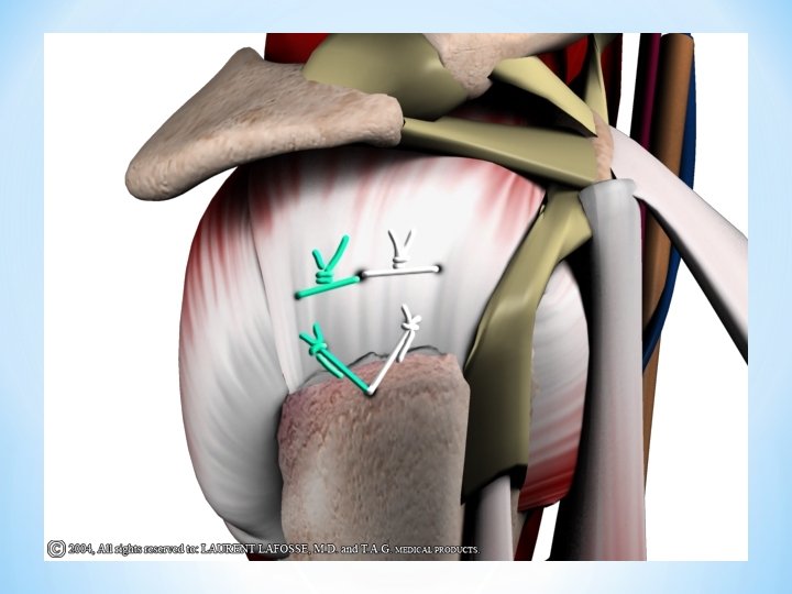
 2 -Casiope Repair (double Bankart row")


- Slides: 59
Shoulder Anatomy and Arthroscopy Mohsen Mardani-Kivi M. D. GUMS
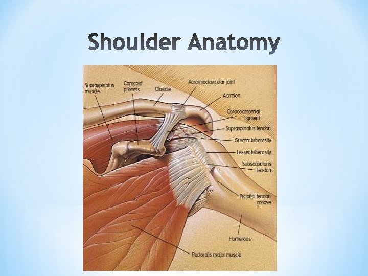
*Greatest ROM *No inherent bony stability *Relies on soft tissues for stability *Many injuries involve the soft tissues (rotator cuff, labrum) *Little glenoid bone stock
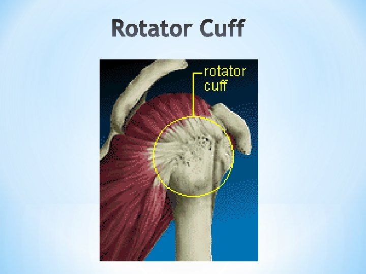
*Rotator Cuff- * dynamic stabilizer * passive muscle tension * ligament tightening * compression of * articular surface *GHL- * static stabilizer
* SGLH * MGHL * IGHL * PIGHL * CAL * CHL
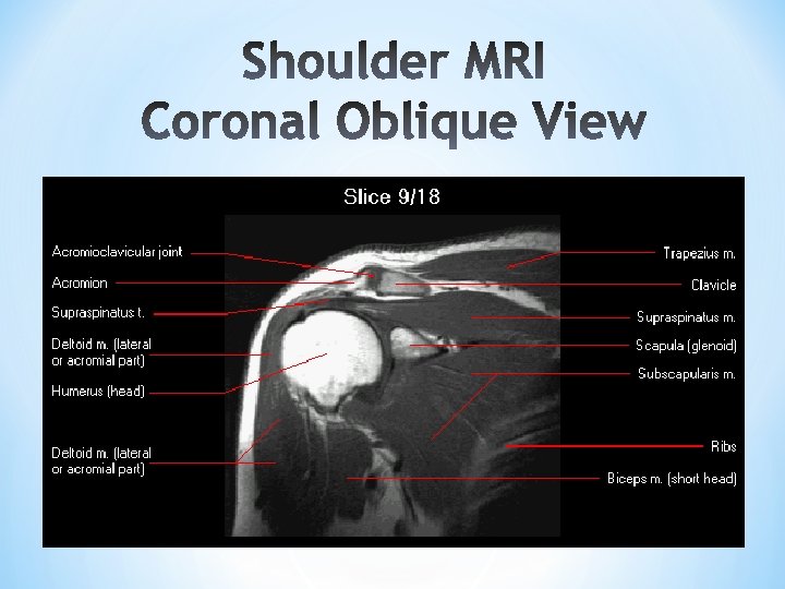
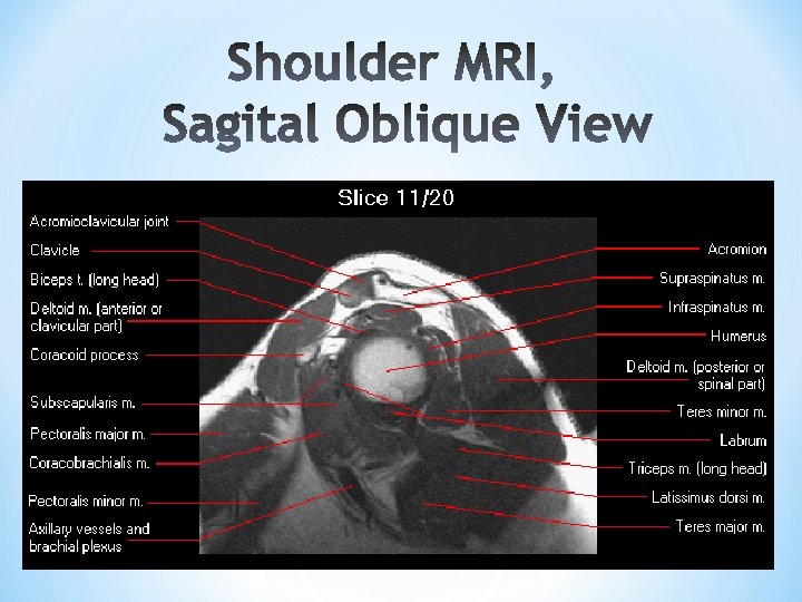
*Axial plane Bankart Lesion, BT *Sagittal oblique plane *Coronal oblique plane Rotator cuff, SLAP
Rotator cuff tear
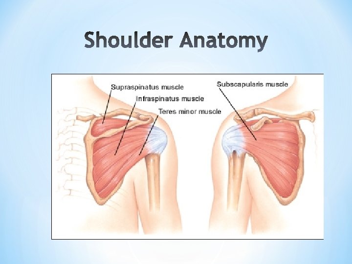
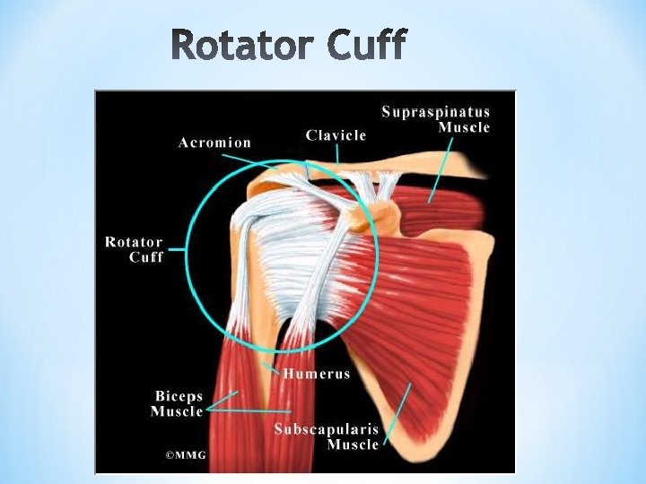
Muscles comprising rotator * cuff Supraspinatus Infraspinatus Teres Minor Subscapularis
Cause: *the rotator cuff can be torn from a single traumatic injury. *A cuff tear may also happen at the same time as another injury to shoulder, such as a fracture or dislocation. *Most tears, however, are the result of overuse of these muscles and tendons over a period of years. People who are especially at risk for overuse are those who engage in repetitive overhead motions. These include participants in sports such as baseball, tennis, weight lifting, and rowing.
* injury to in the * shoulder. 1 or more of the 4 muscles Rotator cuff tears are most common in people who are over the age of 40. * Younger people tend to have rotator cuff tears * following acute trauma or repetitive overhead work or sports activity. Rotator cuff tear may often happen as a result of wear * and tear.
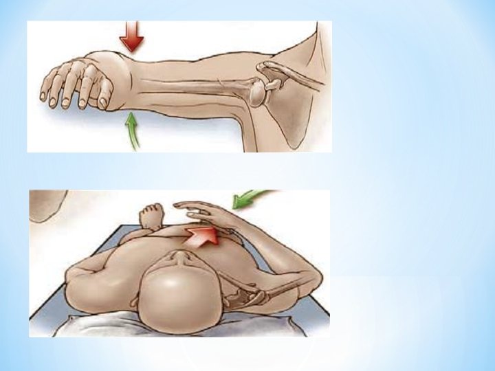
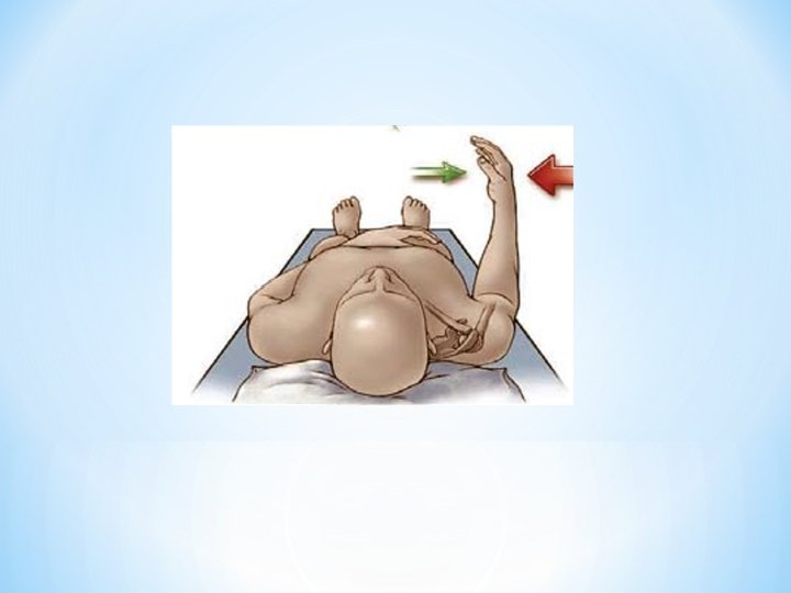
*Pain on the lateral aspect of the shoulder *Often worse at night *When lifting the arm *may radiate to deltoid insertion *Weakness, instability, tenderness *(There may be increase in the pain and weakness experienced when elevating or rotating the arm) *Atrophy or thinning of the muscles about the shoulder
Physical exam:
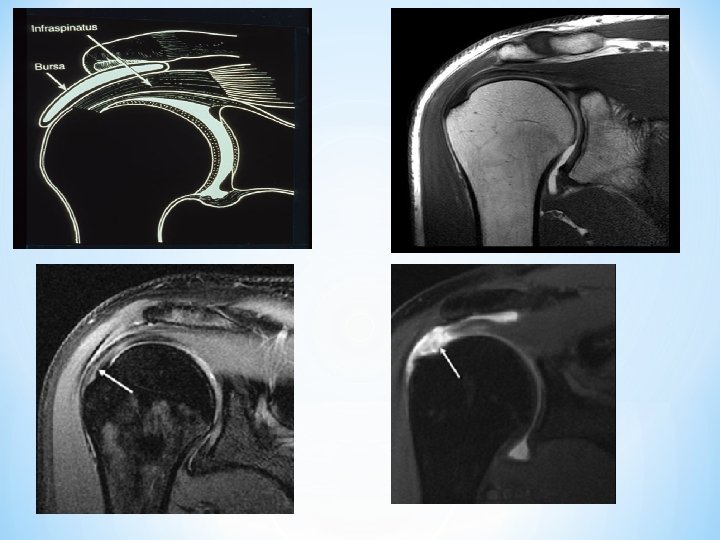
How is it diagnosed? *Diagnosis of a rotator cuff tear is based on the symptoms and physical examination. X-rays, and imaging studies, such as MRI or ultrasound, are also helpful. *An MRI can sometimes tell how large the tear is, as well as its location within the tendon itself or where the tendon attaches to bone. *Shoulder pain is variable and does not always correspond to the size of the tear.
A complete tear of the supraspinatus resulting in a shift upwards of the head of the humerus
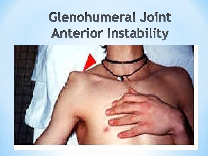
*Mechanism/Etiology *Hill-Sachs Lesion *Bankart Lesion
*Mechanism/Etiology *Hill-Sachs Lesion *Bankart Lesion
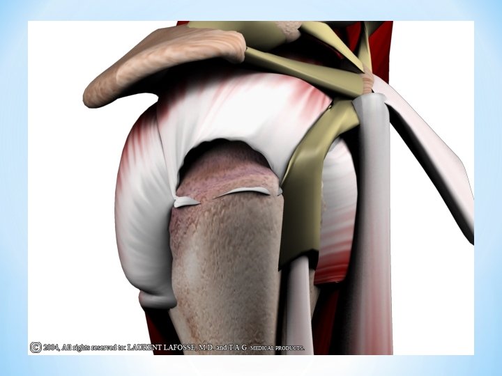
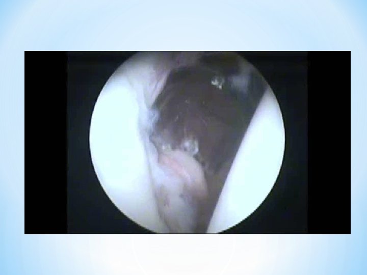
*Traumatic avulsion of anterior/inferior labrum *Cadaver studies have shown that 4 o’clock region is weakest part of labrum
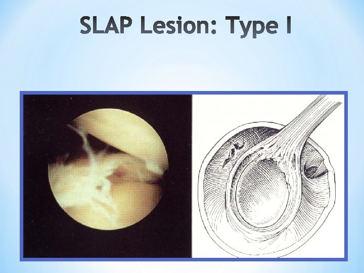
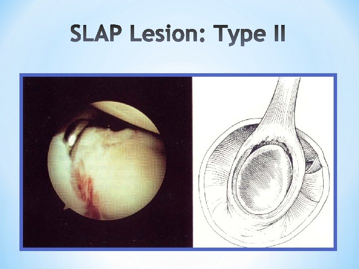
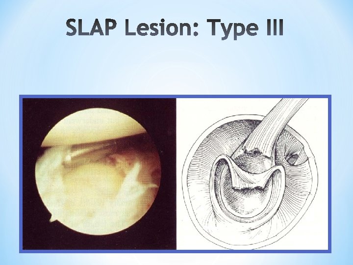
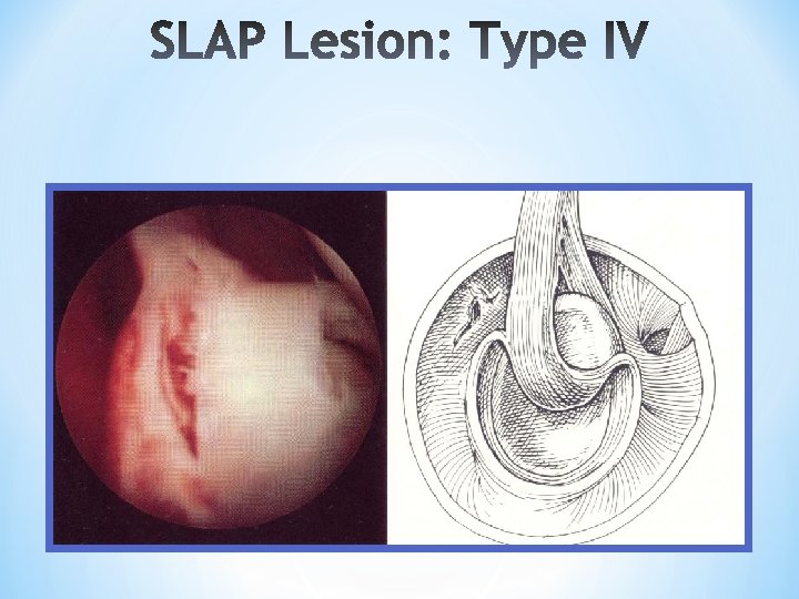
Superior Labrum Anterior to Posterior Tear I. Labral Fraying (21%) II. Labral Avulsion (55%) III. Bucket Handle Tear (9%) I. Bucket Handle Tear into Biceps Tendon (10%)
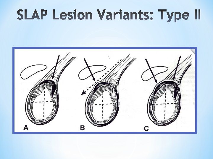
V. VI. Type II + Bankart Type II + Unstable radial or flap tears VII. Type II + Extension into MGHL
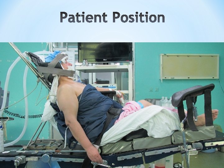
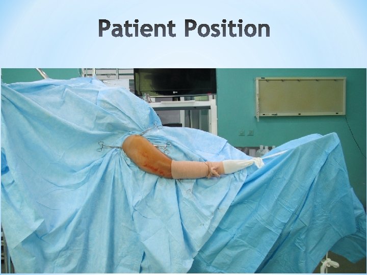
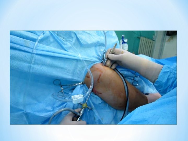
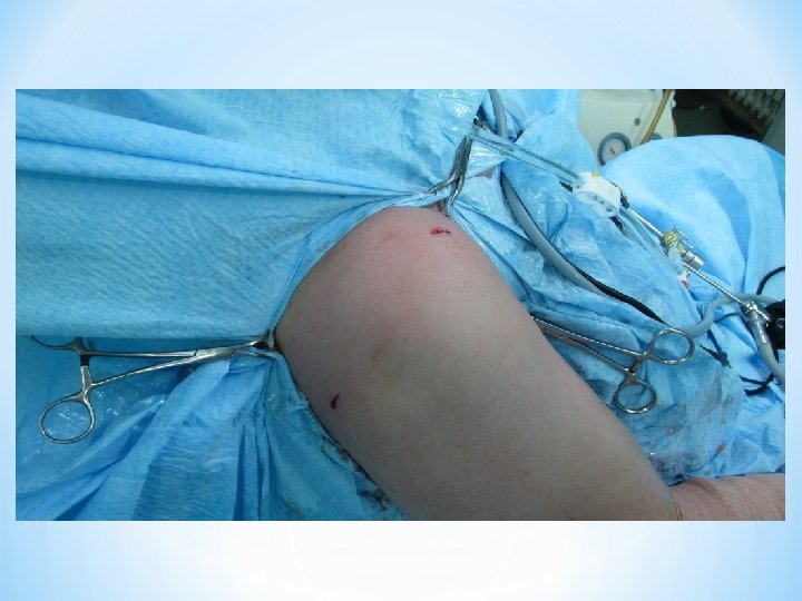
*Beach chair (the most common) *Lateral
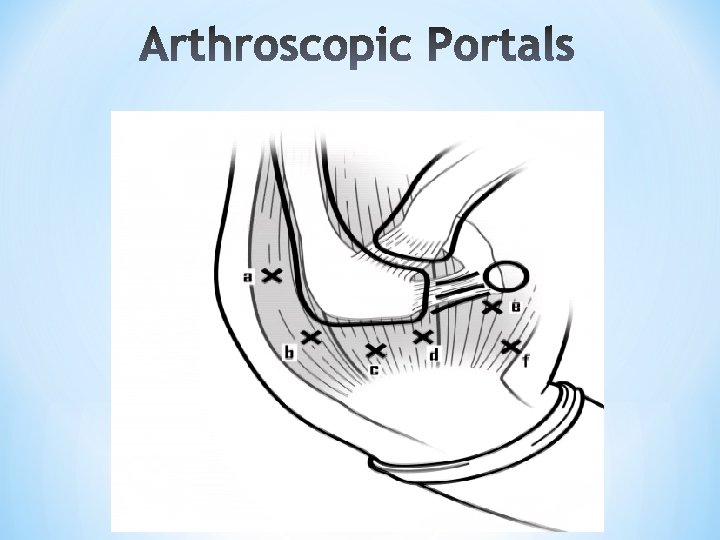
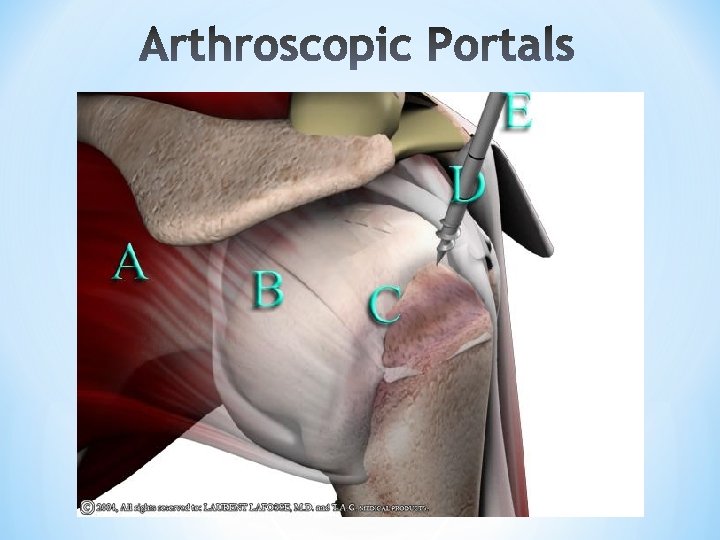
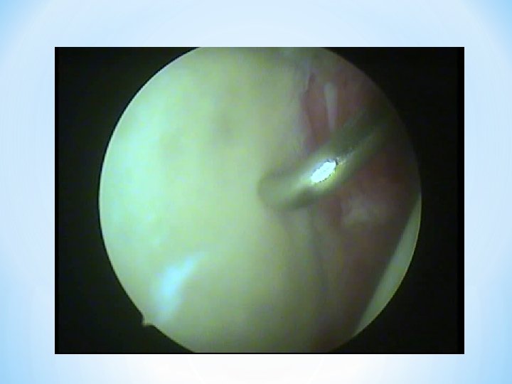
*Arthroscope *30° and 70° scopes *Arthroscope sheath with matching sharp and blunt trochars *Punches, Graspers, Seizers, Probes *Suture passers, Knot pusher
*#11 scalpel blade *Skin marking pencil *18 g. needle *20 cc syringe (if insufflating) *76 mm plastic cannula with a rubber dam *Motorized shaver with soft tissue and bone shaving blades *Suction punch *Suture punch
*Metal Anchor Suture 5, 6. 5 *Bio Anchor Suture 5, 6. 5 *Knotless Anchor Suture; push lock
1 - Arthroscopic Bankart repair (one row repair) 2 -Casiope Repair (double Bankart row repair) 3 -All arthroscopic Latarjet procedure 4 - Capsular shift 5 -Rotator interval closure Repair of HAGEL lesion Repair of SLAP lesion
*Arthroscopic Rotaor cuff Surgery *Arthroscopic Subacromion decompression *Arthroscopic Acropmioplasty *rotator cuff repair -one row -double row
*Latissmos dorsi transfer for rotator cuff deficiency, irreparable tears *Arthroscopic priscapular bursectomy
*Arthroscopic AC arthroplasty *Arthroscopic AC instability reconstruction