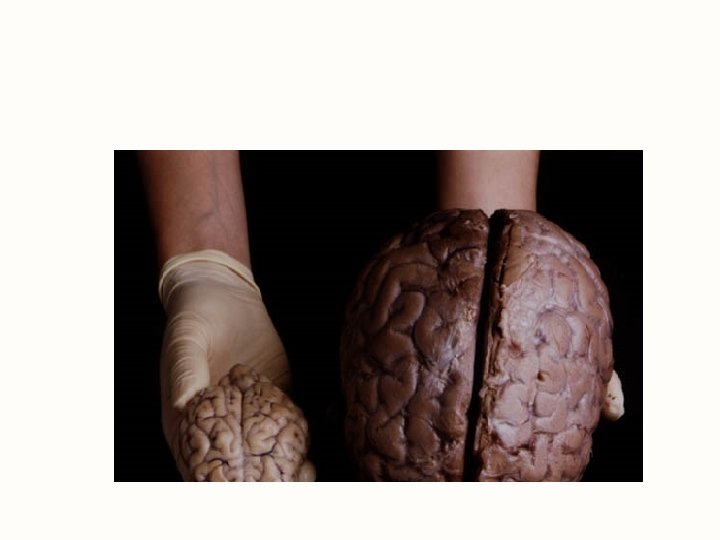
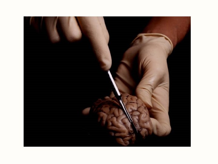
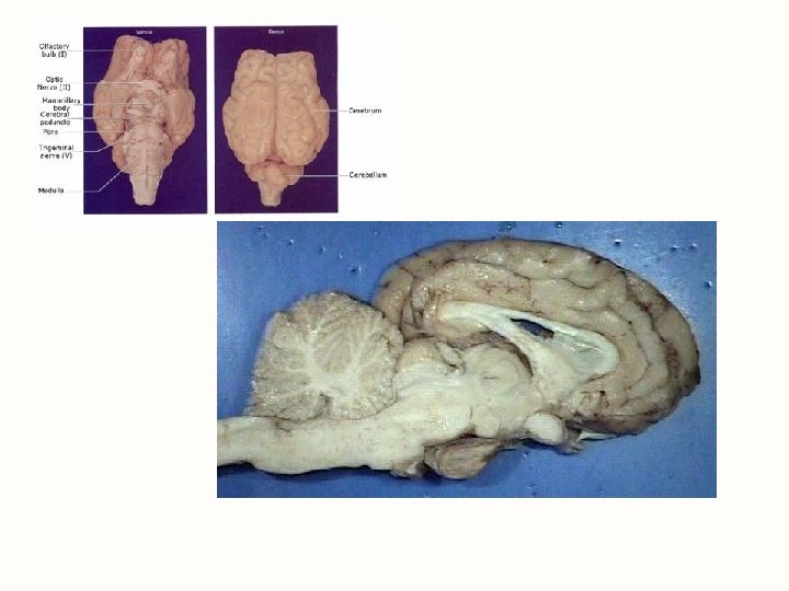
Sheep Brain Dissection http www exploratorium edumemorybraindissectionindex htm

Sheep Brain Dissection http: //www. exploratorium. edu/memory/braindissection/index. htm

Spinal cord

The Central Nervous System • Overview of the central nervous system- brain and spinal cord • Meninges, ventricles, cerebrospinal fluid & blood supply • Spinal cord • Major structures and functions of the brain

Gross Anatomy of the Spinal Cord

I. Spinal Cord • Spinal Cord A. General Structure – Housed in the vertebral column, covered by dura mater – Is an extension of the medulla oblongata – Extends from the foramen magnum to the L 2 vertebrae. – Contains 31 pairs of spinal nerves

B. Obvious enlargements – Cervical enlargements – the nerves that supply the upper body usually named after body regions. – Lumbar enlargements – nerves that supply the lower body – sciatic nerve found in lumbar region. • Conus medullaris – the tapering off area • Cauda equina – means “horse tail” – located at the L 2 vertebrae – a collection of nerves at the end of the spinal cord.

C. Functions of the Spinal Cord 1. Conduction – bundles of fibers passing information up (ascending tracts) & down (descending tracts) spinal cord • Conveys sensory impulses to the brain called ascending tracts. • Conveys motor impulses away from the brain called descending tracts. 2. Locomotion – repetitive, coordinated actions of several muscle groups 3. Reflexes – involuntary, stereotyped responses to stimuli • remove hand from hot stove

Cross-Sectional Anatomy of the Spinal Cord • Central area of gray matter shaped like a butterfly and surrounded by white matter in 3 columns

Meninges of Vertebra & Spinal Cord

II. Protection and coverings A. Vertebral column – protecting spinal cord – part of the axial skeleton. B. CSF (cerebrospinal fluid) – a lubricating fluid, cushioning, contains immune cells C. Meninges – membranes to protect the brain and spinal cord. 1. Dura mater – Latin “tough mother” – tough fibrous covering – the outer layer – forms large cavity for blood. – Epidural space – upon the dura mater – between the vertebrae and dura mater.

2. Arachnoid – a spider-like web structure – delicate – important because of what it secretes into the subarachnoid space. – Subarachnoid space – space between arachnoid and pia mater – contains cerebrospinal fluid. 3. Pia mater – means “gentle mother” – inner most layer, very thin, and highly vascular to nourish the brain and spinal cord…major function is nourishment.

III. Brain Description A. Brain weighs 3 to 3. 5 pounds, 100 billion neurons, 900 billion glia B. Major portions of the brain--brainstem, cerebrum, cerebellum, diencephalon

Brain Longitudinal fissure separates 2 cerebral hemispheres.

C. Cranial Meninges-see notes on spinal meninges 1. Dura mater -- outermost, tough membrane 2. Arachnoid mater is spider web filamentous layer 3. Pia mater is a thin vascular layer adherent to contours of brain

D. Brain Ventricles

D. Ventricles of Brain 1. Internal chambers within the CNS- called ventricles 2. Lined with ependymal cells and containing plexus of capillaries that produce CSF

E. Cerebrospinal Fluid 1. Clear liquid fills ventricles and canals & bathes its external surface (in subarachnoid space) 2. Brain produces & absorbs about 500 ml/day 3. Functions – buoyancy -- floats brain so it neutrally buoyant – protection -- cushions from hitting inside of skull – chemical stability -- rinses away wastes

Flow of Cerebrospinal Fluid
 is tightly joined endothelium – permeable to")
F. Blood-Brain Barrier 1. Blood-brain barrier (BBB) is tightly joined endothelium – permeable to lipid-soluble materials (alcohol, O 2, CO 2, nicotine and anesthetics) – administer drugs through nasal sprays

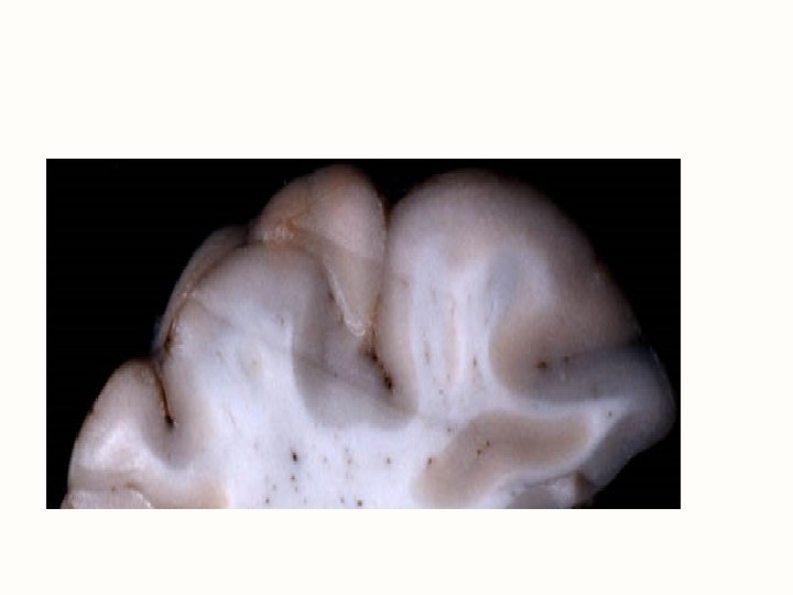
IV. Major subdivisions of the brain A. Cerebrum – divided into right and left hemispheres, also called cerebral cortex or cortex – it is the most prominent in human. • Weight – consists of 80% of the total brain. • Gray matter – unmyelinated fibers – the outer cortex. • White matter – myelinated fibers - interior cortex.

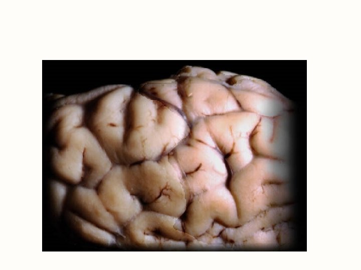
1. Surface features • Sulcus – shallow grooves. • Fissure – deep grooves – most prominent/long. • Longitudinal fissure – down the center of the brain – divides the left and right hemispheres. • Lateral fissure – between the temporal lobe below and the frontal and parietal lobes of the brain. • Gyrus– round ridges – refers to rounded elevations of the brain surface.

Cerebrum -- Gross Anatomy

2. Cerebrum lobes – Parietal lobe – controls sensations for skin, muscles, and joints (somatic senses). – Frontal lobe – controls voluntary movements. – Occipital lobe – controls vision. – Temporal lobe – controls auditory centers – hearing. – Insula – deep within the lateral fissure – relays info between the right and left hemispheres. Each lobe can be divided into functional areas for sensory, motor, and/or association Sensory- feelings/sensations Motor- skills, movement Association- integration/interpretation

cerebrum 3. Hemispheres of the cerebrum – Left – more analytical – math, verbal skills. – Right – artistic, musical, intuitive.

4. Examples of Specialized areas – Pre-motor cortex – learning motor skills – riding a bike, speech patterns. – Primary motor cortex- in the frontal lobe – Prefrontal Cortex- working memory – Broca’s area – motor aspects of speech…how to make the words come out – vocal cords – Sensory cortex – in parietal lobe. – Auditory cortex – in temporal lobe. – Olfactory – special sense for smell – in temporal lobe.

4. CONT – Somatic region – parietal – body positions, equilibrium. – Corpus callosum – internal cerebrum – band of tissue connecting right and left hemisphere. – Limbic system– center in the brain – around corpus callosum – involved in emotions and sense of smell “called the emotional primitive brain” – Hippocampus- part of limbic system that helps to convert short term memory into long term memory • Long-term synaptic potential- frequent nearly simultaneous repeated stimulation of same neurons strengthen synaptic connections aiding in long term memory storage • Converting short term memory into long term memory is called consolidation

Diencephalon

B. Diencephalon – between the mid-brain and cerebrum 1. Thalamus – believed to be a relay center for senses except for smell/olfactory. Has a “dumbbell” shape. Is the gray matter and a mass of many nuclei. Plays part in reflex movements. 2. Hypothalamus – below the thalamus. Is the homeostatic vital center – very small, ¼ ounces but is very important for body temperature, hunger center, and the thirst center. Majority are autonomic functions. Sleep/wake cycle, important for endocrine gland hormones. Secretes nine hormones to the endocrine system.

3. Pineal body – “the biological clock” – is a neuroendocrine structure. Secretes melatonin and serotonin. Works with circadian rhythms – normal sleep/wake cycle, appetite/thirst centers. 4. Pituitary gland- “master gland” of endocrine system, important hormones are secreted by the pituitary gland- 2 parts (anterior (more endocrine functions) and posterior (more neurological functions)

C. Brain stem 1. Midbrain – reflex center, used for visual and auditory centers. 2. Pons – above medulla. Latin for “bridge” – a bridge between the midbrain and medulla. An accessory or helper in respiration. 3. Medulla oblongata – lower portion of the brain stem – vital center for heart rate, respiration, controls coughing, sneezing, vomiting.

Medulla and Pons Olive

D. Cerebellum 1. posterior brain structure 2. 2 nd largest part – gray matter on outer portion, white matter on internal portion called arbor vitae “tree of life” 3. Functions: acts with cerebral cortex for 1) skilled muscle movements, 2) balance, and 3) posture.

Cerebellum

E. Reticular Formation • Clusters of gray matter scattered throughout pons, midbrain & medulla • Regulate balance & posture • Includes cardiac & vasomotor centers • Regulates sleep & conscious attention – injury leads to irreversible coma
- Slides: 39