Sexually Transmitted Diseases STDs n The laboratory diagnosis
 n The laboratory diagnosis of STDs is related to")
Sexually Transmitted Diseases (STDs) n The laboratory diagnosis of STDs is related to the sex of the patient, although some infections are common to both sexes like gonorrhea, syphilis and chlamydial infection but there are differences in the symptoms, the sites and methods of specimens collection in these infections.

Genital infections and STDs in women These include: 1 - Vaginitis : Is caused by a limited number of infectious agents include: Trichomons vaginalis Trichomoniasis: is an infection of urogenital tract and the most common site of infection is the urethra and vagina in women, it is caused by the single-celled protozoan parasite Trichomons vaginalis which classically produce a copious, frothy yellow or yellowgreen discharge. Candida albicans Vulvovaginal Candidiasis: is caused by Candida albicans, squamous epithelial cells of vaginal is invaded and inflamed causing vaginal discharges and pain. Discharge is typically more thick than trichomoniasis and curd like.

2 - Bacterial Vaginosis: is caused by a number of infectious agents include: Gardnerella vaginalis Peptocococcus Mycoplasma 3 - Cervisitis with or without Urethritis: is caused by gonococci or Chlamidia trachomatis. 4 - Uterine sepsis: is caused by S. pyogenes, S. aureus, Clostridium and Mycoplasma. 5 - Genital ulceration: is caused by Tryponema pallidum, Haemophilus ducreyi and Chlamidia. 6 - Tuberculosis of uterus: is caused by Mycobacterium tuberculosis. 7 - Viruses: is caused by viruses like Cytomegalo virus and Herpes.

Genital infections and STDs in men The infections in men are mostly caused by the same organisms as in women, include: 1 - Urethritis: In men C. trachomatis causes urethritis lead to epididymitis and prostatitis. 2 - Prostatitis: caused by gonococci or Chlamydia 3 - Ulceration: caused by Herpes simplex virus, Tryponema pallidum, Haemophilus ducreyi and Chlamydia.

Collection of specimen in men: 1 - Cleanse around the urethral opening using a swab moistened with sterile physiological saline. 2 - When culture is indicated collect a sample of pus on a sterile cotton-wool swab. If possible, before inserting the swab in a container of transport medium if possible inoculated a plate of culture medium. 3 - Inoculated culture plates must be incubated within 30 minutes.

Collection of Sample in women: n Endocervical canal for isolation N. gonorrhoeae: n Use a sterile vaginal speculum to examine the cervix and collected the specimen: 1 - Pass a sterile cotton wool swab 20 -30 mm into the endocervical canal and gently rotate the swab against the endocervical wall to obtain a specimen 2 - When gonorrhea is suspected , before inserting the swab in transport medium, if possible inoculated a plate of culture medium. 3 - Inoculated culture plates must be incubated within 30 minutes. n n n

Collection of vaginal discharge to detect T. vginalis, C. albicans and G. vaginalis: n n n Two preparations are required: 1 - Wet preparation to detect motile T. vaginalis. Use a sterile swab to collect a specimen from the vagina. 2 - Dry smear for Gram staining to detect Candida and examine for clue cells, Gram positive cells and pseudohyphae of C. albicans.

Collection of specimen to detect Treponema pallidum: n n Must be collected before antibiotic treatment 1 - Wearing protective rubber gloves, cleanse around ulcer (chancre) using a swab moistened with physiological saline. 2 - Gently squeeze the lesion to obtain serous fluid collect a drop on a cover glass. 3 - Immediately deliver the preparation to laboratory for examination by dark-field microscopy.

n n n n Collection of specimen to detect Chlamydia granulomatis: Most Chlamydia infections are diagnosed clinically or immunologically by detecting specific Chlamydial antigen. 1 - Cleanse around the ulcerated area using a swab moistened with physiological saline 2 - Pinch off a small piece of tissue from the edge or base of a lesion. Crush this between two microscope slids. Immunological detection of Chlamydial antigen (Monoclonal Ab) to detect by: Direct fluorescence techniques (FAT) Enzyme immunoassays (EIA) Rapid immunochromatographic (IC)
 Thayer Martin medium. For isolation of Neisseria")
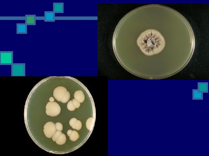
Culture the specimen n n n A) Thayer Martin medium. For isolation of Neisseria gonorrhoeae and incubate in moist carbon dioxide enriched atmosphere at 35 – 37 C for up to 48 hr. Thayer Martin medium contains the antibiotics (Vancomycin, Colistin, Nastatin). B) Blood agar (aerobic and anaerobic) C) Mac. Conkey agar D) Cooked meat medium. When puerperal sepsis or septic abortion is suspected. Incubated specimen in cooked meat medium and incubate at 35 -37 C and then sub culturing as indicated 24 hr, 48 hr, 72 hr E) Chocolate agar F) Sabaroued Agar. For Candida isolation

PH of discharge. n n n The normal reaction of vaginal discharge (puberty to menopause) is PH 3 -3. 5 This to indicate the following: T. vaginalis: yellow-green purulent discharge with PH over 5 C. albicans: White odorless discharge with PH below 5 G. vaginalis: Grey offensive smelling (fishy ammoniacal smell) thin discharge with PH over 5

Gram stain to examine: n Pus cells containing Gram negative diplococci or pus cells have been damaged and the organism seen lying outside the pus cells that could be N. gonorrhoeae. n Vaginal smear from a patient with suspected bacterial vaginosis or candidiasis, looked especially for: Large G+ve yeast cells and Pseudohyphae that could be C. albicans. Clue cells are epithelial cells with adhering G-ve short bacilli and gram variable coccobacilli could be Gardnerella vaginalis. n n n Smear from a patient with suspected puerperal sepsis or septic abortion, looked especially among pus cells for: n Large G+ve rods with straight ends –Clostredium perfringens G+ve Streptococci –S. pyogenes G+ve cocci- Staph. aureus G-ve rods –Bacteroides or coliforms Wet (saline) preparation to detect T. vaginalis To detect motile T. vaginalis trophozoites n n
 n n n Urinary Tract Infection (UTI) is a bacterial")
Urinary Tract Infection (UTI) n n n Urinary Tract Infection (UTI) is a bacterial infection that affects any part of the urinary tract. The main causal agent is Escherichia coli. The most common type of UTI is acute cystitis often referred to as a bladder infection. An infection of the upper urinary tract or kidney is known as pyelonephritis, and is potentially more serious. Women are more prone to UTIs than men. Factors that increase female susceptibility to UTI : Short length of the urethra Urethral contamination by rectal pathogens Introital & vestibular colonization by pathogenic bacteria Decreased urethral resistance after menopause

Urine culture n n n The urine samples routinely culture on Blood agar and Mac. Conkey agar and now culture on Cystine Lactose electrolyte-deficient (CLED) agar. Incubate the plate aerobically at 35 – 37 Cº overnight. CLED agar is now used by most laboratories to isolate urinary pathogens because it gives consistent results and allows the growth of both Gram negative and Gram positive pathogens. (the indicator in CLED agar is bromothymol blue and therefore lactose fermenting colonies appear yellow). .

n n n Possible pathogens: Bacteria: Gram positive: n Staphylococcus saprophyticus Staphylococcus aureus Haemolytic streptococci n Gram Negative: n n E. coli (commonest about 60 – 90 % of UTI) Proteus species(usually in hospitalized patient & with renal stones) Pseudomonas aeruginosa Klebsiella strains Neisseria gonorrhoeae (not pathogen to urinary tract) Mycobacterium tuberculosis(not pathogen to urinary tract) n Fungi: n Candida species(usually in hospitalized patient, in diabetic patient& immunosuppression Parasite: Schistosoma haematobium n n n n

Gastrointestinal Tract Infections n Enteric bacterial infections, causing diarrhoea, dysentery, and enteric fevers are important health problems throughout the world. Diarrhoeal infections are second only to cardiovascular diseases as a cause of death, and they are the leading cause of childhood death.

Etiological agents n n n n The etiological agents which causing gastrointestinal tract infections divided in to: Bacterial infections : The genus Salmonella cause gastroenteritis and typhoid fever Shigella spp. are the main cause of bacterial bacillary dysentery diarrhoea-producing Escherichia coli Vibrio cholerae cause Cholera, Cholera is a typical example of a toxigenic infection. All the symptoms can be attributed to the intestinal fluid loss caused by an enterotoxin released by V. cholerae in the intestine. The stool is voluminous and watery. The main objective of treatment is fluid replacement and antimicrobials have only a secondary role. Campylobacter jejuni and C. coli have emerged as major enteric pathogens that can be isolated as often as Salmonella and Shigella spp. in most parts of the world. Clostridium difficile is the primary cause of enteric disease related to antimicrobial therapy. It produces a broad spectrum of diseases ranging from mild diarrhoea to potentially fatal pseudomembranous colitis.

n n Viral diarrheas: Rotavirus is a major cause of diarrheal disease in children. Parasitic diarrheas: Entamoeba histolytica and Giardia lamblia can cause of diarrheal disease. n Collection and transport of stool specimens n Faecal (stool) specimens should be collected in the early stages of the diarrhoeal disease, when pathogens are present in the highest number, and preferably before antimicrobial treatment is started.

Enrichment of stool specimens n n Enrichment is commonly used for the isolation of Salmonella spp. and Vibrio cholerae from faecal specimens. Selenite or tetrathionate broths are recommended for the enrichment of Salmonella spp. , and alkaline peptone water (APW) for the enrichment of V. cholerae. Enrichment is not required for Shigella spp. , Campylobacter spp. , Yersinia enterocolitica and Clostridium difficile.

Media for enteric pathogens n n For Shigella spp. , Salmonella spp. and Y. enterocolitica, Mac. Conkey agar with crystal violet is recommended as a general purpose medium. Xylose–lysine–deoxycholate (XLD) agar is recommended for the isolation of Shigella and Salmonella. Hektoen enteric agar (HEA) or Salmonella–Shigella (SS) agar are suitable alternatives. For Campylobacter spp. there are several selective media (Blaser, Butzler, Skirrow) containing different antimicrobial supplements used. Thiosulfate citrate bile salts sucrose (TCBS) agar is selective for V. cholerae cefoxitin–cycloserine–fructose agar (CCFA) is selective for Clostridium difficile.

After inoculation of these media with one loopful of the faecal suspension, incubate the agar plates. Incubate the plates for the isolation of Salmonella, Shigella and Yersinia spp. and V. cholerae at 35 C in anaerobic incubator (without CO 2), the plates for Campylobacter spp. at 42 C in an microaerophilic atmosphere with 10% CO 2, and the plates for Clostridium difficile at 35 ∞C in an anaerobic atmosphere.

Purulent exudates, burns, wounds and abscesses n n One of the most commonly observed infectious disease processes is the production of a purulent (sometimes seropurulent) exudate as the result of bacterial invasion of a cavity, tissue, or organ of the body. A smear for Gram-staining and examination should be made for every specimen n Culture n All specimens of wounds, burns, pus or exudate should preferably be inoculated onto aminimum of two culture media: — A blood agar plate for the isolation of staphylococci, streptococci and Clostridium — A Mac. Conkey agar plate for the isolation of Gram-negative rods; All organisms isolated from wounds, pus, or exudates should be considered significant and efforts made to identify them. n n n

Wound swabs n n n n Most common pathogens found in wound swabs Enterobacter Enterococci Peptostreptococcus Proteus Pseudomonas Bacteroides Clostridium Candida Staphylococcus aureus Streptococcus Escherichia coli Fusobacterium Klebsiella

The clinically most significant species is Clostridium perfringens. It is commonly associated with gas gangrene. n C. perfringens grows rapidly in anaerobic broth with the production of abundant n gas. On anaerobic blood agar, colonies of moderate size (2– 3 mm) areseen after 48 hours. n Most strains produce a double zone of haemolysis: an inner zone of complete clear haemolysis, and an outer zone of partial haemolysis. n

Eye and Ear infections n Ocular infection can be caused by bacteria, viruses, or chlamydia and can be detected by culture. Cotton swab will be used to collect the specimen from infected eye.

n n n The culture of ear swab is a lab test. This test checks for germs that can cause infection. The sample taken for this test can contain fluid, pus, wax, or blood from the ear. Cotton swab will be used to collect the specimen from inside the outer ear canal. In some cases, a sample is collected from the middle ear during ear surgery Specimens of eye and ear should be inoculated on to a minimum culture media: -Blood agar plate for the isolation of staphylococci and streptococci. -Mac. Conkey agar plate for the isolation of Gram-negative bacteria. -Chocolate agar plate for the isolation of Neisseria. -Sabouraud dextrose agar plate for the isolation of fungi.
 testing is used to select effective")
Antibiotic susceptibility tests n n n Sensitivity (susceptibility) testing is used to select effective antimicrobial drugs. The standardized discdiffusion method (Kirby–Bauer) is used. Disc diffusion techniques are used by most laboratories to test routinely for antimicrobial sensitivity. A disc of blotting paper is impregnated with a known volume and appropriate concentration of an antimicrobial, and this is placed on a plate of sensitivity testing agar (Mueller– Hinton agar for most bacteria and blood agar for some bacteria) which uniformly inoculated with the test organism. The antimicrobial diffuses from the disc into the medium and the growth of the test organism is inhibited. Strains sensitive to the antimicrobial are inhibited at a distance from the disc whereas resistant strains have smaller zones of inhibition or grow up to edge of the disc.
 should be tested")
n n n All strains of streptococci (such as S. pneumoniae) should be tested on blood agar for susceptibility. All Gram-negative rods and staphylococci were tested on mueller hinton for susceptibility. Strains of H. influenzae and Neisseria should be tested for susceptibility using chocolate agar.

Tha n ky ou
- Slides: 30