SEX DETERMINATION FROM HUMAN SKELETAL REMAINS SKULL PELVIS


 A general")


 of males tends to be slanted back and on females")


 The male mandible tends to have a “square” shape. Mandible of")


 width")


- Slides: 54
SEX DETERMINATION FROM HUMAN SKELETAL REMAINS {SKULL , PELVIS , STERNUM}
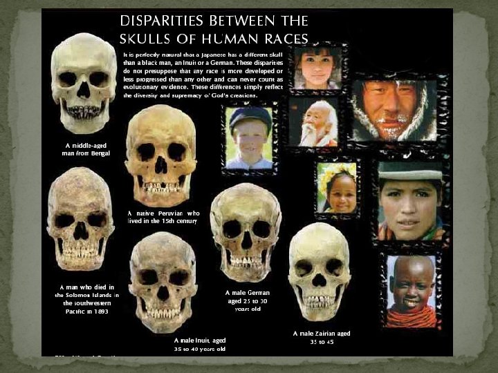
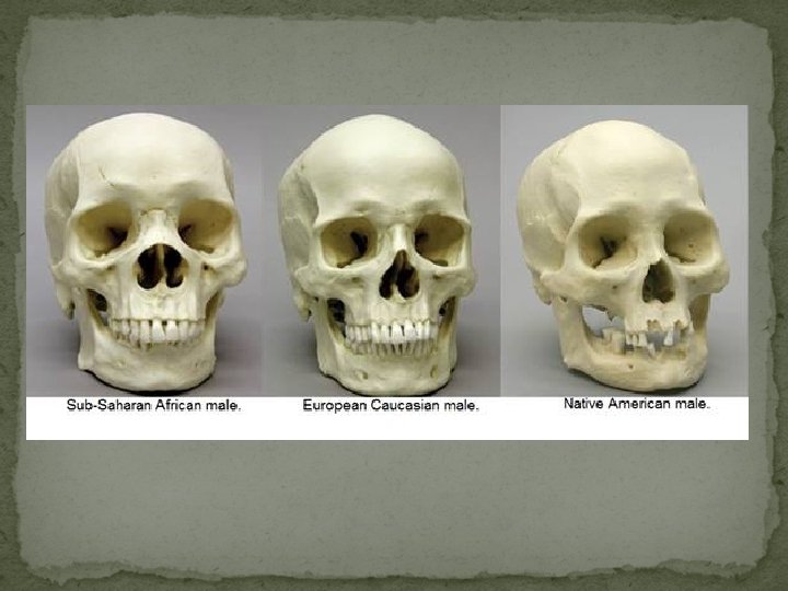
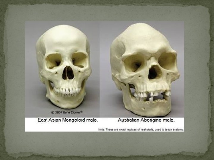
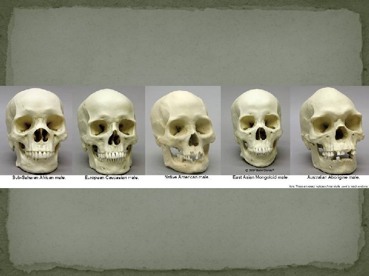
As humans, we have three basic racial groups. These groups are Caucasoid, Mongoloid, and Negroid. With the addition of inter-breeding, many other subgroups can be found. Each group possesses unique skeletal characteristics which allow us to "race" the remains of a person. In addition to race, we can also estimate age, stature, and sex.
Using Bones for Identification It consists of a five phase procedure: 1) A general description and confirmation of the human origins of the material. 2) The estimation and segregation of the minimum number of individuals. 3) The determination of sex. 4) The estimation of age. 5) The estimation of stature.
Introduction Skeleton is an excellent material in living and nonliving population for genetic, anthropological, odontologic and forensic investigations. Skull and bone features vary from male to female and differentiation is usually based on the male features that are typically more pronounced and marked than female features. The human skeleton consists of both fused and individual bone. Fused bones include those of the pelvis and the cranium. At birth a newborn baby has approximately 300 bones, whereas on average an adult human has 206 bones. The difference comes from a number of small bones that fuse together during growth, such as the sacrum and coccyx of the vertebral column.
The determination of sex by examination of the skeleton is based upon the appearances of: 1. Pelvis (innominates + sacrum ) 2. Skull (cranium + mandible ) 3. Long bones (Humerus , Femur ) 4. Sternum 5. Scapula , metacarpal bones Krongman ranks accuracy of sex determination using the pelvis at 95% , the skull at 90%, the pelvis and skull at 98% and long bones at 80%.
SEXUAL DIMORPHISM Differences between men and women include all the features related to reproductive role, notably the endocrine (hormonal) systems and their physical, psychological and behavioral effects.
SEXUAL DIMORPHISM – BASIC PRINCIPLES The evaluation of sexual dimorphism in skeleton is generally based on two factors: 1. Size difference 2. Function related differences.
Determination of sex is based on two methodological approaches: 1. Morphological (based on Shape ) Adult males and females differ in both general size and shape, and this variation is reflected in the skeletal anatomy. 2. Osteometric (based on bone dimensions) Males longer or larger than females.
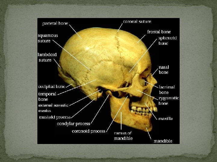
The skull is a bony structure which serves as the general framework for the head. The skull supports the structures of the face and protects the head against injury. The skull can be subdivided into two parts: the cranium and the mandible.
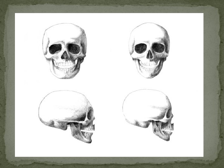
MALE FEMALE When compared, the female skull appears smaller and more gracile. The male skull is usually larger and more rugged.
Supraorbital ridges This is the region directly above the orbit and nose, or the "brow ridge“. Less pronounced=female More pronounced=male
ORBITS SQUARISH WITH ROUNDISH MARGINS MALE ROUNDISH WITH SHARP MARGINS FEMALE
The frontal bone (forehead) of males tends to be slanted back and on females it tends to be more rounded
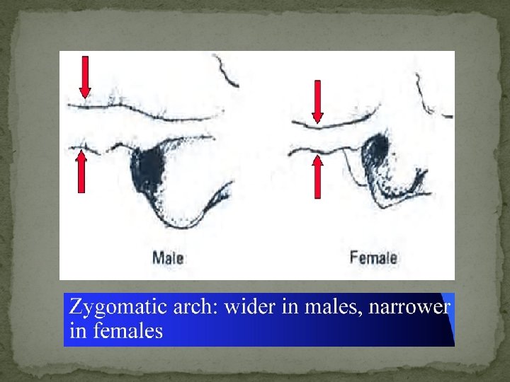
Zygomatic arch and Mastoid process
Zygomatic arch and Mastoid process
Zygomatic arches In females, the zygomatic arch is less pronounced, and tends to not extend posteriorly beyond the external auditory meatus. In males, the zygomatic arch is more pronounced or robust, and tends to extend posteriorly beyond the external auditory meatus.
MASTOID PROCESSES The mastoid processes are located on the inferior portion of the temporal bone, just posterior to the external auditory meatus.
Zygomatic arch and Mastoid process
EXTERNAL OCCIPITAL PROTUBERANCE
MALE FEMALE
The mandible together with the maxilla, the largest and strongest bone of the face. It forms the lower jaw and holds the lower teeth in place.
Chin (anterior mandible) The male mandible tends to have a “square” shape. Mandible of female tends to have a pointed chin.
PALATE LARGER, WIDER AND BROADER MALE SMALLER AND NARROWER FEMALE
Slide 27: Using the previous slides and pictures, identify whether the following skull parts are from a male or female? 1. Mandible 3. Frontal Bone 2. External occipital protuberance
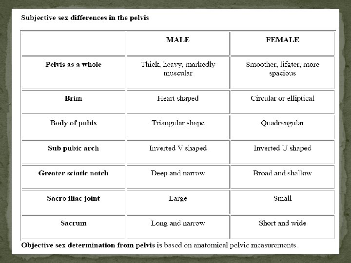
PELVIS The pelvis comprises the two innominates and the sacrum.
HIP BONE The best indicator of sex on the adult skeleton is the shape of the pubic bone of the pelvis. The hip bone (or innominate bone) is a large, flattened, irregularly shaped bone. Together with the sacrum and coccyx, it comprises the pelvis. Components It consists of three parts, the ilium, ischium, and pubis, which are distinct from each other in the young subject, but are fused in the adult.
Sex Determination from Pelvic Morphology The pelvic girdle is the most sexually dimorphic region of the skeleton, and it can be used to determine sex with a high degree of accuracy. The sexual dimorphism of the pelvis is primarily the result of reproductive mechanics, and is not readily apparent until adolescence.
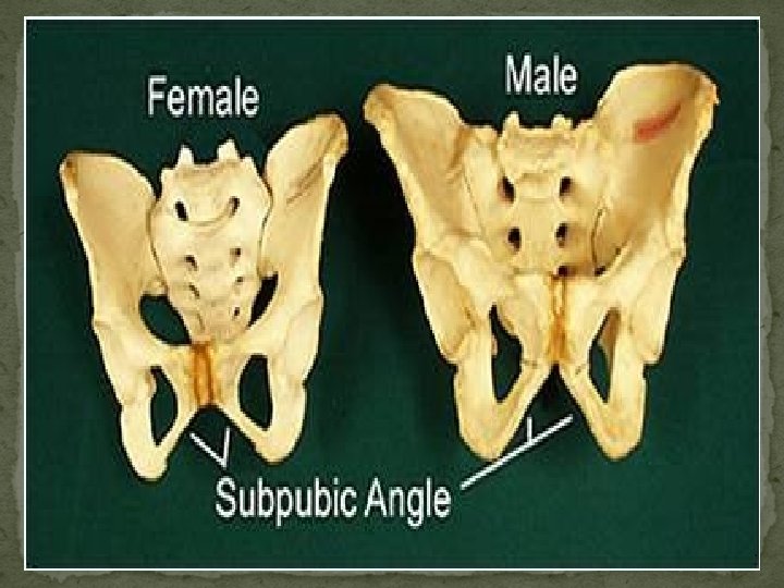
Sex differentiation in the human pelvis 1. Overall appearance between the two pelvic regions. The male pelvis is larger and more robust than the female pelvis. 2. The size of the opening between the hips (A). The female skeleton requires this additional room for birthing. 3. The hips in the male (B) are more vertical than the female's. 4. At the rear of the pelvis is the coccyx (C), the male coccyx is larger than the female's. 5. The angle at the front of hips (D), where both sides join (pubic symphysis) is an obtuse angle in the female, and closer to 90 degrees in the male. The female obtuse angle is in-line with the broader birthing canal.
PELVIS Five features in innominate that indicate sex in pubic region are: (1) width of sciatic notch (inferior ilium) (2) subpubic angle (concavity) (3) ventral arc (on the pubis, near the symphysis, ventral) (4) ischio-pubic ramus (bone connecting pubis and ischium) (5) acetabulum diameter (lateral innominate)
SCIATIC NOTCH
SCIATIC NOTCH Generally, the sciatic notch tends to be wider in the female and narrower in the male.
SCIATIC NOTCH SMALL AND DEEP MALE WIDER AND SHALLOWER FEMALE
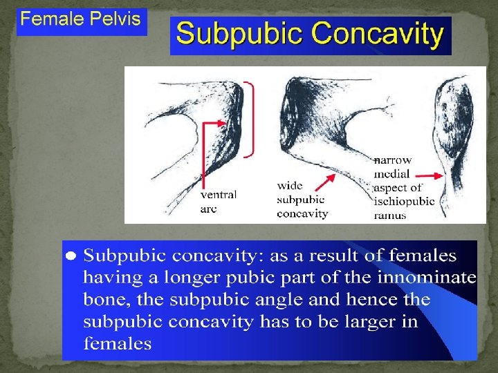
SUBPUBIC ANGLE LESS AND “V” SHAPED MALE WIDE AND TEND TO “U” SHAPED FEMALE The subpubic angle is much wider in females than in males, typically more that 90 degrees and less than 90 degrees, respectively.
It is the curved ridge of bone on anterior surface of the pubic bone. It is common in females and almost never seen in males.
PELVIC INLET The pelvic inlet is the space you see when both innominates and sacrum are articulated. The space in the middle of the pelvic bone (the pelvic inlet) is larger in women to facilitate birthing.
The sacrum is a large, triangular bone at the base of the spine and at the upper and back part of the pelvic cavity. The sacrum articulates with four bones: • the last lumbar vertebra above • the coccyx below • the hip bone on either side
Sexual dimorphism The sacrum is noticeably sexually dimorphic. In the female the sacrum is shorter and wider than in the male. The bone is also directed more obliquely backward; this increases the size of the pelvic cavity. Straighter in males & curved in females. MALE FEMALE
Slide 45: Using the previous slides discussing the pelvis, determine whether each of the pelvic bones pictured corresponds to a male or female. 1. Subpubic angle 3. Sacrum 2. Pelvic Inlet 4. Sciatic Notch
The sternum is an elongated, flattened bone, forming the middle portion of the anterior wall of the thorax. Its upper end supports the clavicles and its margins articulate with the cartilages of the first seven pairs of ribs. It consists of three parts, from above downward: • Manubrium • Body of sternum • Xiphoid process
<125 mm = female >155 mm = male Information from Bass
• The Essentials of Forensic Medicine By : Cyril John Polson , D. J. Gee , Bernard Knight • Encyclopedia of Forensic Sciences • Clinical Anatomy for Medical Students By : Richard S. Snell • Wikipedia • Personal Identification from skeleton or its remains By : G. S. Kaler & N. A. Butt
Male or Female