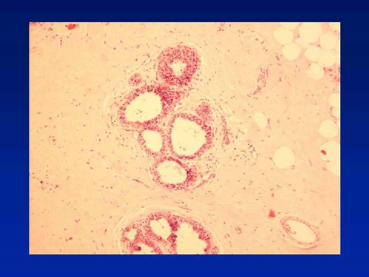
Session 5 Case Diagnosis Sclerosing adenosis with lobular

Session 5

Case

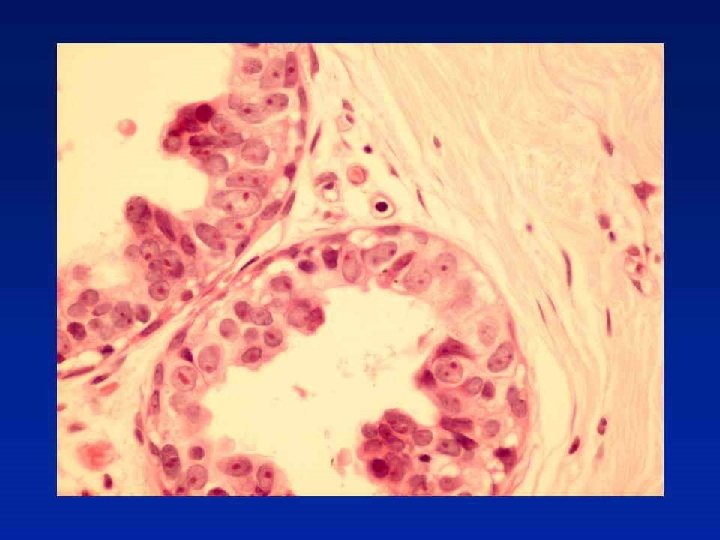
Diagnosis Sclerosing adenosis with lobular neoplasia. No invasion in images provided.

Update - Risk with LISN Meta-analysis 9 studies of 228 patients 15% ipsilateral, 9% contralateral carcinoma Ipsilateral 3 x more likely than contralateral • A “model of premalignancy for ALH intermediate between a local precursor and a generalised risk for both breasts” Page DL. Lancet. 2003; 361: 125 -9

Is there a sub-group of pre-invasive LCIS? LCIS with Microinvasion 6 LCIS with microinvasion described Nemoto T et al. J Surg Oncol 1998; 67; 41 -46

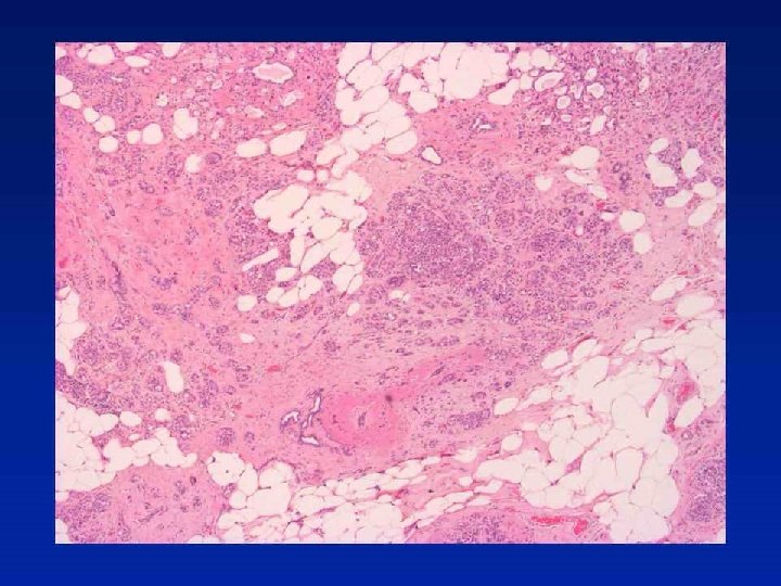
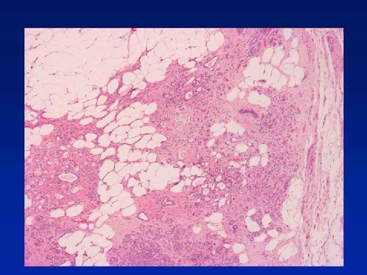
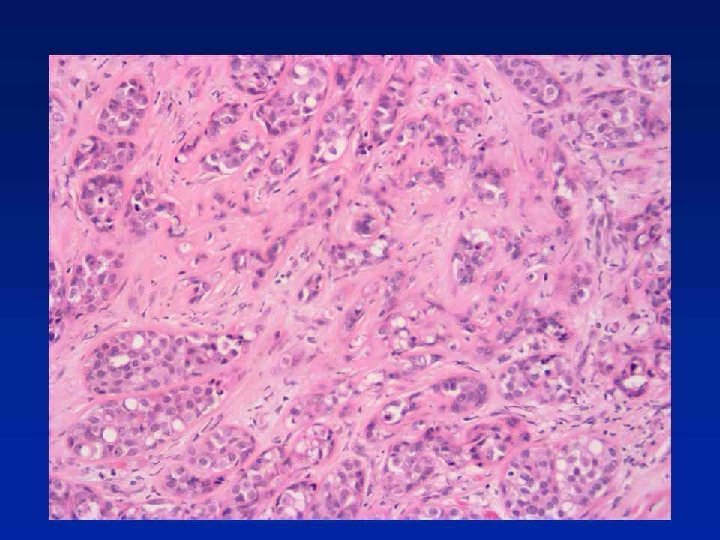
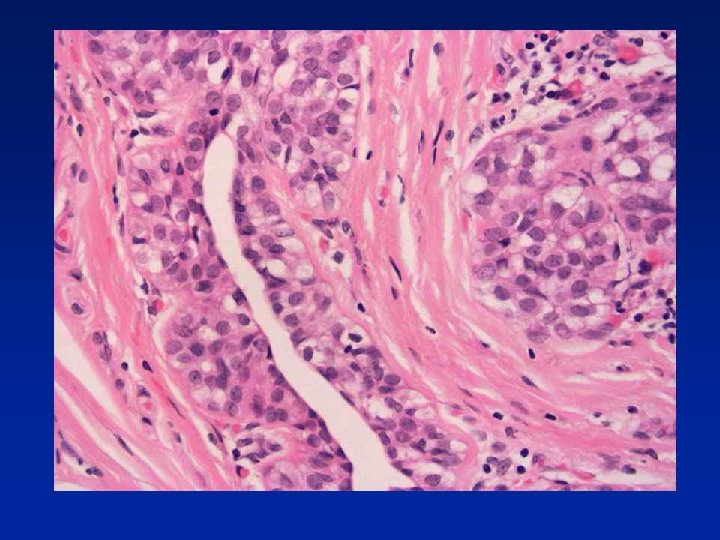
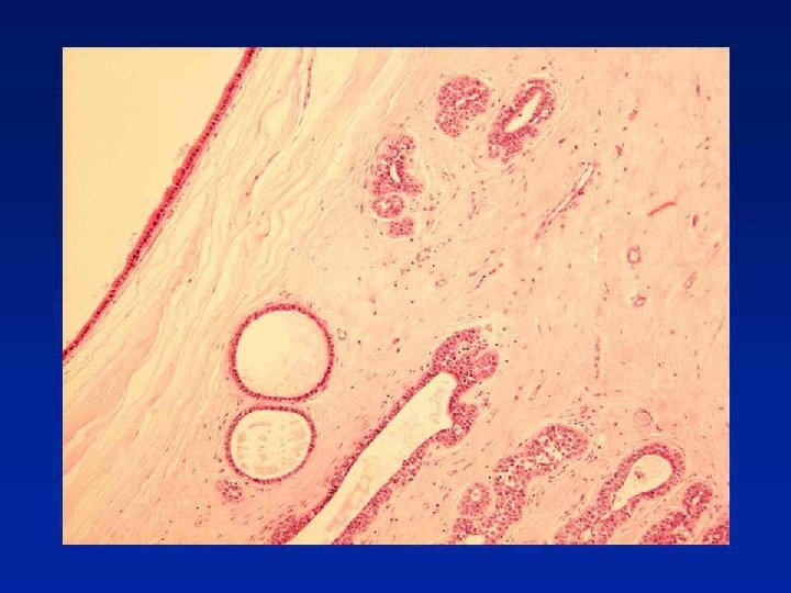
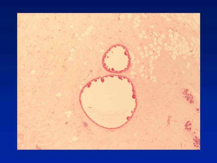
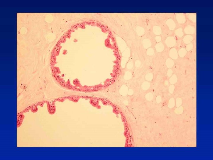
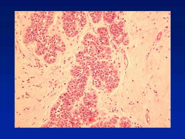
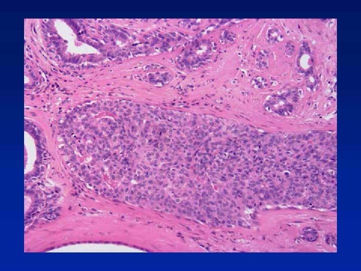
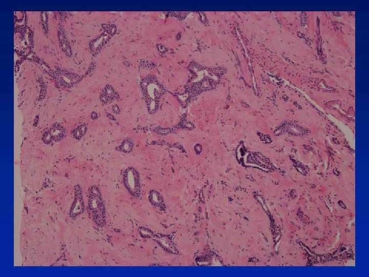
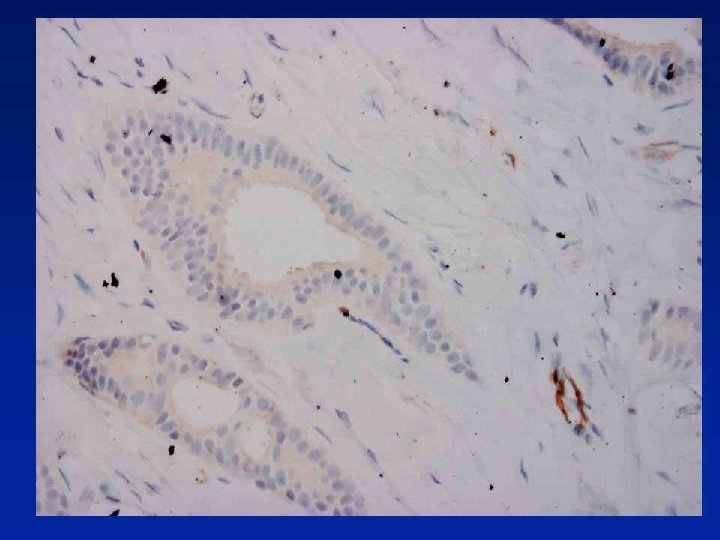
Case 19
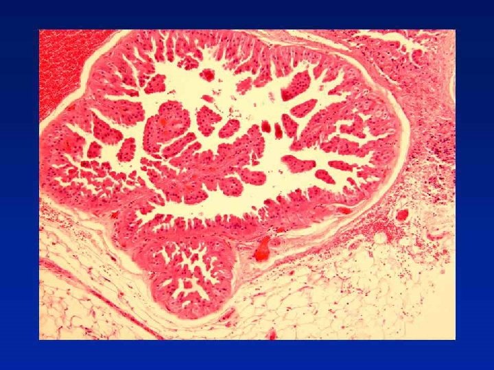
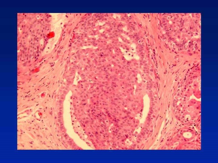

Diagnosis Fibrocystic change with papillomas and florid usual epithelial hyperplasia

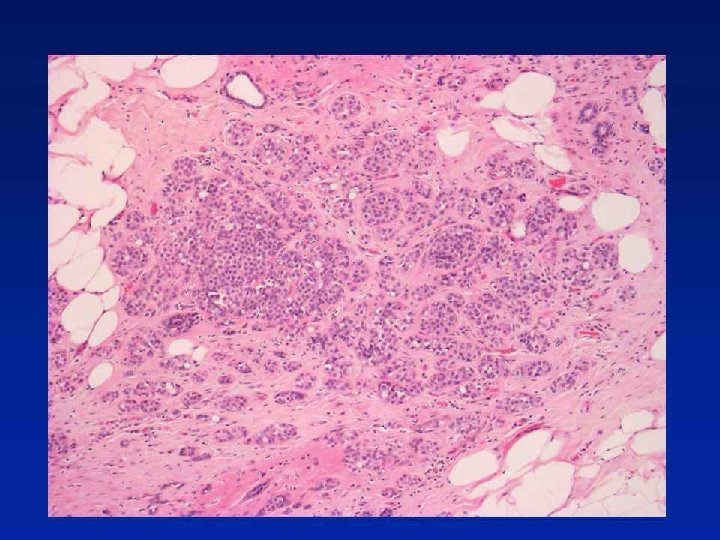
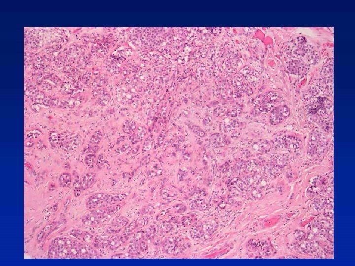
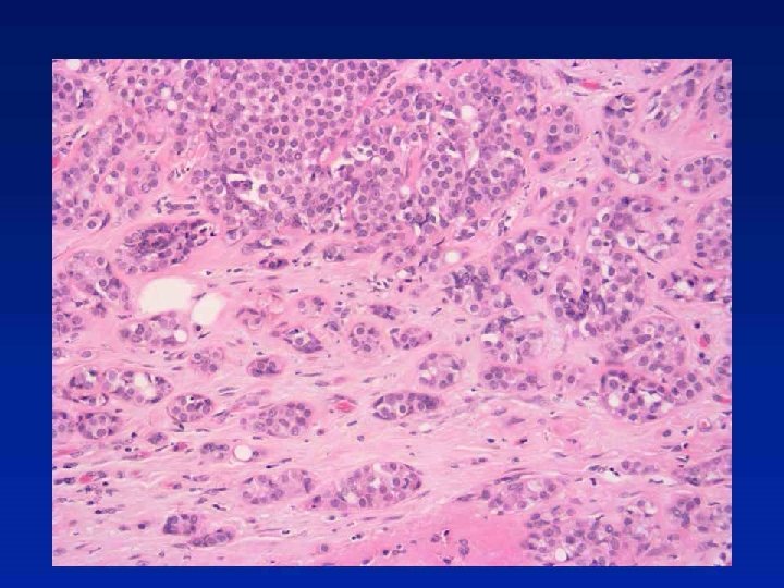
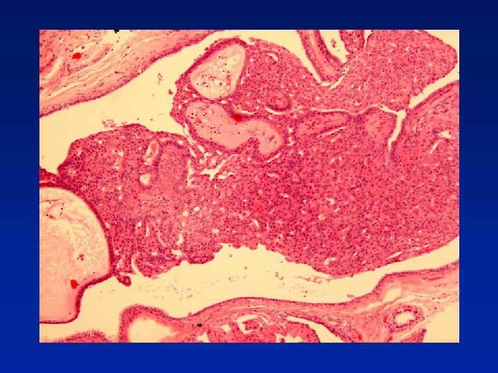
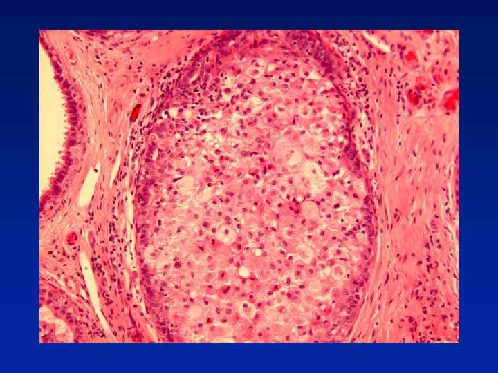
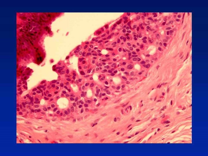
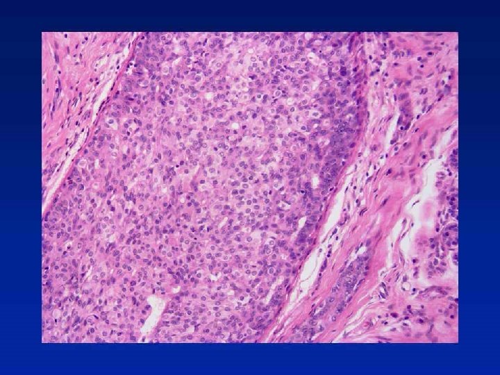
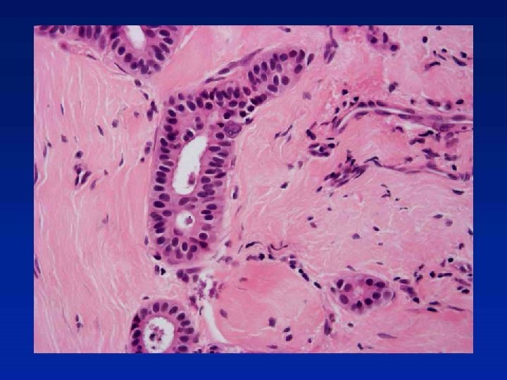
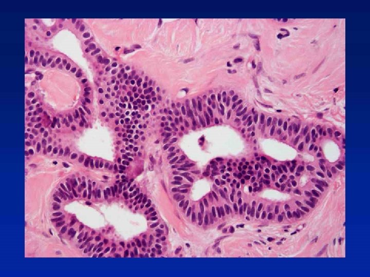
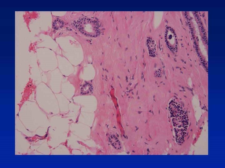
Case 32
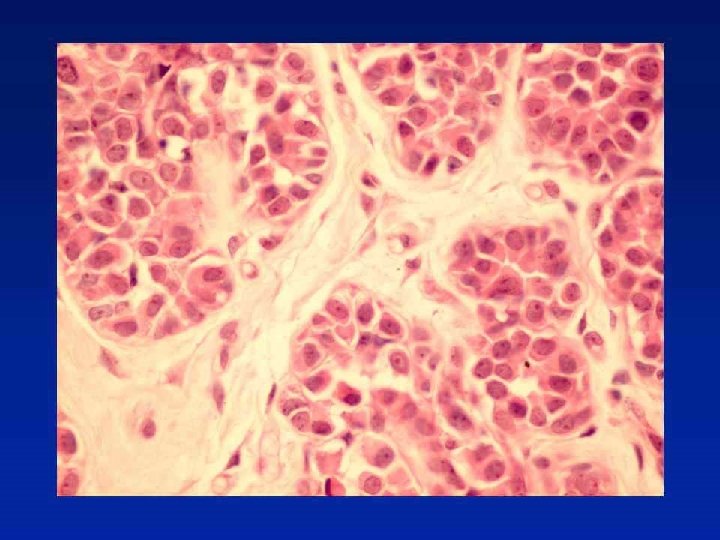
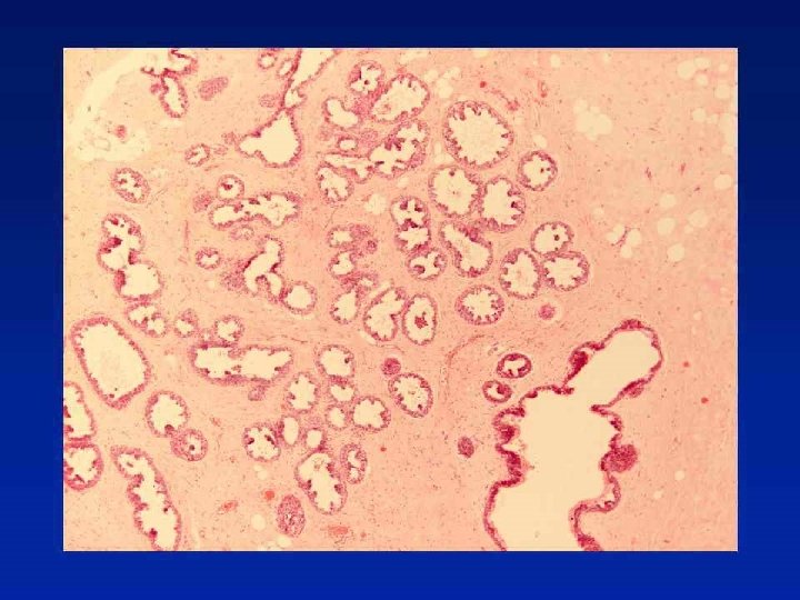
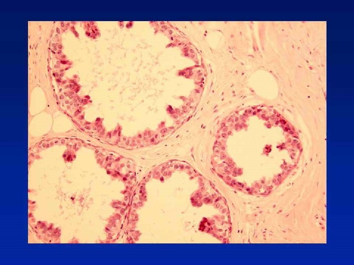
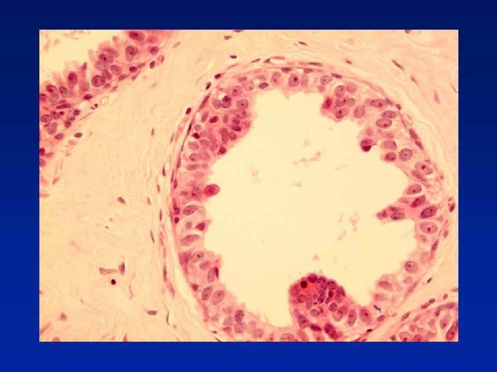
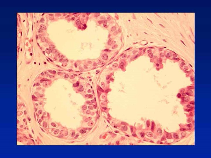

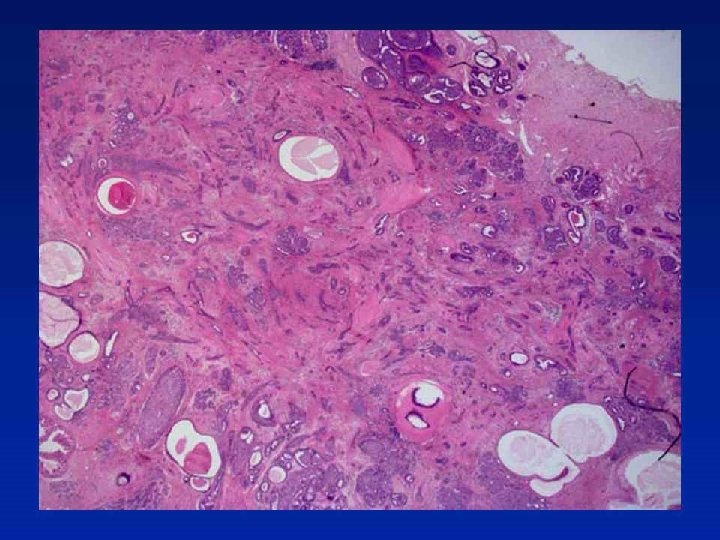
Diagnosis Fibrocystic change with atypical lobular hyperplasia and flat high grade DCIS

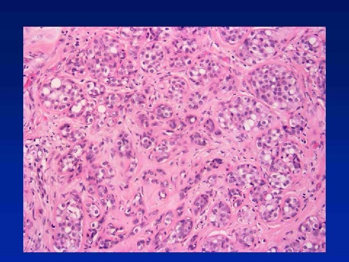
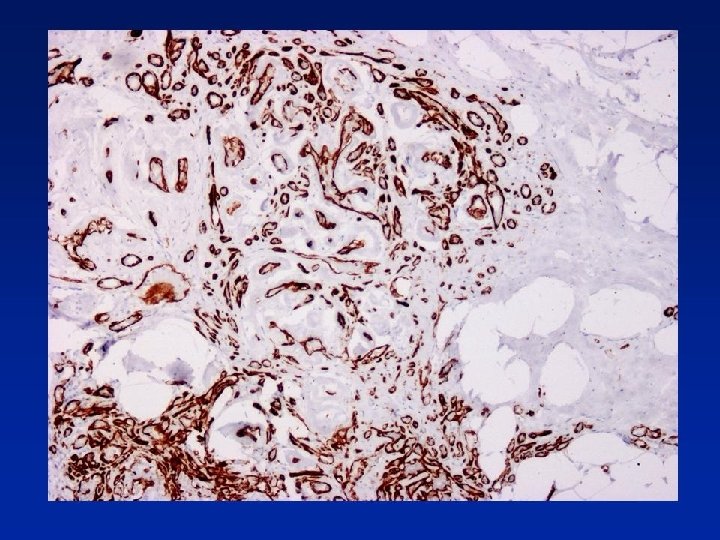
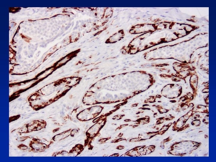
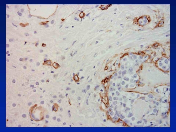
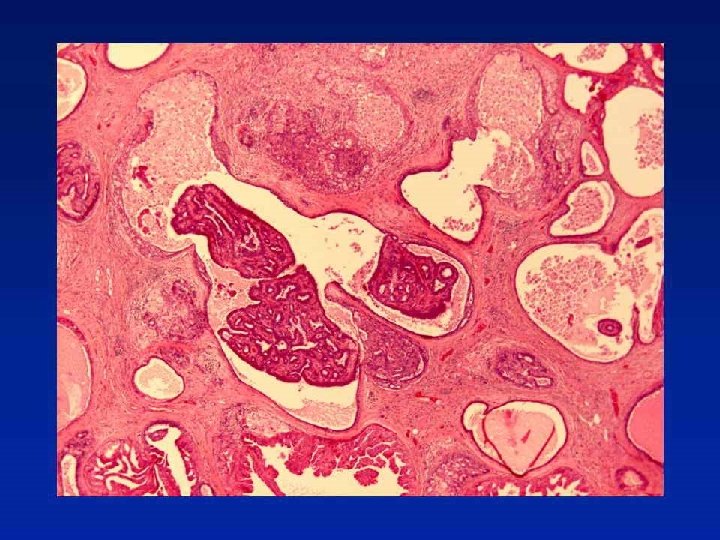
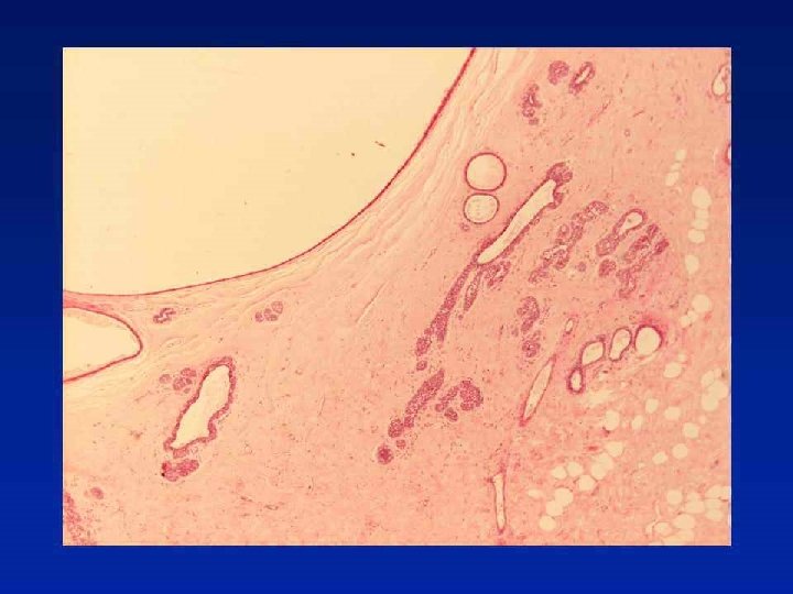
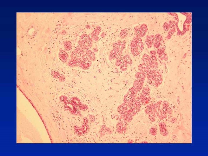
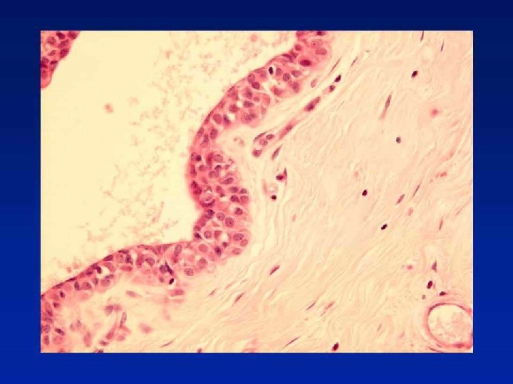
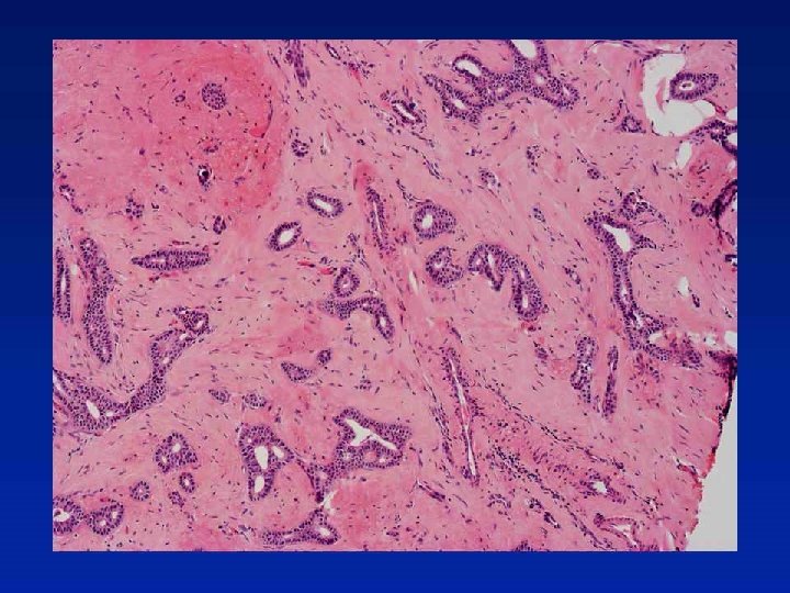
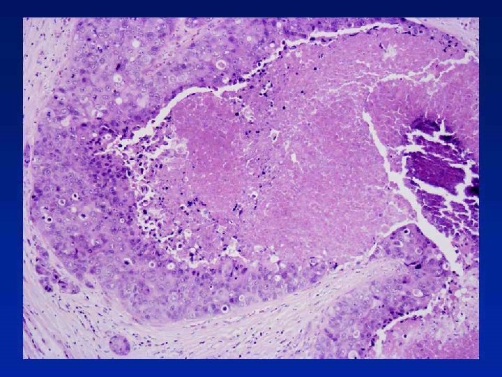
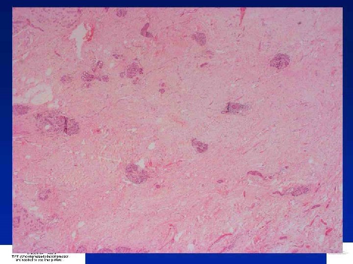
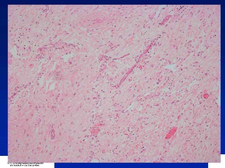
Case 54
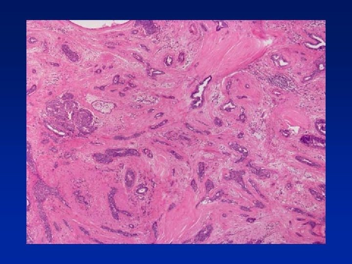
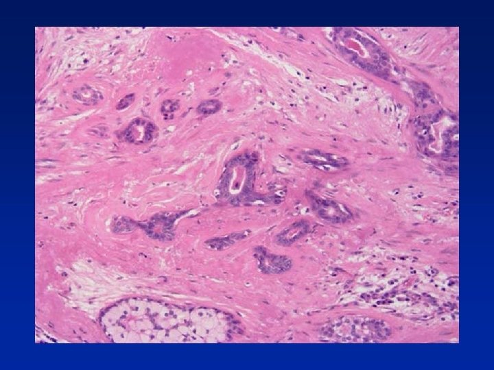

Diagnosis Radial scar with florid usual epithelial hyperplasia

Radial Scar • • • Central fibro-elastosis with entrapped tubular structures Usual epithelial hyperplasia Helpful features – retraction around tubular structures – often seen in radial scars Lack of fibroblastic stromal reaction (commonly, but not invariably seen (!) in tubular carcinoma) Confirm presence of myoepithelial cells and exclude diagnosis of tubular carcinoma (e. g. with smooth muscle myosin or smooth muscle actin or p 63) In the epithelial proliferation look for (a) mixed population, (b) streaming and (c) slit-like peripheral spaces

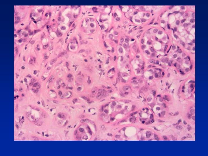
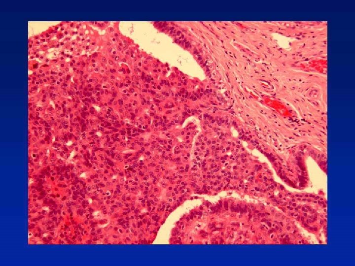
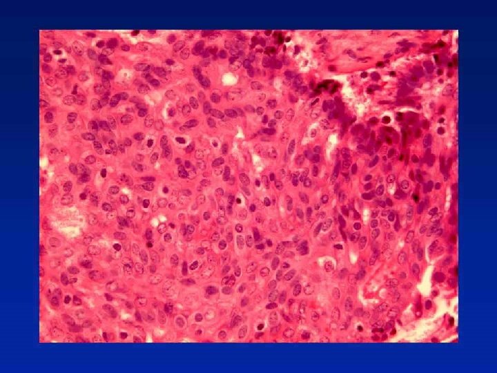
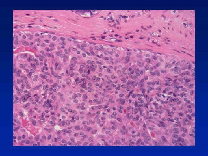
Case 44
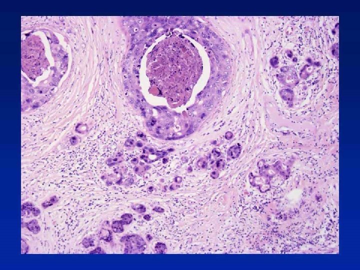
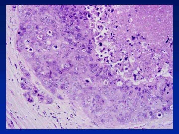

Diagnosis High grade DCIS with comedotype necrosis and cancerisation of lobules. No invasion or microinvasion.

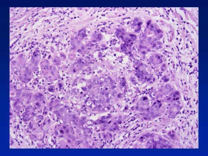
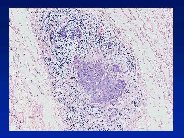
Microinvasion • DCIS with a focus of invasion less than 1 mm in max. dimension • More than one focus, if each less than 1 mm Individual deposits may vary in size from a few islands to 1 mm diameter • In the non-specialised, interlobular or inter-ductal connective tissue - neoplastic islands definitely within interlobular fibrous or adipose tissue Excludes: Ultrastructural or immunocytochemical evidence of breached or discontinuous basement membrane "Cancerisation of lobules"

Microinvasion Observations • Associated with high-grade comedo DCIS more than other types • Increasing risk of axillary node involvement with increasing duct space involvement (>50 ducts)

Microinvasion Problems of interpretation • • Duct boundary poorly defined Periductal fibrosis Indistinct basement membrane zone Tangential cutting of involved duct/lobule

Microinvasion Tips • Outside organoid structures • Involves non-specialised stroma • Host lymphocytic response • No myoepithelial component

Microinvasion Key points • • Restrictive definition Rare Axillary node involvement low Prognosis very good

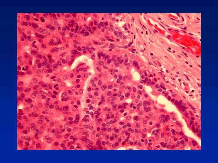
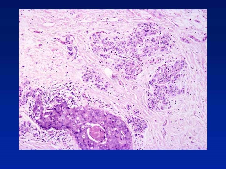
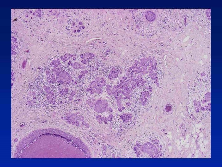
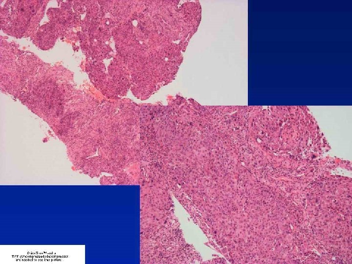
Case 11 a
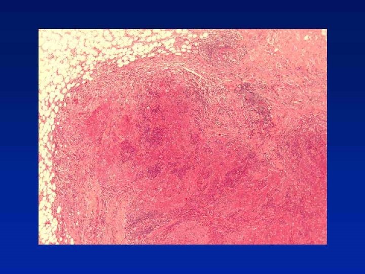
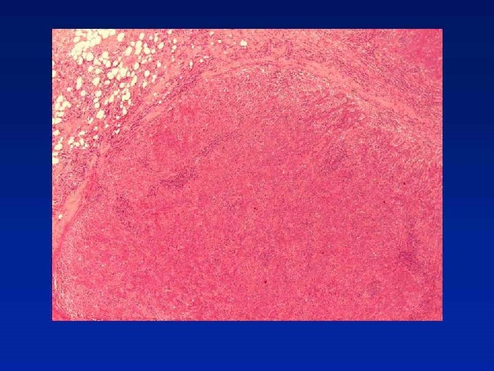
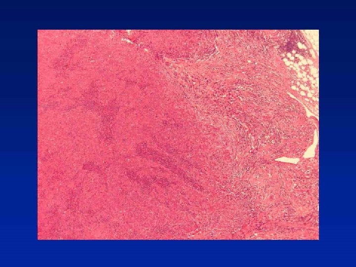
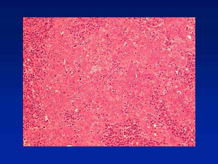

Diagnosis Medullary-like carcinoma

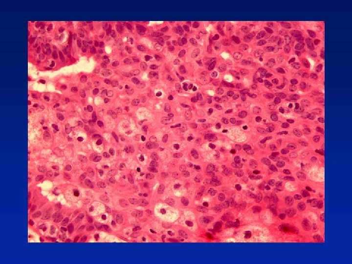
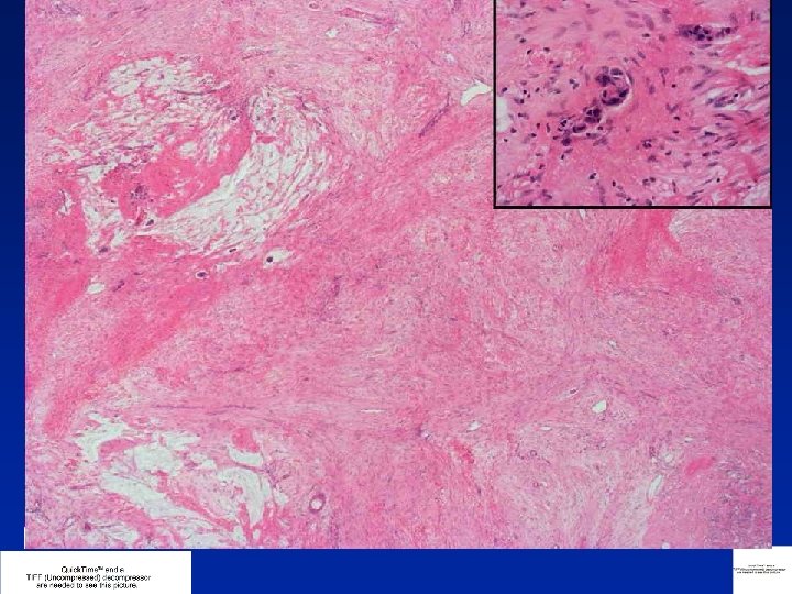
CASE ? • 36 year old patient • Mastectomy and ALND post chemotherapy • 5 months previously had core from 41 mm mobile hypoechoic mobile mass in RUOQ
 • • • grade")
Core Biopsy • B 5, invasive carcinoma of provisional (core) • • • grade 3 (333) and no special type ER = 6/8 PGR = 0/8 HER 2 = negative (score 0)
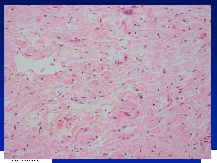
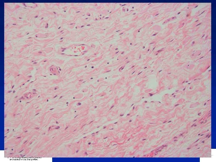
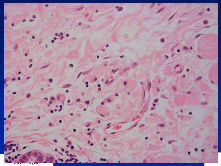
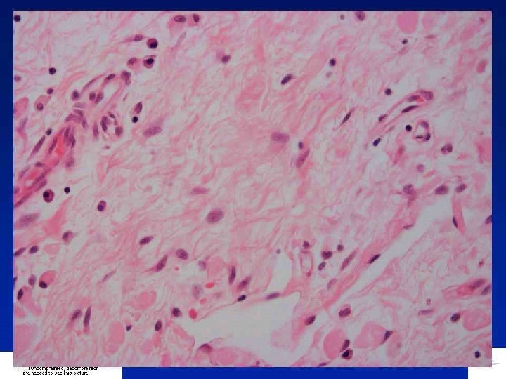
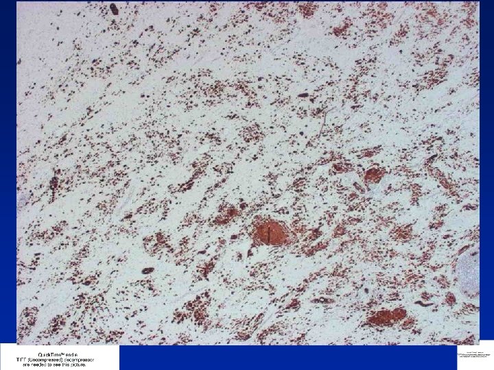
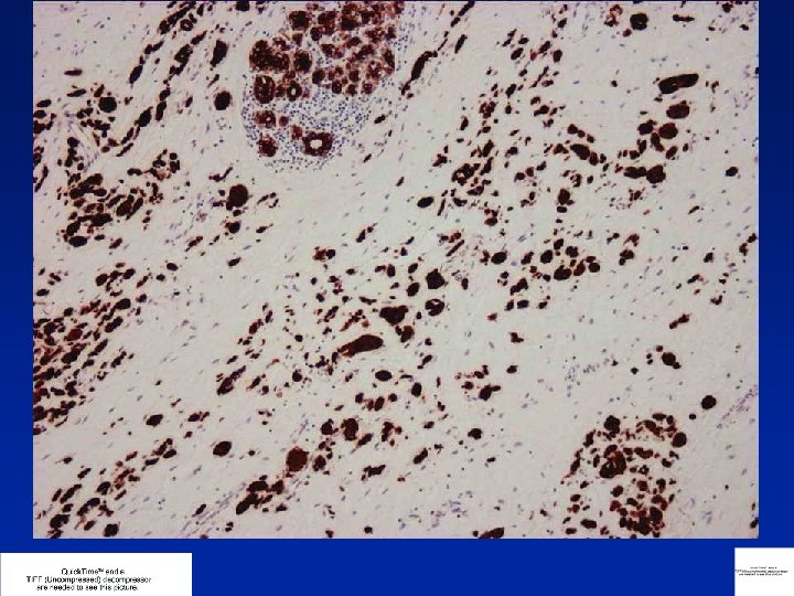
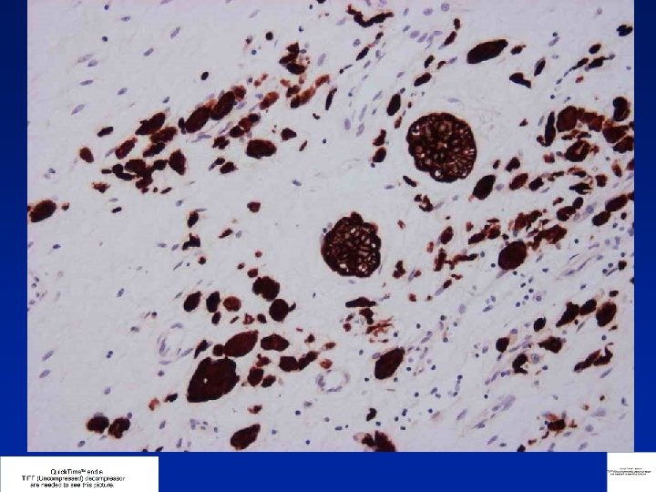
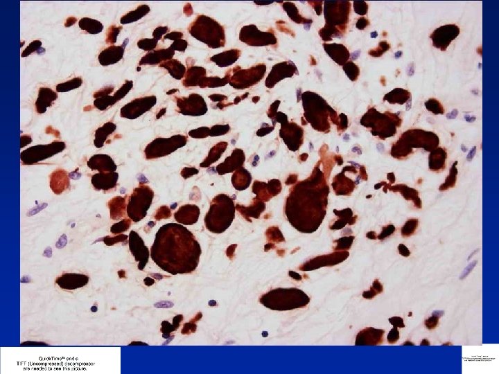
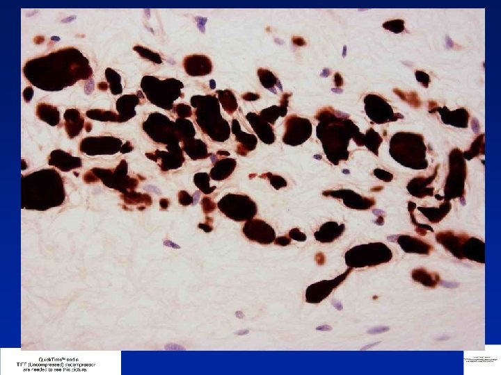

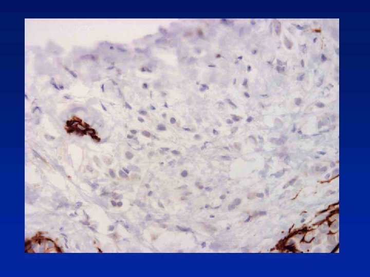
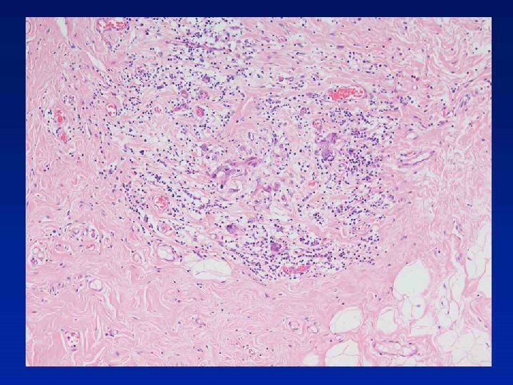
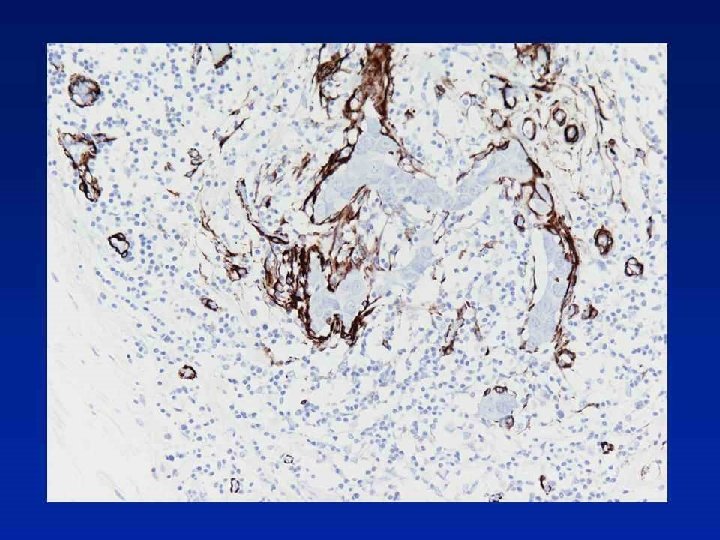
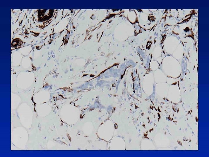
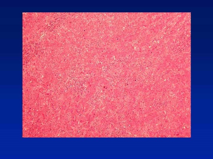
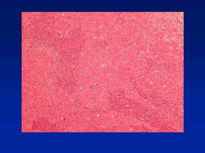
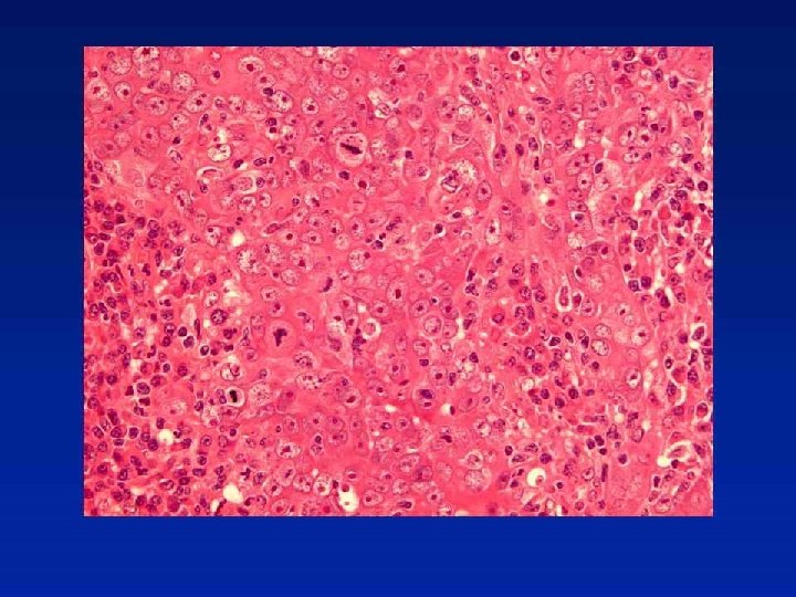
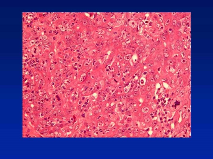
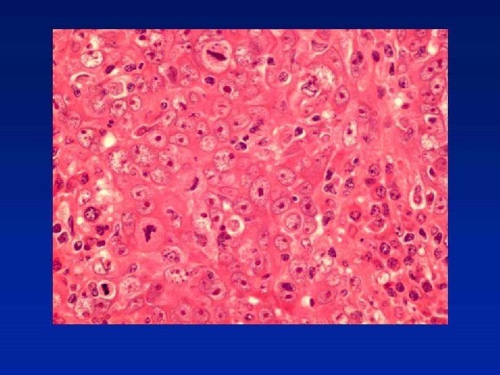
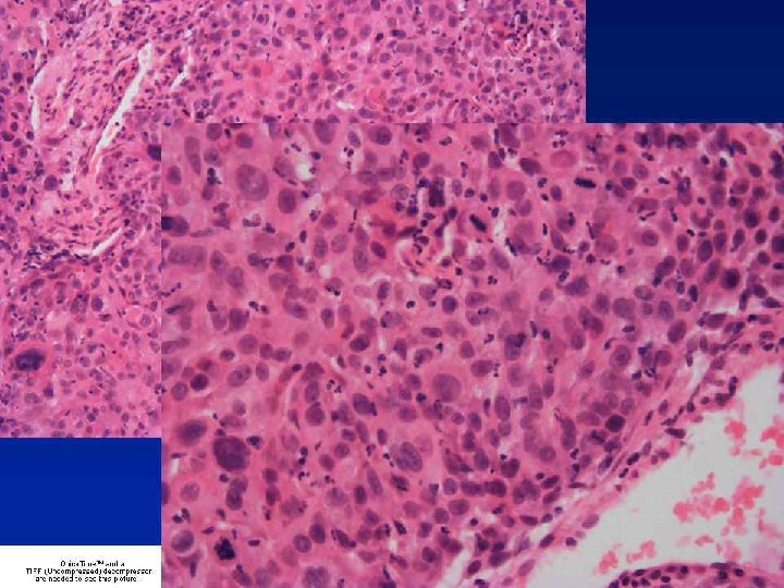
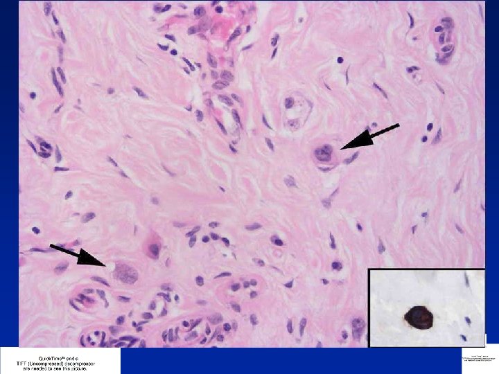
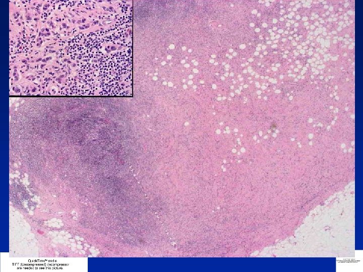
Diagnosis Residual carcinoma cells mimicking histiocytes

Post-Chemotherapy Changes • • • Central fibrotic scarring usually seen Patchy chronic inflammation within and around fibrosis Oedema or mucinous or myxoid changes to stroma or even areas of necrosis • Cancer cells may mimic histiocytes, and vice versa, but collections of macrophages may also be present Cytokeratin IHC invaluable •

Response to Chemotherapy Grade 1 Grade 2 Grade 3 Grade 4 Grade 5 Some alteration to individual cells but no overall reduction in numbers compared to pretreatment core Mild loss of invasive cells, still high cellularity Considerable reduction, up to 90% loss Marked reduction - only small clusters of widely dispersed cells detected No invasive carcinoma, in situ carcinoma or tumour stroma may still be noted Bonadonna G et al. J Natl Cancer Inst. 1990 3; 82: 1539 -45 Smith IC et al. J Clin Oncol 2002; 20: 1456 -66

Response to Chemotherapy 1. Disappearance of all tumour 2. Presence of in situ carcinoma but no residual invasive tumour & no metastatic carcinoma found in the lymph nodes 3. Invasive carcinoma present with stromal changes, such as sclerosis or fibrosis 4. Few modifications of appearance of tumour Chevallier B et al. Am J Clin Oncol 1993; 16: 223– 228

- Slides: 112