Sertoli Cell Structure Functions Sertoli Cell Named after

 They represent 35 - 40")


")

 Rete Testis Seminiferous Tubule Fibrous Trabeculae")
 Duct Mesonephric Tubules Gonad Mesonephric (Wolffian) Duct Gubernaculum Prostate (Male) / Skene")
 Müllerian Duct (Paramesonephric D) 1.")


 Penis (Glans,")
 Androgen Binding Protein (ABP)")


 § A single duct")
")
")


- Slides: 37
Sertoli Cell Structure & Functions
Sertoli Cell § § Named after Enrico Sertoli (1865) They represent 35 - 40 % of Sem. Epith. volume They divide till 1 st meiotic division occurs (15 Y) They have membrane bound FSH receptors & nuclear androgen receptors
Spermatid Sertoli Cell 1 ry Spermatocyte Spermatogonium Sertoli Cell
Sertoli Cell Functions 1. Physical Support of Seminiferous Tubules
Sertoli Cell Functions 1. Physical Support of Seminiferous Tubules 2. Blood-Testis Barrier
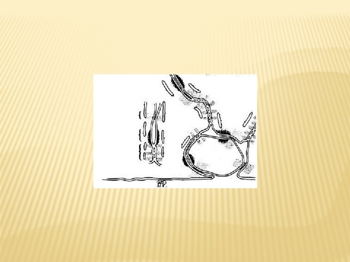
Sertoli-Sertoli Cell Junctions
Lumen of Sem. tubule Sperm Sertoli Cell Spermatids Secondary Spermatocyte Primary Spermatocyte Tight Junction Spermatogonium Sertoli Cell
Sertoli Cell Functions § Inter-Sertoli cell tight & gap junctions to form 2 compartments : • Basal (blood milieu) • Adluminal (special milieu) § § It is a highly dynamic structure that undergo disintegration and reconstruction It is made by membrane proteins (occludins, claudins & junctional adhesion molecules)
Sertoli Cell Functions § It has 2 functions : • Protects spermatocytes and spermatids from the immune system • Creates a specific milieu in the adluminal compartment
Sertoli Cell Functions § It has 2 functions : • Protects spermatocytes and spermatids from the immune system • Creates a specific milieu in the adluminal compartment § Basal compartment contains spermatogonia and young spermatocytes & adluminal compartment contains mature 1 ry spermatocytes onwards
Lumen of Sem. tubule Sperm Sertoli Cell Spermatids Secondary Spermatocyte Primary Spermatocyte Tight Junction Spermatogonium Sertoli Cell
Sertoli Cell Functions 1. Physical Support of Seminiferous Tubules 2. Blood-Testis Barrier 3. Support Spermatogenesis
Sertoli Cell Functions § They support spermatogenesis both : • Physically (cell skeleton structures, cell surface structures & cell adhesion molecules) • Chemically (secretory products)
Sertoli Cell Functions 1. Physical Support of Seminiferous Tubules 2. Blood-Testis Barrier 3. Support Spermatogenesis 4. Facilitate Spermiation
Sertoli Cell Functions 1. Physical Support of Seminiferous Tubules 2. Blood-Testis Barrier 3. Support Spermatogenesis 4. Facilitate Spermiation 5. Secretory Functions
Sertoli Cell Functions • Müllerian Inhibiting Factor (MIF)
Development of Male Genital System
Vas Deferens Epididymis Efferent Ducts (6 - 12) Rete Testis Seminiferous Tubule Fibrous Trabeculae (250 -300 lobules each has 1 – 3 tubule)
Paramesonephric (Mϋllerian) Duct Mesonephric Tubules Gonad Mesonephric (Wolffian) Duct Gubernaculum Prostate (Male) / Skene Gland (Female) Genital Duct Systems
Development of The Duct System Wolffian Duct (Mesonephric D) Müllerian Duct (Paramesonephric D) 1. Efferent Ducts (Meson. T Undergo Regression Under (MIH) except …… of Meson. D) (8 - 12 w) 2. Epididymis 3. VD Under Testosterone (12 th w) 4. SV 5. ED
XX XY Primitive Gonad SRY Testis MIF Leydig Cell Sertoli C. Disappearance of MD 5 α reductase Ovary Female Phenotype Testosterone DHT Int. Genitalia § Epididymis § VD § SV § ED Ext. Genitalia § Penis § Scrotum § Prostate § BU Gland
Glans Area Epithelial Tag Genital Tubercle Urogenital Fold Urogenital Groove Lateral Part of the Tubercle Anal Pit External Genitalia
Glans Area Epithelial Tag Urogenital Fold Urogenital Groove Genital Tubercle Lateral Part of the Tubercle Anal Pit Glans Epithelial Tag Coronal Sulcus 10 th Week Urethral Fold Urogenital Groove Lateral Tubercle (Corpus) Labio-scrptal Swelling Urethral Raphé Anal Tubercle Anus
Body of Clitoris External urethral Orifice Prepuce Glans Penis Glans Prepuce External urethral Orifice Shaft of the Penis Labium Minora Labium Majora Raphe of Penis Perineal Raphé Scrotum Vaginal orifice Anus Male Posterior Commisure Female
Development of External Genitalia Female Male Genital Tubercle Clitoris (Glans & Corpora) Penis (Glans, Corpora cavernosa & Corpus spongiosum) Urogenital Folds § Labia minora § Perineal raphé § Perianal tissue & Ext. anal sphincter § Ventral aspect of penis § Most of penile urethra § Perineal raphé § Perianal tissue & Ext. anal sphincter Labio-scrotal Folds Labia majora Scrotum
Sertoli Cell Functions • • • Müllerian Inhibiting Factor (MIF) Androgen Binding Protein (ABP) Inhibin-B & Activin Transferrin (Iron transport) Retinol Binding Protein (vit-A transport) Glutamyl Transpeptidase (Amino acid transport) SPARC (Calcium transport) Ceruloplasmin (Copper transport) Proteases & Antiproteases Growth Factors & Cytokines () Fluids
Sertoli Cell Functions 1. Physical Support of Seminiferous Tubules 2. Blood-Testis Barrier 3. Support Spermatogenesis 4. Facilitate Spermiation 5. Secretory Functions
Epididymis Functions
Epididymis § Epi = above & didimos = testis (Greek) § A single duct formed of head, body & tail § It is lined by pseudo-stratified ciliated columnar epithelium
Epididymis Functions 1. Sperm Transport (hydrostatic pressure, cilia & myoepithelial cells)
Epididymis Functions 1. Sperm Transport 2. Sperm Maturation § Motility (L-carnitine & α glucosidase) § Fertilizing Ability (increase glycolysis, -ve charges on sperm surface, maturation antigens on sperm surface, sialic acid, zinc, inositol, glycerophosphoryl choline & Dgalactosidase)
Epididymis Functions 1. Sperm Transport 2. Sperm Maturation 3. Sperm Protection § Chemical (antioxidants & proteinase inhibitors) § Immunological (Blood–epididymis barrier & immunosuppressive factors)
Epididymis Markers § L-carnitine § α glucosidase § Glycerophosphoryl choline
Capsule Summary § § § Sertoli cell forms the Blood-Testis barrier and has many secretory functions that support spermatogenesis Blood-Testis barrier divides the ST into Basal & Adluminal compartments Epididymis helps in sperm Transport, Maturation as well as Protection
Thank You