Sensory systems Chapter 50 Five senses Touch Taste


 Located")


 Slow breathing")


 Distinguish thousands of")


 Eustachian tube Small")


- Slides: 45
Sensory systems Chapter 50
Five senses Touch Taste Smell Sound Vision
Sensory systems Sensory info is received Nerve impulse or action potential All or nothing response Response depends on part of brain that receives the info
Sensory information Sensory info to CNS 1. Sensory reception 2. Transduction Graded potential Ion channels open or close Receptor potential Change in membrane potential Depolarization
Sensory information 3. Transmission Goes to CNS via afferent pathway 4. Interpretation Perception by the brain
Sensory receptors Nerve endings Specialized neurons or epithelial cells Associated with sensory organs -eyes All stimuli is a form of energy
Sensory receptors Stimuli-outside body Heat, light, pressure & chemicals Stimuli-inside body BP, body position, body temperature
Types of sensory receptors Mechanoreceptors – Pressure, stretch, touch Chemoreceptors – chemicals Electromagnetic receptors (photoreceptors) Nociceptors (pain) Thermoreceptors
Receptors Cutaneous receptors: Skin Heat, cold, pressure, pain & touch Thermoreceptors Heat/Cold Hypothalamus – Regulates temp of blood (core temp)
Receptors Mechanoreceptors Touch Close to surface of skin Hair follicle receptors Pressure Deeper
skin Heat Gentle touch Pain Cold Hair Epidermis Dermis Hypodermis Nerve Connective tissue Hair movement Strong pressure
Receptors Nociceptors: Pain Severe temperature change Tissue damage Free nerve endings (naked dendrites) Located in the epidermis
PAIN
Receptors Proprioceptors Give info on animal’s body parts Position Movement Stretch receptors on muscle Prevent over stretch
Receptors Baroreceptors: Detect tension or stretch in blood vessel walls Internal carotids Aortic arch Drop in BP Stimulation to increase HR & vasoconstriction
Receptors Chemoreceptor Aorta & carotid Medulla oblongata p. H (blood & CSF) Slow breathing Increased CO 2 Lowers p. H Causes an increased respiration rate
Taste buds Collections of receptor cells Epithelial cells Papillae Raised areas on tongue Taste buds located
Taste buds contain 50 -100 taste cells Food dissolves in saliva Contact taste cells Taste salty, sweet, sour, bitter
Taste Chemoreceptors Salt: Na+1 Sour: H+1 Directly through ion-channel Sweet: receptor proteins for sugar Bitter: K+channels are closed by receptor proteins
TONGUE
Sweet Sugar molecule G protein Sweet receptor Tongue Taste pore Taste bud Sugar molecule Phospholipase C SENSORY RECEPTOR CELL PIP 2 Sensory receptor cells IP 3 (second messenger) Sensory neuron Nucleus ER IP 3 -gated calcium channel Ca 2+ (second messenger) Sodium channel Na+
Smell Olfactory receptors Chemoreceptors Located upper portions of nasal passages Dendrites are in cilia Axon goes directly to cerebral cortex Odorant or odorous substance binds proteins Second messenger response in receptor cell
Smell Opens membrane to Ca & Na Causes impulse (action potential) Distinguish thousands of odors Very accurate Single odorant molecule
NOSE
Nose Brain Action potentials Olfactory bulb Odorants Nasal cavity Bone Epithelial cell Odorant receptors Chemoreceptor Plasma membrane Odorants Cilia Mucus
Hearing Outer ear: Pinna, canal Middle ear: Tympanic membrane (ear drum) Eustachian tube Small bones (malleus, incus, stapes) Inner ear: Cochlea, auditory nerve
EAR
Ear
Hearing Vibrations move in canal Cause eardrum to move Vibrations pass through the bones Stapes pass vibration to inner ear Causes pressure waves in fluid in cochlea Basilar membrane of the cochlea vibrates
Hearing Hair cells on membrane vibrate Leads to change in membrane potentials in sensory neurons Sound interpreted Humans hear 20 -20, 000 hertz Age decreases higher frequencies Dogs hear sounds at 40, 000 hertz
Ears Inner ear Body position & balance Two chambers near the cochlea Utricle & saccule Filled with fluid Hair cells in chambers respond to changes in head positions
Ears Utricle: horizontal motion Saccule: vertical motion Different movement causes different sensory neurons to be stimulate Labyrinth system Spin around become dizzy
Equilibrium Semicircular canals Flow of fluid Vestibular nerve Cupula Hairs Hair cells Axons Vestibule Utricle Saccule Body movement
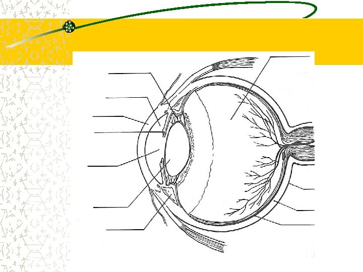
Eye Sclera: White outer layer of connective tissue Conjunctiva: Epithelial layer Covers outer surface of sclera Under surface of the eyelid Cornea: Clear part of sclera, light passes through
Eye Choroid Pigmented layer under the sclera Iris Color part of eye formed by the choroid Pupil Opening at the center of the iris Controlled by iris Lens Behind the pupil, held in place by ligaments
Eye Retina Back of eye where image is focused Optic nerve Sensory neurons Vitreous humor Jellylike substance behind the lens Aqueous humor Thinner fluid Fills smaller chamber in front of the lens
EYE
Eye Light enters eye through cornea Passes through pupil to lens Lens focuses images on retina Photoreceptor cells of retina transduce light energy Action potentials pass via sensory neurons in the optic nerve
Eye Rods & cones Photoreceptors of eyes Rods: black and white vision in dim light Cones: high visual acuity & color vision Located in center of retina
EYE
Rods/cones Retina Light Photoreceptors Neurons Retina Choroid Cone Rod To brain Optic nerve Light Ganglion cell Amacrine cell Optic nerve axons Horizontal cell Bipolar cell Pigmented epithelium
Eyes Binocular vision Axons of ganglion cells form optic nerves Optic nerves meet at the optic chiasm (base of the cerebral cortex) Visions from the right visual field go to the left side of the brain and vise versa Thalamus Cortex
Vision Right visual field Optic chiasm Right eye Left visual field Optic nerve Lateral geniculate nucleus Primary visual cortex
Eyes Nearsightedness: longer eyeball Farsightedness: shorter eyeball Asitgmatism: problems with lens or cornea Light rays converge unevenly Colorblindness: inherited lack of one or more types of cones