Sensory Systems Chapter 45 Sensory Receptors Exteroceptors vs


 aid in “taste” Salty and sour are tasted directly")


- Slides: 30
Sensory Systems Chapter 45
Sensory Receptors • Exteroceptors vs Enteroceptors • Three categories of receptors – Mechanoreceptors- examples touch hearing and balance – Chemoreceptors- examples taste and smell – Energy-detecting receptors- sight ( some special cases as well)
Steps in Conveying sensory information • 1. Stimulation • 2. Transduction- energy is transformed into graded potentials in the dendrites of sensory neurons. • 3. Transmission- Action potentials develop are moved to the CNS • 4. Interpretation- The brain creates a sensory perception from the information given by the afferent stimulation.
Mechanoreceptors • Touch– Merkle cells- located near the skin surface detect slight pressure – Meissner Corpuscle- Located below merkle cellssensitive to fine touch – Ruffini Corpuscle- Located below Meissner Corpuscle- sensitive to touch and pressure duration – Pacinian Corpuscle- Deep below skin- sensitive to pressure
• http: //porpax. bio. miami. edu/~cmallery/1 50/neuro/c 7. 49. 3. skin. jpg
Thermoreceptors • Found in the epidermis as well as in the Hypothalamus • Different receptors for hot and cold
Some Interoreceptors that are also mechanoreceptors • Proprioceptors • Baroreceptors
More Mechanoreceptors • Hearing, vibration , and detection of Body Position • Fish- can detect positions of things using vibrations, they can also hear • Humans and other vertebrates- Can only hear
Fish • Lateral Line System • Fish Hearing- Use Otiliths
Humans • http: //www. infj. ulst. ac. uk/~pnic/Human. E ar/Andy's%20 Stuff/MSc. Project/workingc ode_Local/humanear. jpg
Transduction Occurs in The Cochlea
Body Position • Statocysts and statoliths
Chemorecptors • Taste, Smell, and p. H
Taste • Taste buds (papillae) aid in “taste” Salty and sour are tasted directly • Salt taste due to Na+ ions • Sour due to H+ • Sweet, bitter and Umami are idirectly trasnferred to CNS by G protiein receptors
• Flies taste with their feet • Fish taste with their scales
Smell • Only animals exposed to Air can smell • We can discern thousands of different smells, but only a few tastes • Smell using Olfactory Bulb
Internal Chemoreceptors • Regulate p. H and Glucose Levels • Hypoventilation vs Hyperventilation
Energy Detecting Receptors • • Vision Infared Radiation in Snakes Electrical Currents in some Fish Magnetic Fields in Birds
Evolution of Vision • Invertebrated eyes- have photoreceptors clustered in eyespots • Cannot detect and image, just the presence or absence of light – Example flatworms
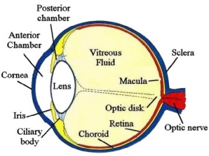
Structure of the Vertebrate Eye
Focusing the Human Eye • To see close- Ciliary Muscle constricts, syspensory ligament relaxes , lens curves • To see far- Ciliary muscle relaxes, suspensory ligament constricts, lens flattens
Cones and Rods • Rods- Shaped like rods- black and white vision • Cones- shaped like cones, used for color images – Colors that you can see depend on the rods you have. Most humans have 3 colored rods
Retina Made of of 3 layers of cells • Rods and cones are closes to the surface of the eye • Bipolar cells are next • Then there are the ganglion cells • Light must first go though the ganglion cells then the bipolar cells to reach the photoreceptors.
• 1. Light passes through the cornea which helps focus • 2. Behind the cornea is a colored ring-shaped membrane called the iris. The iris has an adjustable circular opening called the pupil, which can expand or contract depending on the amount of light entering the eye.
• 3. A clear fluid called the aqueous humor fills the space between the cornea and the iris. • 4. Situated behind the pupil is a colorless, transparent structure called the crystalline lens. Ciliary muscles surround the lens. When the muscles relax, they pull on and flatten the lens, allowing the eye to see objects that are far away. To see closer objects clearly, the ciliary muscle must contract in order to thicken the lens.
• 5. The interior chamber of the eyeball is filled with a jelly-like tissue called the vitreous humor. After passing through the lens, light must travel through this humor before striking the sensitive layer of cells called the retina. • 6. The retina is the innermost of three tissue layers that make up the eye. The outermost layer, called the sclera, is what gives most of the eyeball its white color. The cornea is also a part of outer layer. The middle layer between the retina and sclera is called the choroid. The choroid contains blood vessels that supply the retina with nutrients and oxygen and removes its waste products.
• 7. Embedded in the retina are millions of light sensitive cells, which come in two main varieties: rods and cones. Rods are good for monochrome vision in poor light, while cones are used for color and for the detection of fine detail. Cones are packed into a part of the retina directly behind the retina called the fovea. • 8. When light strikes either the rods or the cones of the retina, it's converted into an electric signal that is relayed to the brain via the optic nerve. • 9. The brain then translates the electrical signals into the images we see.
Visual Processing Takes Place in the Cerebral Cortex • Binocular Vision • Color Blindness
Echolocation • Aquanetta