Sensory Receptors Figure 50 3 a Receptor is

 Receptor is afferent neuron. (b) Receptor regulates afferent neuron. To")
 Single sensory receptor activated Gentle pressure Sensory receptor Low")
 Multiple receptors activated Sensory receptor Gentle pressure Fewer receptors")

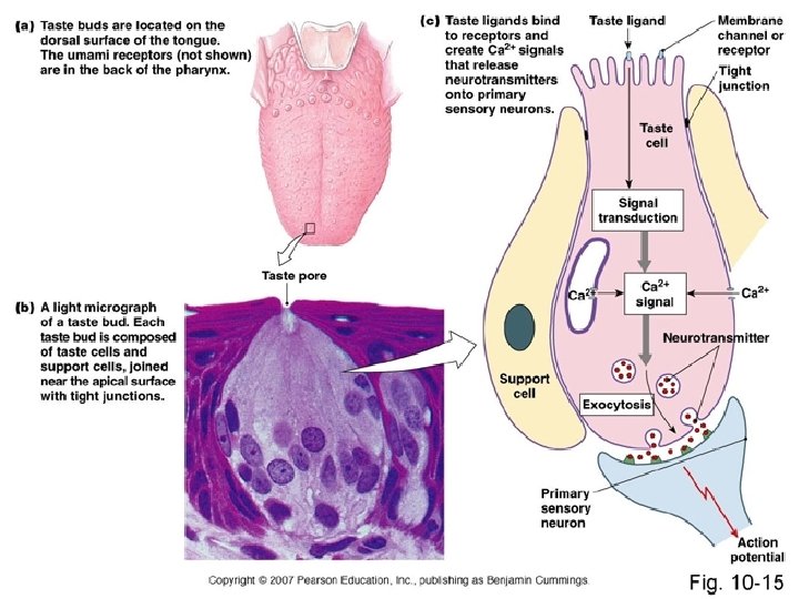
 Tongue Key Taste bud Sweet Salty Sour")


- Slides: 21
Sensory Receptors
Figure 50. 3 (a) Receptor is afferent neuron. (b) Receptor regulates afferent neuron. To CNS Afferent neuron Receptor protein Neurotransmitter Sensory receptor Stimulus Sensory receptor cell Stimulus leads to neurotransmitter release. Stimulus
Figure 50. 4 a (a) Single sensory receptor activated Gentle pressure Sensory receptor Low frequency of action potentials per receptor More pressure High frequency of action potentials per receptor
Figure 50. 4 b (b) Multiple receptors activated Sensory receptor Gentle pressure Fewer receptors activated More pressure More receptors activated
Figure 50. 5 Gentle pressure, vibration, and temperature Connective tissue Hair Pain Epidermis Dermis Strong pressure Hypodermis Nerve Hair movement
Figure 50. 24 Papillae Taste buds (a) Tongue Key Taste bud Sweet Salty Sour Bitter Umami Taste pore Sensory neuron (b) Taste buds Sensory receptor cells Food molecules
Figure 50. 25 Brain Action potentials Olfactory bulb Odorants Nasal cavity Bone Epithelial cell Receptors for different odorants Chemoreceptor Plasma membrane Odorants Cilia Mucus
Figure 50. 17 aa Sclera Choroid Retina Suspensory ligament Fovea Cornea Iris Optic nerve Pupil Aqueous humor Lens Vitreous humor Optic disk Central artery and vein of the retina
Figure 50. 17 ab Retina Neurons Optic nerve fibers Photoreceptors Rod Cone Amacrine Horizontal cell Bipolar Ganglion cell Pigmented epithelium
Figure 50. 17 bb CYTOSOL Retinal INSIDE OF DISK Opsin Rhodopsin
Figure 50. 18 INSIDE OF DISK Light Active rhodopsin Phosphodiesterase EXTRACELLULAR FLUID Disk membrane Plasma membrane CYTOSOL c. GMP Transducin GMP Na Dark Light Membrane potential (m. V) Inactive rhodopsin 0 40 Hyperpolarization 70 Time Na
Figure 50. 10 a Outer ear Middle ear Skull bone Inner ear Stapes Incus Malleus Semicircular canals Auditory nerve to brain Cochlea Pinna Oval Auditory window canal Round Tympanic window membrane Eustachian tube
Figure 50. 10 b Cochlear duct Bone Auditory nerve Vestibular canal Tympanic canal Organ of Corti
Figure 50. 10 c Tectorial membrane Hair cells Axons of sensory Basilar neurons membrane To auditory nerve
1 m Figure 50. 10 d Bundled hairs projecting from a hair cell
Figure 50. 12 C B Apex A Cochlea Point B Tympanic membrane Basilar membrane Base Round window Tympanic canal Relative motion of basilar membrane Axons of sensory neurons Oval window Vestibular Stapes canal A 6, 000 Hz 3 Point C C 0 3 1, 000 Hz 0 3 100 Hz 0 10 30 20 0 Distance from oval window (mm) Point A (a) B (b)
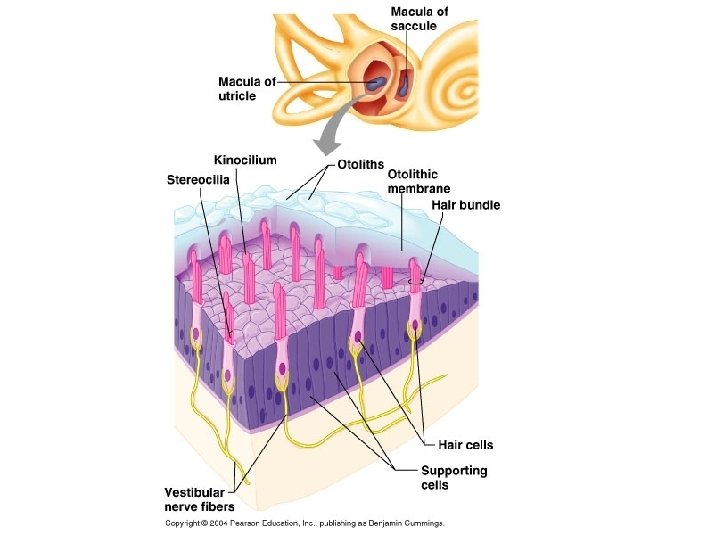
Figure 50. 13 Semicircular canals PERILYMPH Vestibular nerve Cupula Fluid flow Hairs Hair cell Vestibule Utricle Saccule Nerve fibers Body movement