SENSORY ORGANS Organ of vision Organ of balance

SENSORY ORGANS Organ of vision, Organ of balance and hearing and accessory organs
 - anterior segment of eye - posterior")
Organ of vision Ø Eyeball (bulbus oculi) - anterior segment of eye - posterior segment of eye Ø Eyelid (palpebra) Ø Tear gland (gl. lacrimalis)

Anterior segment Posterior segment
 Tunica media (vasculosa) Tunica interna (nervosa) anterior")
The wall of eyeball Tunica externa (fibrosa) Tunica media (vasculosa) Tunica interna (nervosa) anterior segment cornea iris corpus ciliare pars caeca retinae posterior segment sclera choroidea pars optica retinae

Anterior segment of eye

Anterior segment of eye – cornea anterior epithe lium Bowman‘s m embrane substantia propria co Descemet‘s membrane posterior epithelium rnae

Anterior segment of eye – corpus ciliare

Anterior segment of eye – iris dorsal corneal epithelium continues to ventral surface of iris as ventral epithelium of iris irid oco r stroma iridis [pink strip] dorsal epithelium ne al a ngl e

Dorsal segment of eye

Posterior segment of eye – choroidea Lamina chorocapillaris - with capillaries Lamina vasculosa - with vessels Lamina vitrea Lamina suprachoroidea

Posterior segment of eye – retina membrana limitans interna layer of nerve fibers layer of ganglionic cells inner plexiform layer inner granular layer outer plexiform layer outer granular layer membrana limitans externa layer of rods and cones pigmented epithelium (stratum pigmenti retinae)
 eyellash dorsal side (conjunctiva)")
Palpebra ventral side (skin) eyellash dorsal side (conjunctiva)

Palpebra – conjunctival side pseudostratified columnar epithelium goblet cells

Gl. lacrimalis
 auditory meatus (meatus acusticus")
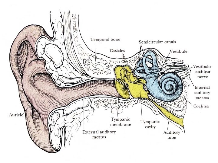
Organ of balance and hearing Ø Outer ear auricle (auricula) auditory meatus (meatus acusticus ext. ) ear drum (membrana tympani) Ø Middle ear tympanic cavity (cavum tympani) Ø Inner ear sacculus – macula sacculi utriculus – macula utriculi ductus semicirculares – cristae ampullares ductus cochlearis – organon Corti
 Bony labyrinth 1) vestibulum 2)")
Inner ear (in pyramid of ossis petrosum ossis temporalis) Bony labyrinth 1) vestibulum 2) canales semicirculares 3) cochlea Membranous labyrinth pars statica (1, 2) pars acustica (3) perilymphatic space spatium perilymphaceum 1) sacculus utriculus saccus endolymphaticus 2) ductus semicirculares 3) ductus cochlearis endolymph
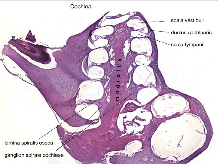

organ of Corti

Cochlea – organ of Corti hair cells inner outer inner phalangeal columnes outer phalangeal cells of Corti cells of Hensen
 Elastic cartilage Ear lobule")
Auricle (auricula) Elastic cartilage Ear lobule

SENSORY ORGANS Slides: 88. Anterior segment of the eye 89. Posterior segment of the eye 90. Fasciculus opticus 91. Palpebra 92. Gl. lacrimalis 93. Ductus cochlearis 94. Auricula

Development of eye 3 rd brain ventricle ectoderm stalk of optic cup intraretinal space of optic cup lens placode diencephalon mesenchyme
 outer layer of cup")
Development of optic cup and lens neuroectoderm (wall of diencephalon) outer layer of cup intraretinal space inner layer of cup ectoderm stalk of optic cup lens vesicle lumen of stalk of optic cup

intraretinal space diencephalon wall lens vesicle lumen of stalk of optic cup

pigmented layer of retina nervous layer of retina anterior epithelium of lens intraretinal space ectoderm eyelid mesenchyme

+ retinal pigmented layer retinal nervous layer camera oculi ant. eyelid ectoderm conjunctival sac mesenchyme

Development of inner ear otic placode otic pit mesenchyme ectoderm dorsal aorta wall of rhombencephalon otic pit otocyst the 1 st ectodermal cleft

Development of membranous labyrinth utricular part of otocyst saccular part of otocyst tubular process of sacculus

Development of semicircular canals *central regions of developing semicircular canals *these regions perforate later

Development of middle ear ossicles rhombencephalon wall of inner ear utriculus sacculus Meckel‘s cartilage primitive cavum tympani 1 st ectodermal cleft wall of inner ear

Development of external ear the 1 st ectodermal cleft basis of eye auricular tubercles
- Slides: 32