Sense Organs Sensory Receptors They make it possible

Sense Organs

Sensory Receptors • They make it possible for the body to respond to stimuli caused by changes occurring in our internal/external environment

• Receptor Response 1. General function-responds to stimuli by converting them to nerve impulses

2. Different types of receptors respond to different stimuli

3. Receptor Potential a. The potential that develops when an adequate stimulus acts on a receptor; it is a graded response

b. When a threshold is reached, an action potential in the sensory neuron’s axon is triggered

c. Impulses travel over sensory pathways to the brain and spinal cord where they are either interpreted as a particular sensation or they initiate a reflex action

d. Sensory projection- a brain function that pinpoints the area of the body from which the receptor potential was initiated

4. Adaptation- A functional characteristic of receptors; receptor potential decreases over a period of time in response to a continuous stimulus,

which leads to a decreased rate of impulse conduction and decreased intensity of sensation.

Classification of receptors-there are 5 categories based on the types of stimuli that activate them

1. Mechanoreceptors-activated by mechanical stimuli that change the position of the receptor, resulting in the generation of a receptor potential

2. Chemoreceptors-activated by the amount or the changing concentration of certain chemicals, e. g. , taste and smell

3. Thermoreceptors-activated by changes in temperature

4. Nociceptors-Activated by intense stimuli that result in tissue damage; the sensation produced is pain

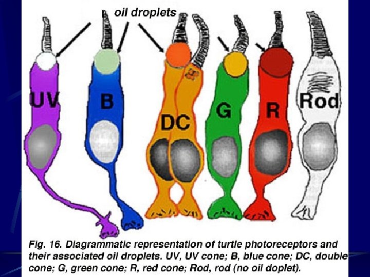
5. Photoreceptors- found only in the eye; respond to light stimuli if the intensity is great enough to generate a receptor potential

Distribution of Receptors 1. Receptors for special senses of smell, taste, vision, hearing, and equilibrium are grouped into localized areas or into complex organs

2. General sense organs of somatic senses are microscopic receptors widely distributed throughout the body in the skin, mucosa, conn. tissue, muscles, tendons, joints, viscera

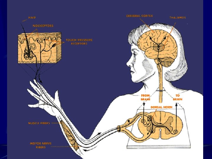
Somatic Senses • Detect sensations such as pain, temperature, pressure, touch, body position, tension in muscles, hunger, thirst, etc.

The three types of somatic sense receptors are: 1. exteroceptors-located on the body surfaces

2. Proprioceptors-located in muscles and joints 3. Visceroceptors-located in internal visceral organs
 1. are widely")
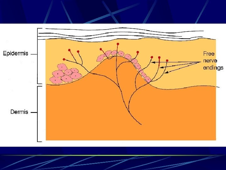
Pain and Temperature Receptors A. Pain Receptors (also known as nociceptors) 1. are widely distributed exteroceptors and visceroceptors

2. Are free nerve endings that respond to a number of different stimuli;

There are two types of nerve fibers that conduct pain impulses from free nerve endings to the brain:
 fibers-mediate sharp, intense, localized pain sensations b. Chronic (B) fibers-associated with")
a. Acute (A) fibers-mediate sharp, intense, localized pain sensations b. Chronic (B) fibers-associated with dull, aching pain

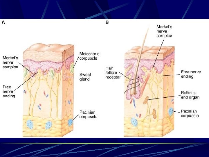
Touch and Pressure Receptors

A. Exteroceptors that respond to stimuli that change their shape or placement

B. Meissner’s corpuscles and Krause’s end bulb-involved with sensations of touch, low frequency vibrations, and twopoint discrimination; found on areas devoid of hair

C. Ruffini’s corpuscles- sense of deep pressure and continuous touch; located in the dermis of the skin and are numerous in the fingers

D. Pacinian corpuscles-respond quickly to sensations of deep pressure, high frequency vibration and stretch; found in deep dermis of the hands & feet & in joint capsules

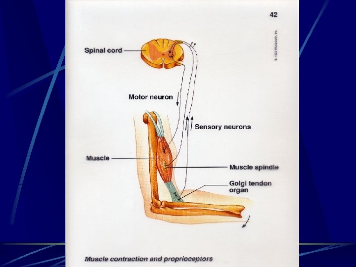
Stretch Receptors • The two most important stretch receptors are associated with muscles and tendons; proprioceptors

Muscle spindles-provide body with information regarding muscle length; stimulated if a relaxed muscle is stretched beyond a certain limit; causes the muscle to contract

Golgi tendon receptors-provide body with info regarding strength of muscle contraction

• Stimulated by excessive muscle contraction; located near points of attachment of tendon to bone; causes muscle to relax

Special Senses-Characterized by receptors grouped closely together or grouped in specialized organs; smell, taste, hearing, equilibrium, vision

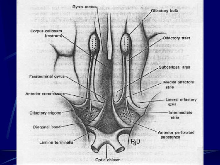
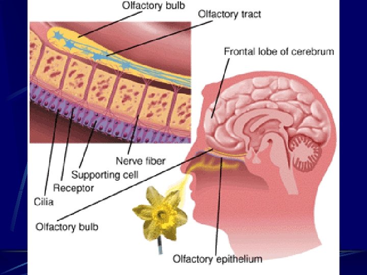
Sense of Smell-Olfactory receptors • Olfactory sense organs consist of epithelial cells and specialized olfactory receptor neurons

a. olfactory cilia-located on olfactory receptor neurons that touch the olfactory epithelium lining the upper surface of the nasal cavity

b. Olfactory cellschemoreceptors; gas molecules or chemicals dissolved in the mucus covering the nasal epithelium stimulate the olfactory cells

c. Olfactory epithelium-located in the most superior portion of the nasal cavity

d. Olfactory receptors-extremely sensitive and easily fatigued
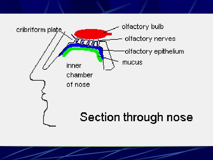


Olfactory pathway-when the level of odor-producing chemicals reaches a threshold level, the following occurs:

1. Receptor potential, and then action potential, is generated and passed to the olfactory nerves in the olfactory bulb

2. The impulse then passes through the olfactory tract & into the thalamic & olfactory centers of the brain for interpretation integration, & memory storage
 stimuli: associated with")
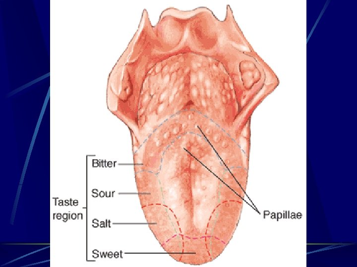
Sense of Taste • Taste buds-sense organs that respond to gustatory(taste) stimuli: associated with papillae

1. Chemoreceptors that are stimulated by chemicals dissolved in the saliva

2. Gustatory cells-specialized cells found in taste buds; gustatory hairs extend from each gustatory cell into the taste pore

3. Sense of taste depends on the creation of a receptor potential in gustatory cells due to tasteproducing chemicals in the saliva

4. Taste buds are similar structurally; functionally, each taste bud responds most effectively to one of 4 primary taste sensations: sour, sweet, bitter, and salty

5. Primary tastes to which a taste bud responds is related to its placement on the tongue

Tip of the tongue-sweet and salty Sides of the tongue-sour Back of the tongue-bitter
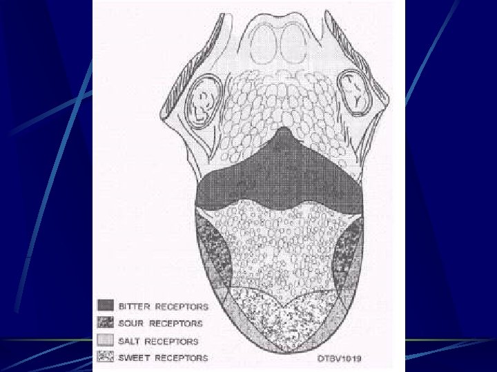

6. Adaptation and sensitivity thresholds are different for each of the primary taste sensations

Neuronal Pathway for Taste 1. Taste sensation begins with a receptor potential in the gustatory cells of a taste bud;

Generation and propagation of an action potential then transmits the sensory input to the brain

2. Nerve impulses from the anterior 2/3 of the tongue travel over the facial nerve;

Those from the posterior 1/3 of the tongue travel over the glossopharyngeal nerve; vagus nerve plays a minor role in taste

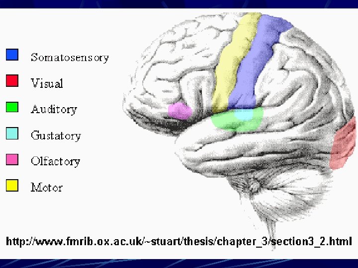
3. Nerve impulses are carried to the medulla oblongata, relayed into the thymus, and then into the gustatory area of the cerebral cortex in the parietal lobe of the brain

Sense of Hearing and Balance: The Ear

External ear-two divisions: 1. Auricle or pinna-the visible portion of the ear

2. External auditory meatus-tube leading from the auricle into the temporal bone and ending at the tympanic membrane (ear drum)

Middle Ear 1. Tiny, epithelia-lined cavity hollowed out of the temporal bone
 a. malleus(hammer)-attached to the inner surface of the")
2. Contains 3 auditory ossicles (bones) a. malleus(hammer)-attached to the inner surface of the tympanic membrane
-attached to the malleus and stapes c. stapes(stirrup)-attached to the incus")
b. incus(anvil)-attached to the malleus and stapes c. stapes(stirrup)-attached to the incus
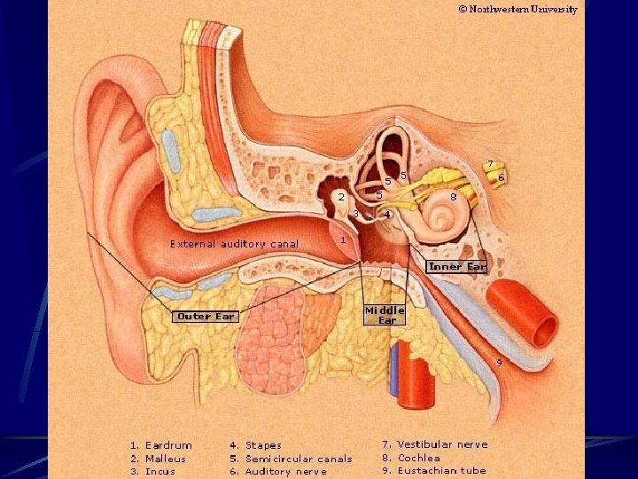

3. Openings into the middle ear

a. Opening from the external auditory meatus covered with tympanic membrane

b. Oval window-opening into inner ear; stapes fits here

c. Round window-opening into inner ear; covered by a membrane
 tube")
d. Opening into the auditory (eustachian) tube

C. Inner ear 1. Structure

a. Bony labyrinth-made up of the vestibule, cochlea, & semicircular canals

b. Membranous labyrinth-made up of utricle & saccule inside the vestibule, cochlear duct inside the cochlea, & the membranous semicircular canals inside the bony ones

c. Vestibule and semicircular canals are involved with balance

d. cochlea-involved with hearing

e. endolymph-clear, K+ rich fluid filling the membranous labyrinth

f. perilymph-similar to csf, surround the membranous labyrinth, filling the space between the membranous tunnel and its contents & the bony walls that surround it

2. Cochlea & cochlear duct a. cochlea-bony labyrinth

c. Cochlear duct 1. Lies inside the cochlea; only part of the internal ear concerned with hearing

2. Vestibular membrane-the roof of the cochlear duct 3. Basilar membrane-floor of the cochlear duct

4. Organ of Corti-rests on the basilar membrane; consists of supporting cells & hair cells

5. Axons of the neurons that begin around the organ of Corti, extend in the cochlear nerve to the brain to produce the sensation of hearing

Sense of Hearing a. Sound is created by vibrations b. Ability to hear sound waves depends on volume, pitch, & other acoustic properties

c. Sound waves must be of sufficient amplitude to move the tympanic membrane and have a frequency capable of stimulating the hair cells in the organ of Corti

d. Basilar membrane is not the same width & thickness throughout its length

High frequency sound waves vibrate the narrow portion near the oval window, low frequencies vibrate the wider, thicker portion near the apex of the cochlea

This fact allows different hair cells to be stimulated & different pitches of sound to be perceived

e. perception of loudness is determined by the amplitude of movement of the basilar membrane; the greater the movement, the louder the perceived sound

f. hearing-results from stimulation of the auditory area of the cerebral cortex

g. pathway of sound 1. Enter the external auditory canal 2. Strike the tympanic membrane, causing vibrations

3. Tympanic vibrations move the malleus, which in turn moves the incus and then the stapes

4. The stapes moves against the oval window, which begins the fluid conduction of sound waves.

5. The perilymph in the cochlea begins a “ripple” that is transmitted through the vestibular mem. to the endolymp, then the basilar membrane, then the organ of Corti

6. From the basilar membrane, the ripple is transmitted through the perilymph & then expends itself against the round window


Sense of Balance-the sense organs involved in balance or equilibrium are found in the vestibule & semicircular canals

The sense organs located in the vestibule function in static equilibrium

Static equilibrium-a function needed to sense the position of the head relative to gravity or to sense acceleration or deceleration of the body.

Changing head position produces a change of pressure, which stimulates the hair cells that, in turn, stimulate the receptors of the vestibular nerve.

The vestibular nerve conducts impulses to the brain & produce a sensation of the position of the head & also a sensation of a change in the pull of gravity.

Righting reflexes-muscular responses to restore the body & its parts to their normal position when they have been displaced

The sense organs associated with the semicircular canals function in dynamic equilibrium

Dynamic equilibrium-a function needed to maintain balance when the head or body itself is rotated or suddenly moved.

The Eye • Three layers of tissue compose the eyeball

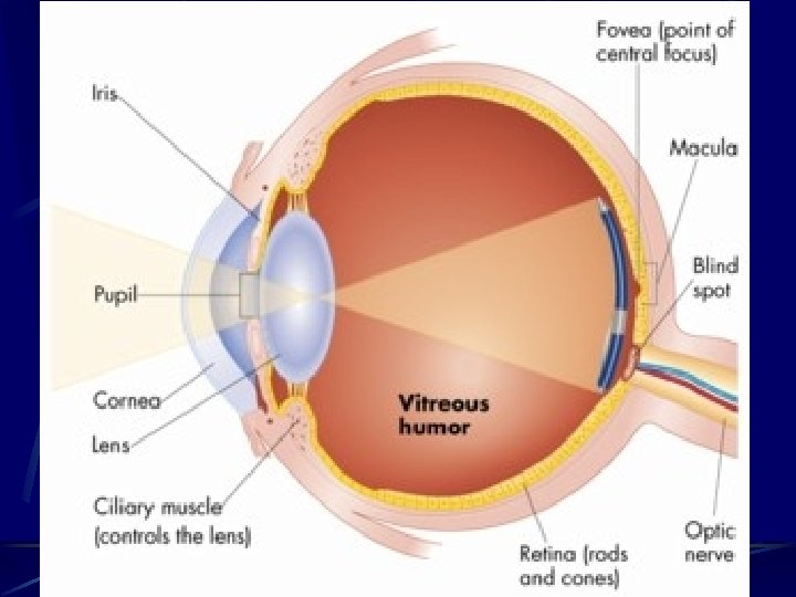
1. Sclera-outer coat a. tough, white fibrous tissue b. cornea-transparent anterior portion that lies over the iris

2. Choroid-middle coat a. contains many blood vessels & a large amount of pigment

Anterior portion contains: 1. Ciliary body-controls the shape of the lens

2. Suspensory ligament-holds lens in place 3. Iris-colored part of the eye. Contains circular and radial muscles. Controls amount of light into the eye.

4. pupil-hole in the middle of the iris. Allows light to enter the eye. 5. retina-lining on the inside of the posterior eye. Contains rods and cones.

5. rods-use the photopigment rhodopsin to see in dim light. See in grays. Detect minute movement.
. Allows for color vision.")
6. Cones-Uses 3 photopigements (red, green, blue). Allows for color vision.

7. Macula lutea-yellowish area near the center of the retina. Contains the fovea centralismost concentrated area of cones. (both lack rods)

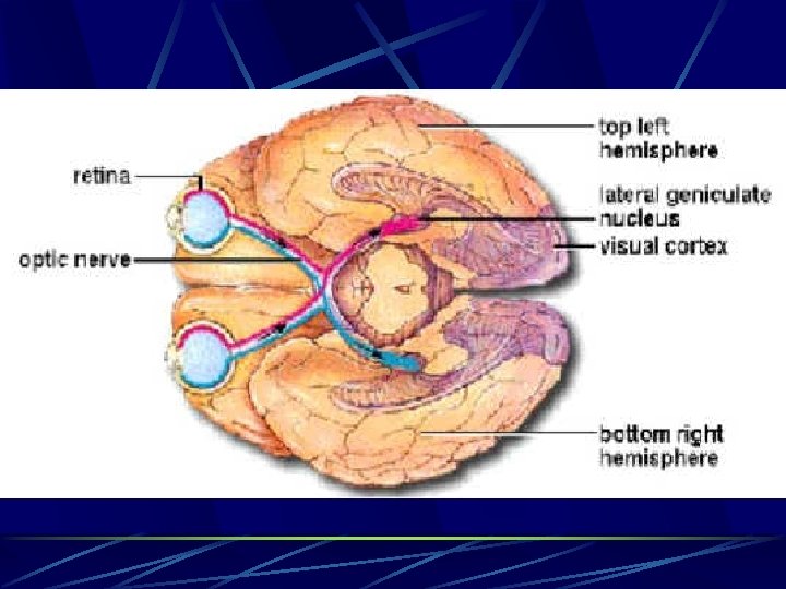
8. Optic nerve-CNII-connects to the back of the eye. Carries visual info to the brain.
-area in front of where the optic nerve connects. No")
9. Optic disc (blind spot)-area in front of where the optic nerve connects. No rods or cones. Vision is not possible.
 and posterior chamber")
10. Anterior cavity-contains the anterior chamber (in front of the lens) and posterior chamber (directly behind the iris but in front of the lens)

Contains aqueous humor-a clear and watery fluid.

11. Posterior cavity-behind the lens. Contains vitreous humor (gelatinous fluid that maintains pressure and shape of the eyeball)

12. Extrinsic muscles-skeletal muscles on the outside of the eye which move the eye. Superior, inferior, medial, lateral rectus, superior and inferior oblique
-ciliary muscles")
13. Intrinsic muscles (smooth muscles inside the eye)-ciliary muscles

Accessory Structures Eyebrows-divert sweat away from the eye Eyelashes-keep things out of the eyes Eyelids-protect and moisten the eye

Lacrimal apparatus-gland, ducts, sacs-produce tears to moisten and clean the eye. Drain into the nose

For vision to occur: 1. An image must be formed on the retina to stimulate the rods and cones, the impulse must be conducted to the occipital lobe for interpretation.
 Accomidation-the changing of the")
Refraction-bending of light rays (cornea, aqueous humor, lens, vitreous humor) Accomidation-the changing of the shape of the lens to focus. Rounds up for close vision.
")
Visual acuity-the clearness or sharpness of visual perception Eye chart at 20 ft. (20/20) 20/100 means you see at 100 ft what 20/20 person sees at 100 ft

Constriction-constriction of the pupil prevents divergent light rays from the object from entering the eye from the periphery.
 at")
Convergence-movement of the eyeballs inward so that their visual axes come together (converge) at the object viewed.

Otosclerosis-inherited disorder caused by structural irregularities of the stapes and blocks conduction. Tinnitus-ringing in the ears

Otitis-ear infection Presbycusis-progress hearing loss due to nerve damage Myopia-nearsightedness, elongated eye causes the image to form in front of the retina

Hyperopia-farsightedness, shorter eye causes the image to form behind the retina Astigmatism-irregular curvature of the cornea or lens

Cataracts-cloudy spots that develop in the lens Trachoma-bacterial infection caused by chlamydia. Put antibiotics in newborns’ eyes

Retinal detachment-retina detaches from the back of the eye Glaucoma-build up of pressure in the eye Colorblindness-inability to distinguish certain colors

Nyctalopia-night blindness, caused by degeneration of the retina or lack of vit A
- Slides: 148