Sense of Vision Introduction The human eye is


")


 § Light causes rhodopsin to change shape")


. They also undergo")


- Slides: 66
Sense of Vision
Introduction • The human eye is the organ which gives us the sense of sight and allowing us to observe and learn more about the surrounding world than we do with any of the other four senses. We use our eyes in almost every activity we perform, whether reading, working, watching television or writing etc. • The eye is able to detect bright light or dim light, but it cannot sense objects when light is absent.
Cont… • 70 percent of all sensory receptors are in the eyes • The vision is a photoreceptor sense
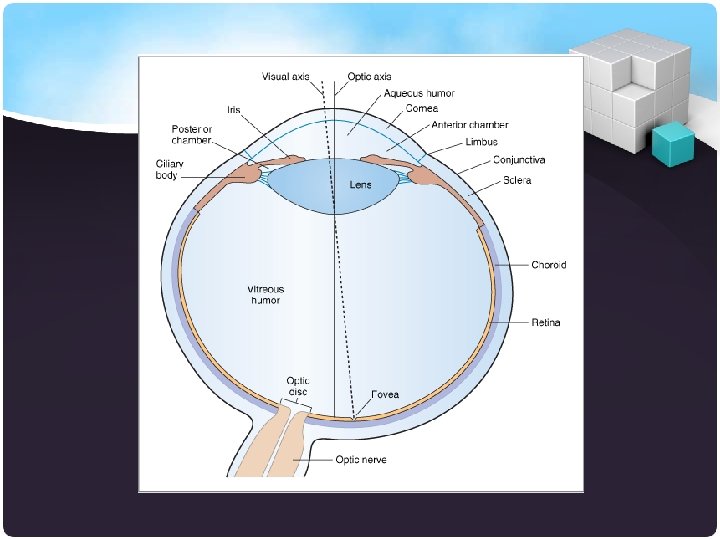
Anatomy of EYE • The adult eye is a sphere about 1 inch (2. 5 cm) in diameter. • Only 1/6 th of the eye’s anterior surface can normally be seen. • Protection for the eye § The rest of the eye is enclosed in a bony orbit. § A cushion of fat surrounds most of the eye. Ø It has two basic structures § Eyeball § Accessory structures
Structure of the Eyeball
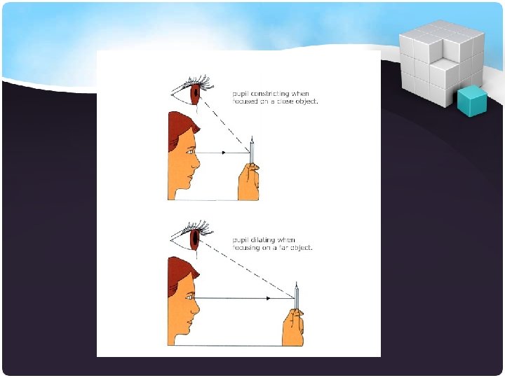
Pupil • Pupil is a central opening of the iris • Regulates the amount of light entering the eye during: § Close vision and bright light – pupils constrict § Distant vision and dim light – pupils dilate § Changes in emotional state – pupils dilate when the subject matter is appealing or requires problem-solving skills
Pupil Dilation and Constriction
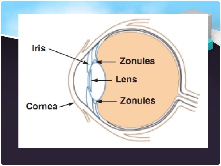
Lens • Biconvex crystal-like structure • Held in place by a suspensory ligament attached to the ciliary body • Light passing through a convex lens is bent so that the rays converge to a focal point • When a convex lens forms an image, the image is upside down and reversed right to left
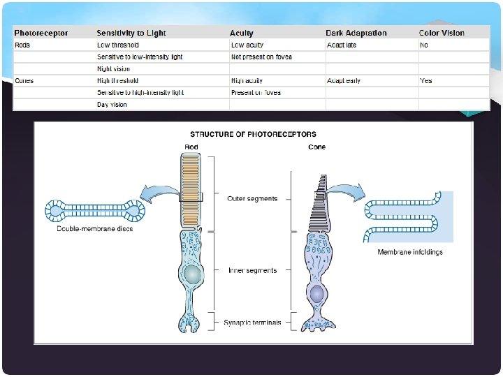
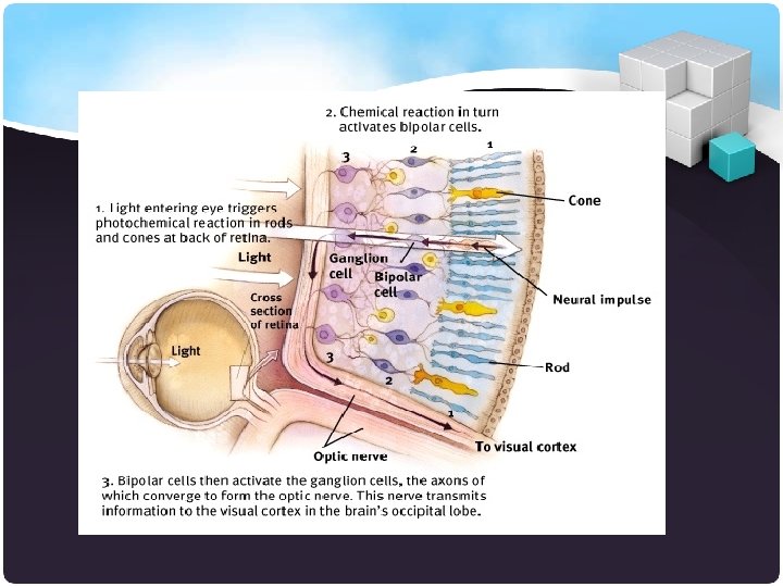
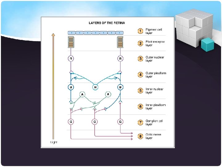
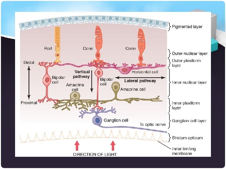
Retina • Extends anteriorly only to the ciliary body. • Contains receptor cells (photoreceptors) § Rods § Cones • Signals leave the retina toward the brain through the optic nerve. • No photoreceptor cells are at the optic disk, or blind spot
Retina
Visual Receptors Rods Hundreds of times more sensitive to light than cones Provide vision in poor light. Produce colorless (black and white) vision. Nerve fibers converge so impulses produce more general outlines. § Concentration of rods increases in areas away from fovea centralis. § §
That means that your peripheral vision is better in the dark than your direct vision.
Visual Receptors Cones § Provide vision in good light. § Produce colored and sharp vision § Nerve fibers do not converge as much so impulses produce more detailed images. § Concentration of cones greatest in fovea centralis. § Concentration of cones decreases in areas away from fovea centralis. § Fovea centralis – area of the retina with only cones found laterally to each blind spot, this is the area of greatest visual acuity, or point of sharpest vision
Cone Sensitivity § There are three types of cones § Different cones are sensitive to different wavelengths § Color blindness is the result of lack of one cone type
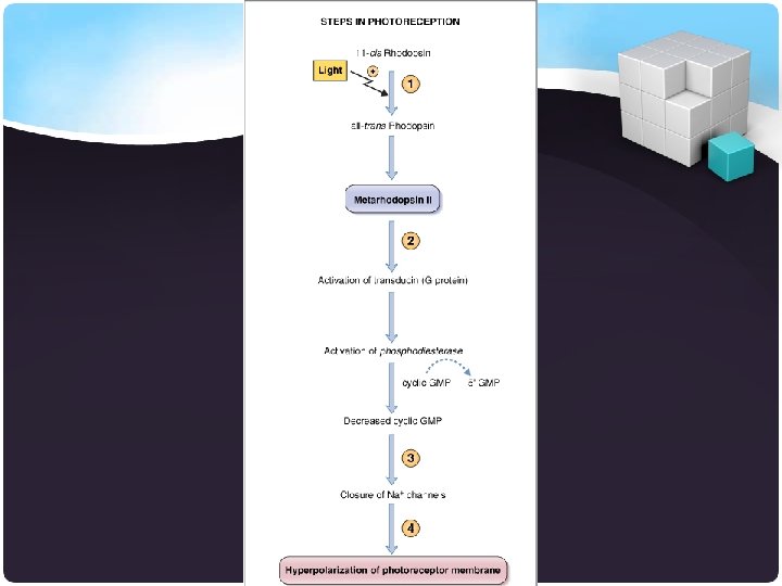
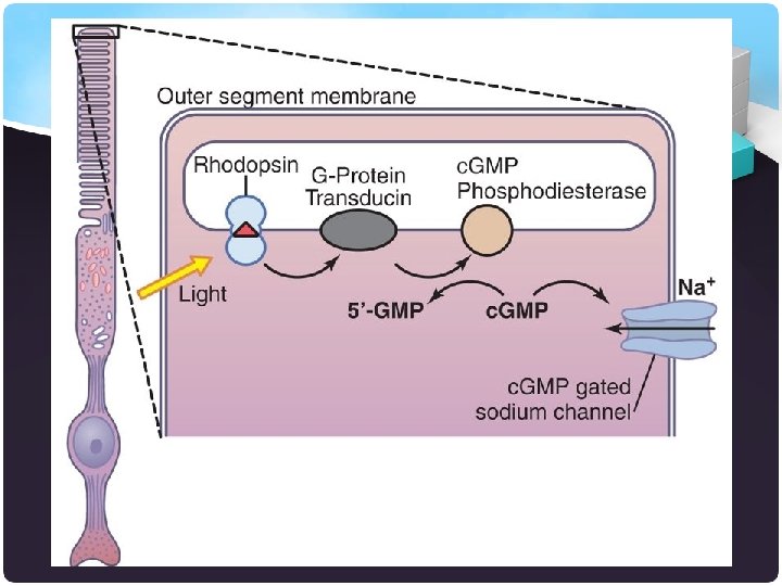
Visual Pigments Rods contain rhodopsin (visual purple) § Light causes rhodopsin to change shape and release opsin which acts as an enzyme in further reactions. § Net result is hyperpolarization directly proportional to intensity of light stimulus. § Rhodopsin replenished in dim light. § In dim light, a rhodopsin-replenished eye is said to be dark-adapted (can see in dark).
Visual Pigments Cones contain iodopsins § A group of pigments sensitive to light waves of different frequencies.
Eye Cavities • The internal space of the eye is subdivided by the lens into two separate cavities. § Anterior cavity § Posterior cavity
Cont… • The anterior cavity is the space anterior to the lens and posterior to the cornea • The iris of the eye subdivides the anterior cavity further into two chambers. § anterior chamber is between the iris and cornea § posterior chamber is between the lens and the
Cont… • Posterior cavity is posterior to the lens and anterior to the retina. • Transparent, gelatinous vitreous body which completely fills the space between the lens and the retina.
Physiology of EYE • Light waves from an object enter the eye first through the cornea, The light then proceed to the pupil • Fluctuations in incoming light change the size of the pupil. When the light entering the eye is bright enough, the pupil will constrict due to the pupillary light response.
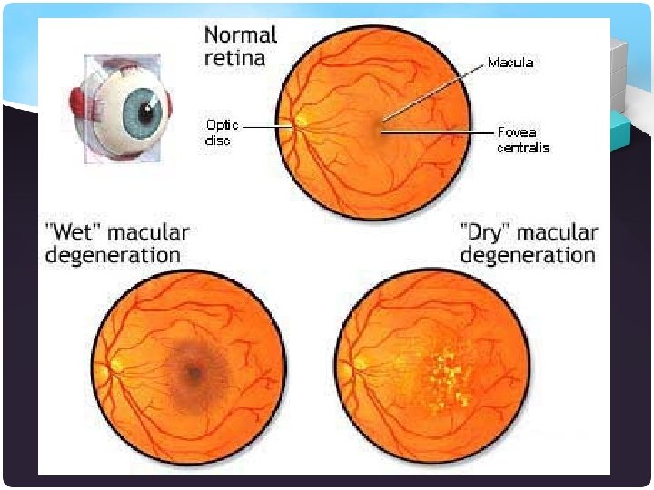
Cont… • The light continues through the vitreous humor then, ideally, back to a clear focus on the retina, behind the vitreous. The small central area of the retina is the macula, which provides the best vision of any location in the retina. • Within the layers of the retina, light impulses are changed into electrical signals. Then they are sent through the optic nerve, along the visual pathway, to the occipital cortex. Here, the electrical signals are interpreted or “seen” by the brain as a visual image.
• Actually, we do not “see” with our eyes but, rather, with our brains. Our eyes merely are the beginnings of the visual process
It is not that easy …! • To see every thing clearly three processes must talk place v. The Refraction or bending of light by the lens and cornea. v. The change in shape of the lens Accommodation. v. The Constriction or narrowing of the pupil
Refraction • Images focused on the retina are inverted (upside down). They also undergo light to left reversal. • 75% of the refraction occurs at the cornea. • 25% occurs at the lens, which also changes the focus to view either distant or near objects.
Refraction Abnormality • Myopia or nearsighted is results from a lens that is too strong or a long eyeball, when the image focuses in front of the retina, corrected by concave a lens. • Hyperopia or farsighted is results from a lens that is too lazy or a too short eyeball, when the image focuses behind the retina, corrected by convex a lens.
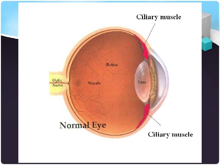
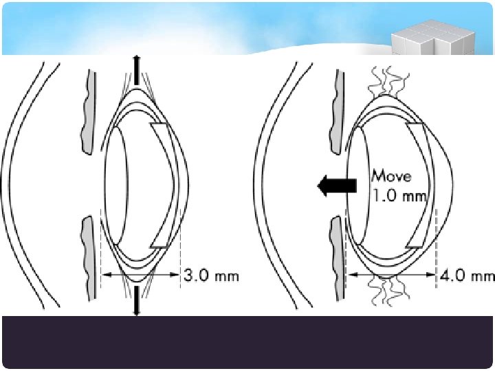
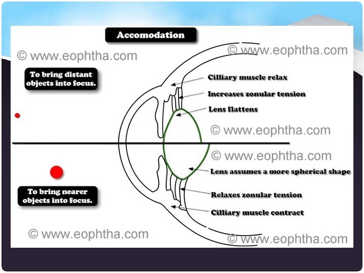
Accommodations • When the eye is focusing on a close object, the lens becomes more curved, causing greater refraction of the light rays. • This increase in the curvature of the lens is called Accommodation.
Near & Far Accommodation • When viewing distant objects, the ciliary muscle is relaxed and the lens is flatter because the taught zonular fibers are stretching it in all directions. • When viewing close objects, the ciliary muscle contracts, which pulls the ciliary processes towards the lens. This releases tension on the lens and zonular fibers. The lens is elastic and then becomes more spherical.
Constriction of the Pupil • Constriction of the pupil is a narrowing of the diameter of the hole through which light enters the eye due to contraction of the circular muscles of the iris. • This occurs automatically during accommodation to prevent light from entering at the periphery of the lens. • The pupil also constricts in bright light.
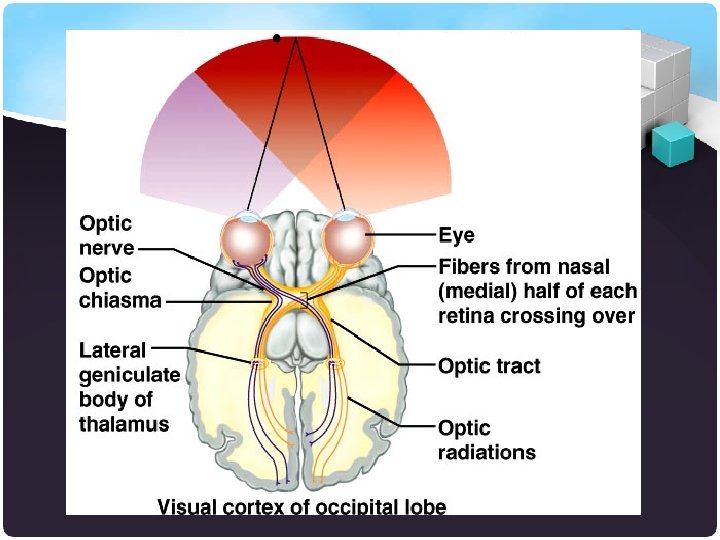
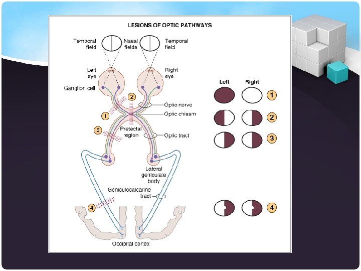
Visual Pathway 1. Axons of all retinal ganglion cells in one eye exit the eyeball at the optic disc and form the optic nerve on that side. 2. At the optic chiasm, axons from the temporal half of each retina do not cross but continue directly to the lateral geniculate nucleus of the thalamus on the same side. 3. Axons from the nasal half of each retina cross the optic chiasm and continue to the opposite hypothalamus.
Visual Pathway 4. Each optic tract consists of crossed and uncrossed axons that project from the optic chiasm to the thalamus on one side. 5. Axon collateral extend to the midbrain to govern pupil constriction and to the hypothalamus to govern patterns of sleep and other circadian rhythms relevant to light and darkness. 6. Axons of thalamic neurons form the optic radiations and project to the primary visual area of the cortex on the same side.
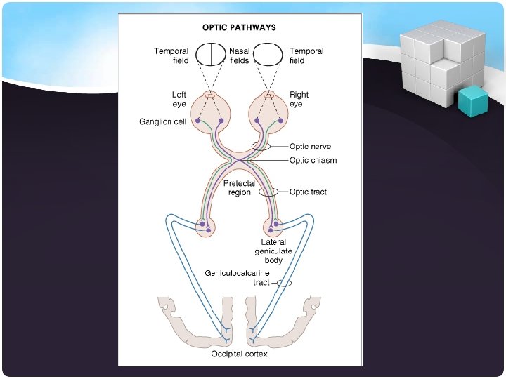
• Optic nerve. Cutting the optic nerve causes blindness in the ipsilateral (same side) eye. Thus, cutting the left optic nerve causes blindness in the left eye. All sensory information coming from that eye is lost because the cut occurs before any fibers cross at the optic chiasm. • Optic chiasm. Cutting the optic chiasm causes heteronymous (both eyes) bitemporal (both temporal visual fields) hemianopia. In other words, all information is lost from fibers that cross. Thus, information from the temporal visual fields from both eyes is lost because these fibers cross at the optic chiasm. • Optic tract. Cutting the optic tract causes homonymous contralateral hemianopia. As shown in the figure, cutting the left optic tract results in loss of the temporal visual field from the right eye (crossed) and loss of the nasal visual field from the left eye (uncrossed). • Geniculocalcarine tract. Cutting the geniculocalcarine tract causes homonymous contralateral hemianopia with macular sparing (the visual field from the macula is intact). Macular sparing occurs because lesions of the visual cortex do not destroy all neurons that represent the macula.
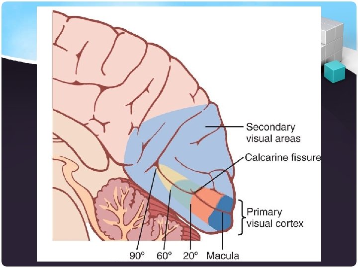
Visual cortex • The visual cortex is the largest system in the human brain and is responsible for processing the visual image. It lies at the occipital lobe of the brain above the cerebellum. The region that receives information directly from the LGN is called the primary visual cortex, (also called V 1 and striate cortex). Visual information then flows through secondary visual areas, These areas include V 2, V 3, V 4 and area V 5/MT.
Visual cortex
How we can test our sense of vision ?
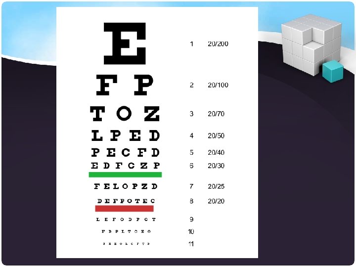
A. Test of visual acuity • A clear focused image requires adaptation on the focal length of the optic lens by alteration of its curvature to suit the varying of distance • It is a measure of the smallest retinal images which can be appreciated • By using a Snellen’s chart we can recode the visual acuity for distance and for near
Cont… • A visual acuity test is a measure of how well you see or the sharpness and clarity of your vision. Your eye doctor will ask you to read letters on a chart while standing 20 feet away. The smallest letters you are able to read will be recorded as your acuity. • Your visual acuity may be written as 20/20 or (6/6) if your vision is normal. If your vision is reduced, it might be recorded as less than 20/20, such as 20/100.
Cont… • If you have 20/100 vision, it means that you must be as close as 20 feet to see what a person with normal vision can see at 100 feet. Someone with 20/60 vision would need to move up to 20 feet away to read what a person with normal vision could read from 60 feet away
B. Test of visual filed • The normal visual filed extends 160 degree horizontally and 130 degree vertically 60 > 90 60 60 70 Right Eye >90 60 70 Left Eye
Cont… • Sit directly facing the pt about 1 meter away • The pt field of vision is compared with that of the examiner having a normal field of vision • First examine both eye together , then You should test each eye separately for the Peripheral visual filed • To test the central visual filed we use a red hatpin to one eye qualitatively at a time
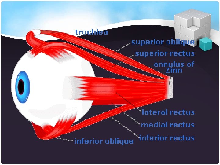
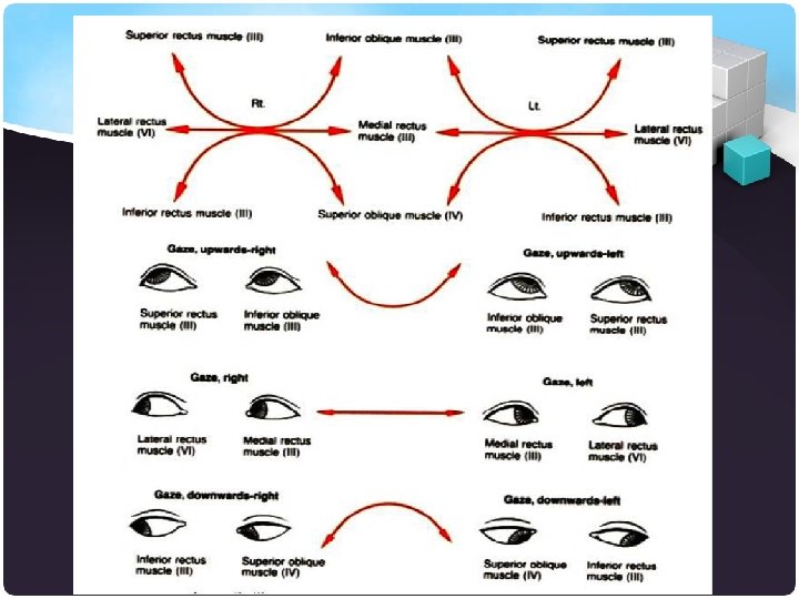
C. Ocular Alignment To test the Extraocular muscles functions ( check cranial nerves 3, 4, 6) 1. Stand or sit 3 to 6 feet in front of the patient. 2. Ask the patient to follow your finger with their eyes without moving their head. 3. Check gaze in the six cardinal directions using an “H” pattern of motion (i. e. move your finger from the center out (as if you were checking visual fields and then up and down, drawing a big “H”) 4. Record whether your volunteer can follow your fingers in all directions and whether the pursuit was smooth
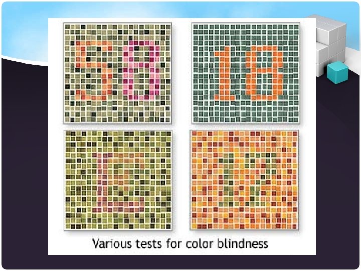
D. Test of Color Vision • Red and Green color vision can be assessed using Ishihara test plate. • These coloured spots forming numbers which the patient reads out
E. Pupillary Examination • Examine the pupil for the shape and symmetry • Ask the pt to fix the eye on a distant point straight ahead • Bring a bright torchlight from the side to shine on the pupil • Look for constriction of that pupil ( direct light reflex) and for the constriction of the opposite pupil ( consensual light reflex )
Cont…
Cont… • With the pt vision fixed on a distant point , present an object about 15 cm in front of the eye and ask the pt to focus on it. • look for pupil constriction and convergence ( Accommodation reflex)