Seeing much suffering much and studying much are


 thin filaments of keratin epithelial invaginations of")

 all over the body. coiled")


 encapsulated mechanoreceptors present in the dermal papilla /")


 encapsulated mechanoreceptors in the papillary layer dermis oral")
- Slides: 23
“Seeing much, suffering much and studying much are three pillars of learning. ” –B Disraeli
Integumentary system Skin
Introduction: • largest organ of the body 15 -20% • The skin and its derivitives constitute the integumentary system • made up of multiple layers of epithelial tissues covering CT, muscles and organs Functions • Protection • Sensation • Heat regulation • Control evaporation • Aesthetic and communication • Storage and synthesis • Excecretion • Absorption
Skin structure • Skin is composed of three primary layers -Epidermis which provides waterproofing and serves as a barrier to infection -Dermis appendages of the skin - Hypodermis (superfecial facia) subcuteneous adipose layer • The thickness of the skin varies from less than 1 mm to more than 5 mm. Thick skin (hairless skin) contains thick epidermis e. g palms of hands soles of feet. It has sweat glands, no hair follicles and no sebaceous glands. Thin skin (thin epidermis) contains hair follicles, sweat glands and sebaceous glands.
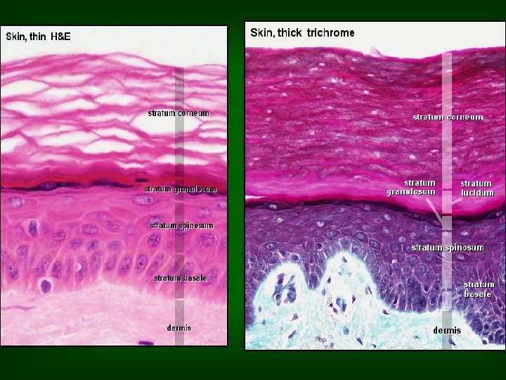
Epidermis Consists of a keratinized stratified squamous epithelium. layers of keratenocytes differentiate and become filled with keratin migrates upward through layers (keratinization), desquamated Sub-layers • stratum germinativum (also stratum basale or basal cell layer) single layer simple cuboidal to low columnar basophilic cytoplasm active mitosis scattered intermediate filaments 10 - 25% are melanocytes • stratum spinosum multi-layered arrangement of cuboidal cells adjacent cells are joined by desmosomes spiny appearance (it is called prickle cell layer) structural support, resist abrasion intermediate filaments (light microscope) called cytokeratins
• stratum granulosum 1 to 3 rows of squamous cells with many small basophilic granules keratohyalin granules(lamellar granules) soft keratin ▪stratum lucidum a thin, clear layer of dead skin cells only found on the thick skin non nucleated cells and immature keratin ▪ stratum corneum dead squamous cells that lack nuclei and filled with keratin protein that helps keep the skin hydrated also absorb water thickness varies Pruning, calluses
Skin pruning Palm calluses
Other cells of the epidermis include 1. Melanocytes 2. Langerhans cells 3. Merckle cells Melanocytes • melanin gives the brown colour component of the skin (melanin) • ectodermal in oirgin, neural crest • dendritic cells with no desmosomes with keratenocytes • melanosomes for melanogenesis • mainly in the epidermis basal layer, could not be identified in LM • found in skin, eye and hair • melanogenesis under the effect of - race - hormones - exposure to light - age Albinism recessive hereditory condition
Langerhans' cells -dendretic cells -contain large granules called Birbeck granules -important in immune reactions of the epidermis/ macrophages (antigenpresenting cells) -no desmosomes Merkel cells large oval cells numerous cytoplasmic granules commonly associated with free nerve endings sensory mechanoreceptors / neuroendocrine cells
Dermis Papillary region loose areolar CT fingerlike projections called papillae very well-vascularized friction ridges occur in patterns (fingerprint) arterio-venous anastomoses /thermoregulation P Reticular region much thicker dense irregular connective tissue collagenous, elastic, and reticular fibers strength, extensibility, and elasticity aging thickening of the collagen and lose the elasticity roots of the hair, sebaceous glands, sweat glands, receptors, and blood vessels R
Appendages of the Skin Hair (hard keratin) thin filaments of keratin epithelial invaginations of the epidermis Shaft, root, sheath, hair follicle, bulb, arrector pili muscle
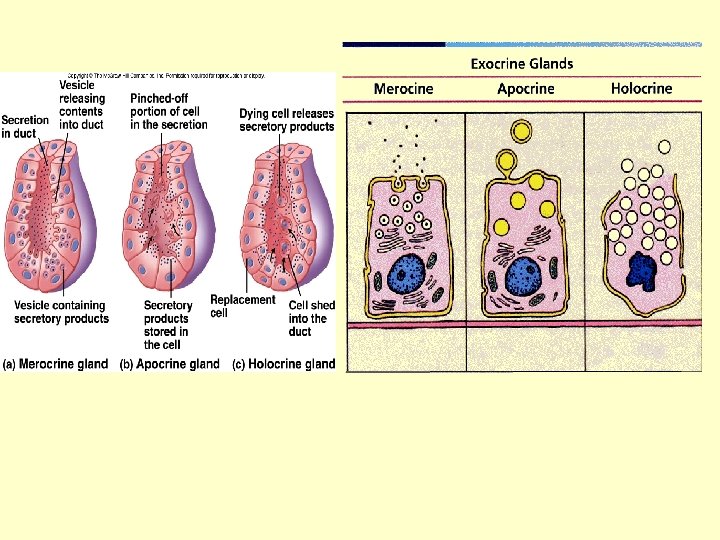
Sebaceous glands • dermal exocrine glands associated with hairs • holocrine secretion • secrete an oily substance (sebum) for hair flexibility • the sebum secretion is influenced by sex hormones/ adolescence acne
Sweat glands • Eccrine sweat glands (Merocrine sweat glands) all over the body. coiled tubular glands body temperature regulation. cholinergic innervations/ respond to heat. • Apocrine sweat glands large /axilla /external genitalia and anus functional at puberty adrenergic innervation/ respond to emotional and sensory stimuli
Nails plates of keratin nail plate, nail bed, nail groove, nail root, dorsal and ventral matrix, lunula eponychium or cuticle, hyponychium
Nerve supply -Free such as free endings, Merkel’s disc and hair follicle receptors. - Encapsulated terminals such as tactile (Meissner’s) corpuscles, bulboid corpuscles (Krause’s end bulb), lamellar (Pacinian) corpuscles, and Ruffini’s corpuscles
Free nerve endings no complex sensory structures/ nonencapsulated/ epidermis/ sense pain and temperature.
Merkel disk receptor non- encapsulated mechanoreceptors/ surrounding hair follicles/ touch/ basal layer/ the most sensitive of the main types of mechanoreceptors to vibrations at low frequencies Hair follicle receptors these mechanoreceptors sense the presence and direction of hair displacement.
Meissner’s corpuscles (or tactile corpuscles) encapsulated mechanoreceptors present in the dermal papilla / sensitivity to light touch (sensitive to low frequency stimuli) / do not detect pain/ their number drops with age
Pacinian corpuscles dermis of thick skin of fingers /respond to pressure and vibration. large receptive field on the skin's surface with an especially sensitive center. larger and fewer than others
Ruffini’s corpuscles – are encapsulated mechanoreceptors /deep in the reticular layer where they respond to distortion or stretching of the skin (sustained or continuous stress)
Krause end bulbs (bulboid corpuscles) encapsulated mechanoreceptors in the papillary layer dermis oral mucosa of the oral cavity and tongue / respond to cold and low-frequency vibration