
Section 5 Stereochemistry Amino Acids Peptides and Proteins

- Slides: 32
Section 5: Stereochemistry, Amino Acids, Peptides, and Proteins • Stereochemistry Handedness - Optical Activity • Stereochemistry involves the study of the relative spatial arrangement of atoms that form the structure of molecules and their manipulation. An important branch of stereochemistry is the study of chiral molecules 1
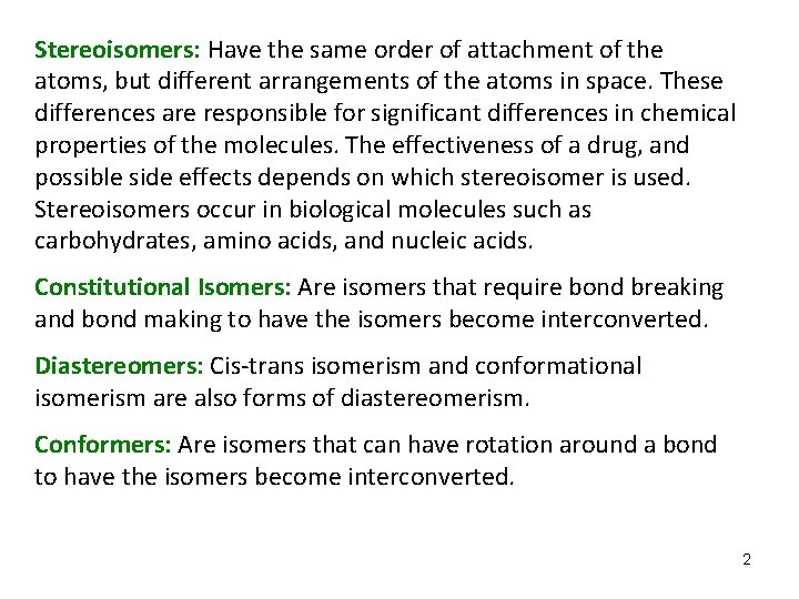
Stereoisomers: Have the same order of attachment of the atoms, but different arrangements of the atoms in space. These differences are responsible for significant differences in chemical properties of the molecules. The effectiveness of a drug, and possible side effects depends on which stereoisomer is used. Stereoisomers occur in biological molecules such as carbohydrates, amino acids, and nucleic acids. Constitutional Isomers: Are isomers that require bond breaking and bond making to have the isomers become interconverted. Diastereomers: Cis-trans isomerism and conformational isomerism are also forms of diastereomerism. Conformers: Are isomers that can have rotation around a bond to have the isomers become interconverted. 2
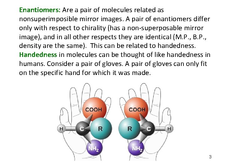
Enantiomers: Are a pair of molecules related as nonsuperimposible mirror images. A pair of enantiomers differ only with respect to chirality (has a non-superposable mirror image), and in all other respects they are identical (M. P. , B. P. , density are the same). This can be related to handedness. Handedness in molecules can be thought of like handedness in humans. Consider a pair of gloves. A pair of gloves can only fit on the specific hand for which it was made. 3
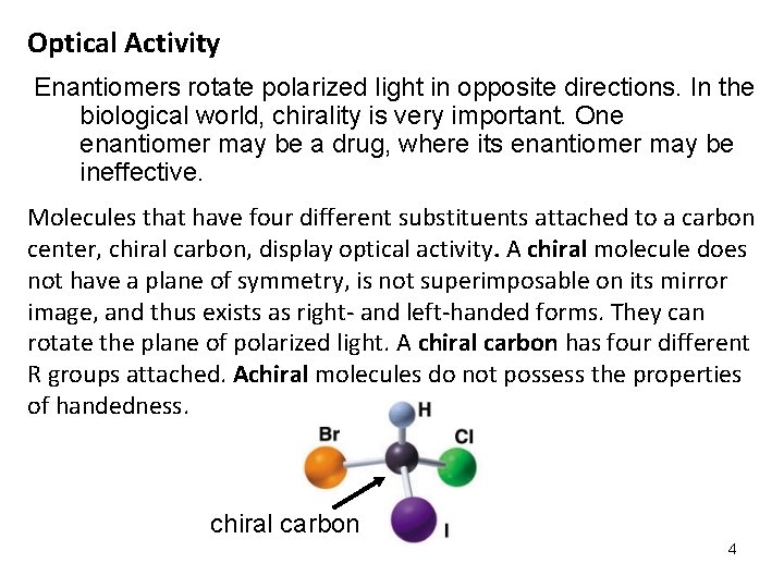
Optical Activity Enantiomers rotate polarized light in opposite directions. In the biological world, chirality is very important. One enantiomer may be a drug, where its enantiomer may be ineffective. Molecules that have four different substituents attached to a carbon center, chiral carbon, display optical activity. A chiral molecule does not have a plane of symmetry, is not superimposable on its mirror image, and thus exists as right- and left-handed forms. They can rotate the plane of polarized light. A chiral carbon has four different R groups attached. Achiral molecules do not possess the properties of handedness. chiral carbon 4
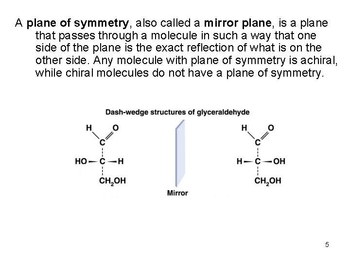
A plane of symmetry, also called a mirror plane, is a plane that passes through a molecule in such a way that one side of the plane is the exact reflection of what is on the other side. Any molecule with plane of symmetry is achiral, while chiral molecules do not have a plane of symmetry. 5
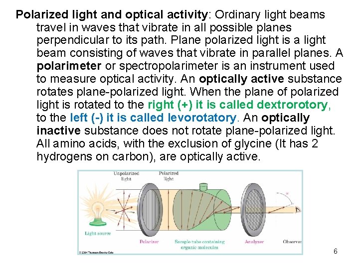
Polarized light and optical activity: Ordinary light beams travel in waves that vibrate in all possible planes perpendicular to its path. Plane polarized light is a light beam consisting of waves that vibrate in parallel planes. A polarimeter or spectropolarimeter is an instrument used to measure optical activity. An optically active substance rotates plane-polarized light. When the plane of polarized light is rotated to the right (+) it is called dextrorotory, to the left (-) it is called levorotatory. An optically inactive substance does not rotate plane-polarized light. All amino acids, with the exclusion of glycine (It has 2 hydrogens on carbon), are optically active. 6
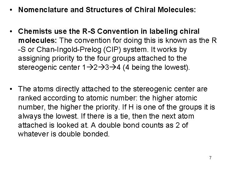
• Nomenclature and Structures of Chiral Molecules: • Chemists use the R-S Convention in labeling chiral molecules: The convention for doing this is known as the R -S or Chan-Ingold-Prelog (CIP) system. It works by assigning priority to the four groups attached to the stereogenic center 1 2 3 4 (4 being the lowest). • The atoms directly attached to the stereogenic center are ranked according to atomic number: the higher atomic number, the higher the priority. If H is one of the groups it is always the lowest. If there is a tie, then the next atom attached is looked at. A double bond counts as 2 of whatever is double bonded. 7
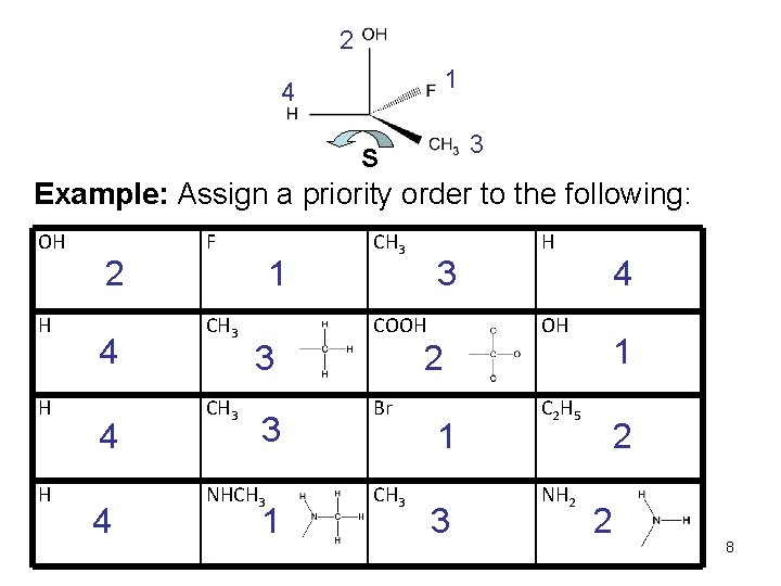
2 1 4 3 S Example: Assign a priority order to the following: OH H 2 4 4 4 F CH 3 1 CH 3 3 COOH 3 3 NHCH 3 1 H 4 OH 1 2 Br CH 3 1 3 C 2 H 5 NH 2 2 2 8
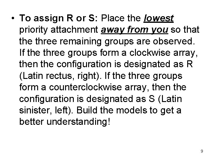
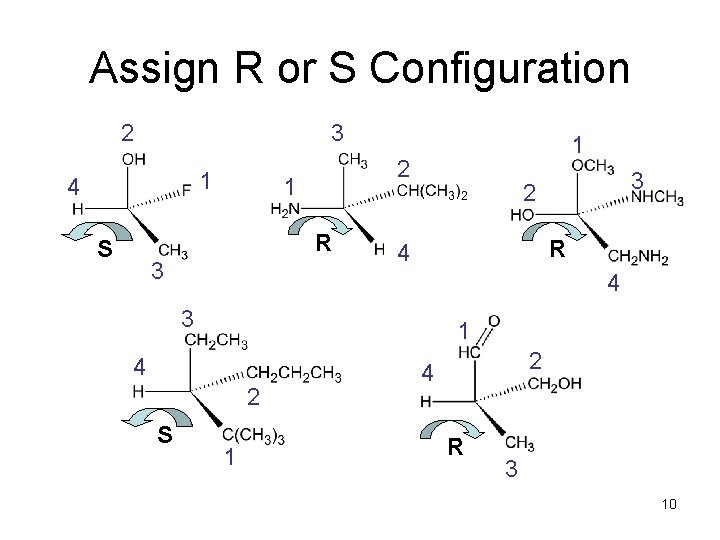
• To assign R or S: Place the lowest priority attachment away from you so that the three remaining groups are observed. If the three groups form a clockwise array, then the configuration is designated as R (Latin rectus, right). If the three groups form a counterclockwise array, then the configuration is designated as S (Latin sinister, left). Build the models to get a better understanding! 9
Assign R or S Configuration 2 3 1 4 2 1 R S 3 1 R 4 4 3 1 4 2 S 3 2 1 2 4 R 3 10
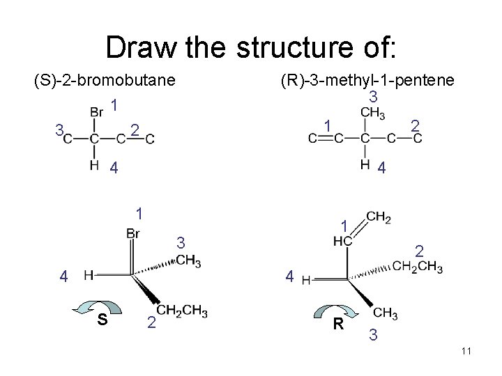
Draw the structure of: (S)-2 -bromobutane (R)-3 -methyl-1 -pentene 3 1 2 2 4 4 1 1 3 4 2 4 S 2 R 3 11
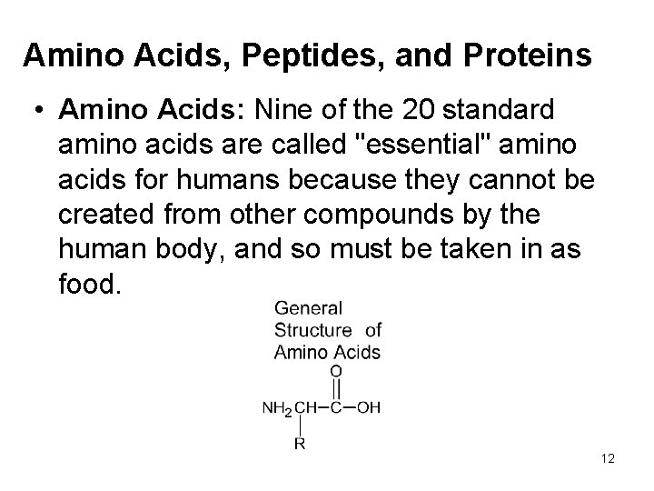
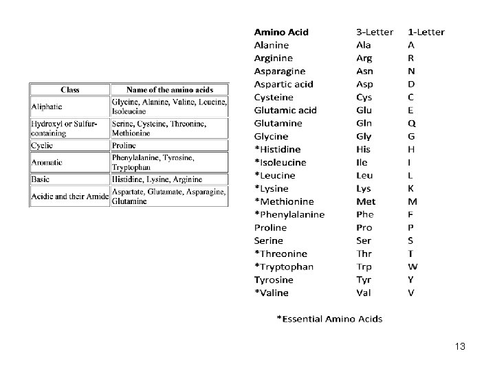
Amino Acids, Peptides, and Proteins • Amino Acids: Nine of the 20 standard amino acids are called "essential" amino acids for humans because they cannot be created from other compounds by the human body, and so must be taken in as food. 12
13
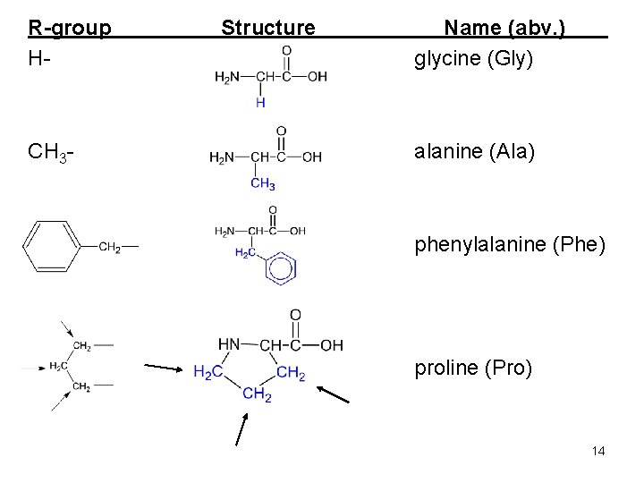
R-group H- CH 3 - Structure Name (abv. ) glycine (Gly) alanine (Ala) phenylalanine (Phe) proline (Pro) 14
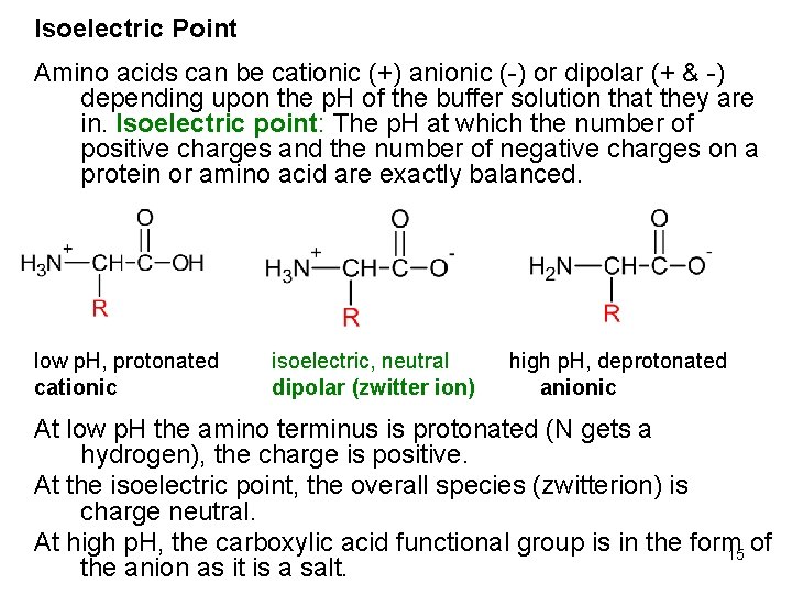
Isoelectric Point Amino acids can be cationic (+) anionic (-) or dipolar (+ & -) depending upon the p. H of the buffer solution that they are in. Isoelectric point: The p. H at which the number of positive charges and the number of negative charges on a protein or amino acid are exactly balanced. low p. H, protonated cationic isoelectric, neutral dipolar (zwitter ion) high p. H, deprotonated anionic At low p. H the amino terminus is protonated (N gets a hydrogen), the charge is positive. At the isoelectric point, the overall species (zwitterion) is charge neutral. At high p. H, the carboxylic acid functional group is in the form of 15 the anion as it is a salt.
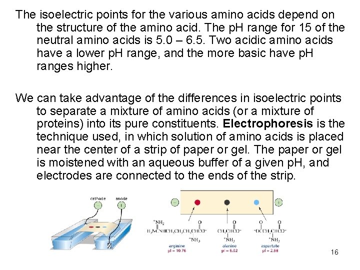
The isoelectric points for the various amino acids depend on the structure of the amino acid. The p. H range for 15 of the neutral amino acids is 5. 0 – 6. 5. Two acidic amino acids have a lower p. H range, and the more basic have p. H ranges higher. We can take advantage of the differences in isoelectric points to separate a mixture of amino acids (or a mixture of proteins) into its pure constituents. Electrophoresis is the technique used, in which solution of amino acids is placed near the center of a strip of paper or gel. The paper or gel is moistened with an aqueous buffer of a given p. H, and electrodes are connected to the ends of the strip. 16
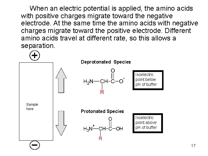
When an electric potential is applied, the amino acids with positive charges migrate toward the negative electrode. At the same time the amino acids with negative charges migrate toward the positive electrode. Different amino acids travel at different rate, so this allows a separation. Deprotonated Species Isoelectric point below p. H of buffer Sample here Protonated Species Isoelectric point above p. H of buffer 17
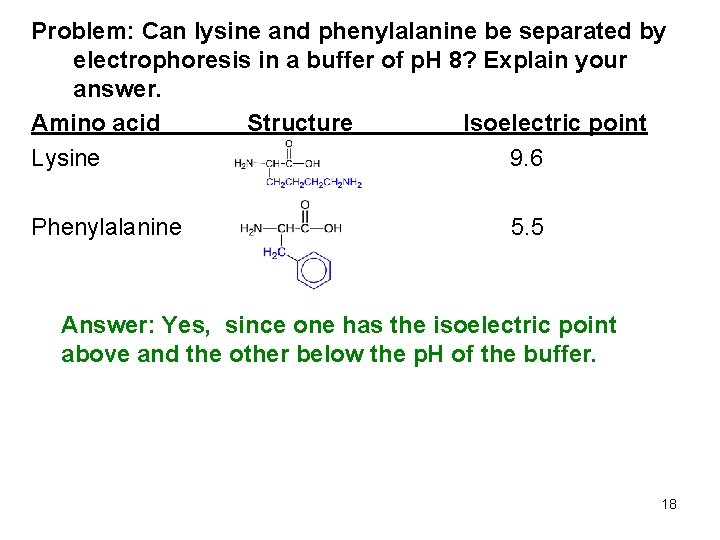
Problem: Can lysine and phenylalanine be separated by electrophoresis in a buffer of p. H 8? Explain your answer. Amino acid Structure Isoelectric point Lysine 9. 6 Phenylalanine 5. 5 Answer: Yes, since one has the isoelectric point above and the other below the p. H of the buffer. 18
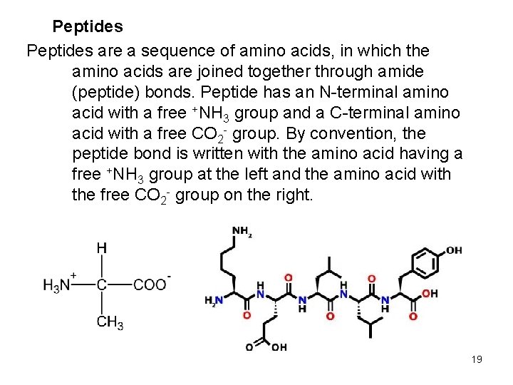
Peptides are a sequence of amino acids, in which the amino acids are joined together through amide (peptide) bonds. Peptide has an N-terminal amino acid with a free +NH 3 group and a C-terminal amino acid with a free CO 2 - group. By convention, the peptide bond is written with the amino acid having a free +NH 3 group at the left and the amino acid with the free CO 2 - group on the right. 19
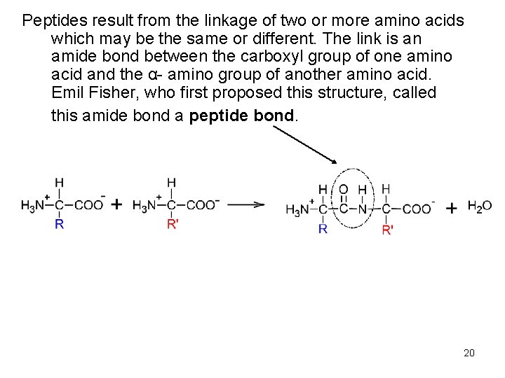
Peptides result from the linkage of two or more amino acids which may be the same or different. The link is an amide bond between the carboxyl group of one amino acid and the α- amino group of another amino acid. Emil Fisher, who first proposed this structure, called this amide bond a peptide bond. 20
Peptides are classified according to the number of amino acids that are linked together. The size boundaries which distinguish peptides, polypeptides, and proteins are arbitrary. 1. 2. 3. 4. Dipeptides: Two amino acids Tripeptides: Three amino acids Polypeptides: Generally four or more amino acids Protein: Over eight amino acids 21
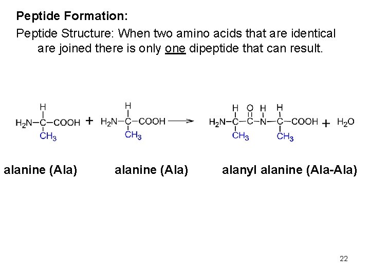
Peptide Formation: Peptide Structure: When two amino acids that are identical are joined there is only one dipeptide that can result. alanine (Ala) alanyl alanine (Ala-Ala) 22
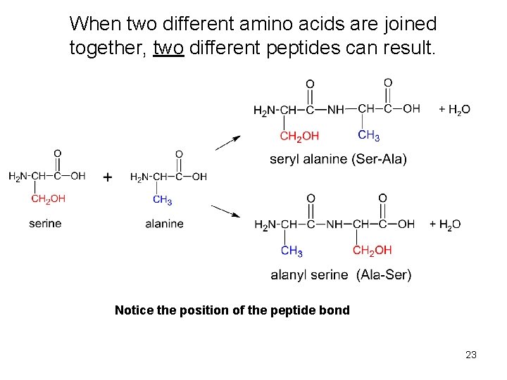
When two different amino acids are joined together, two different peptides can result. Notice the position of the peptide bond 23
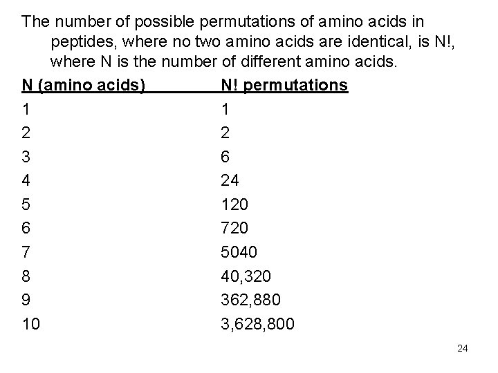
The number of possible permutations of amino acids in peptides, where no two amino acids are identical, is N!, where N is the number of different amino acids. N (amino acids) N! permutations 1 1 2 2 3 6 4 24 5 120 6 720 7 5040 8 40, 320 9 362, 880 10 3, 628, 800 24
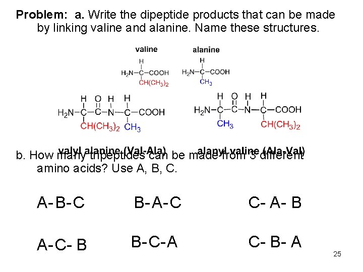
Problem: a. Write the dipeptide products that can be made by linking valine and alanine. Name these structures. b. How many tripeptides can be made from 3 different amino acids? Use A, B, C. A- B- C B- A- C C- A- B A- C- B B- C- A C- B- A 25
Proteins are biopolymers composed of many amino acids connected to one another through amide, or peptide, bonds. Some proteins are major components of biological structural tissue such as muscle, skin, nails, and hair. Other proteins transport molecules from one part of a living system to another, while others serve as catalysts (such as enzymes) for many biological reactions. Protein Structure: The primary structure of peptides and proteins is how many amino acids are present and what is their sequence is in the peptide or protein chain. The secondary, tertiary, and quaternary structures examine three-dimensional aspects of peptides and protein structures. 26
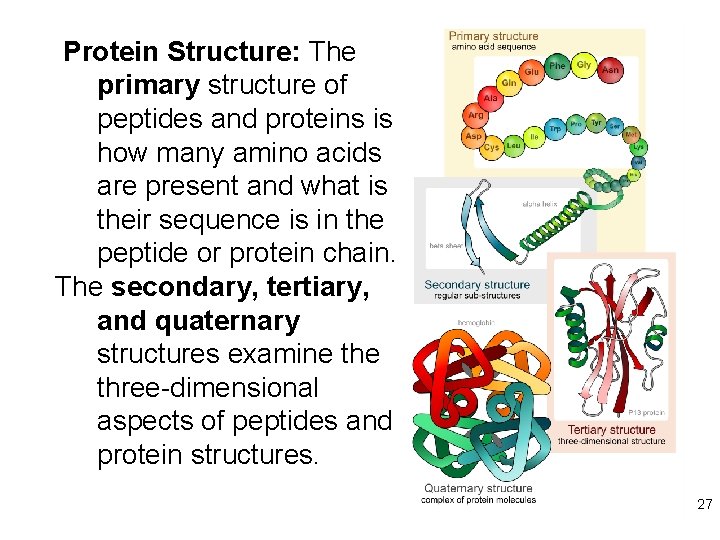
Protein Structure: The primary structure of peptides and proteins is how many amino acids are present and what is their sequence is in the peptide or protein chain. The secondary, tertiary, and quaternary structures examine three-dimensional aspects of peptides and protein structures. 27
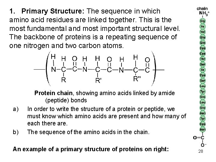
1. Primary Structure: The sequence in which amino acid residues are linked together. This is the most fundamental and most important structural level. The backbone of proteins is a repeating sequence of one nitrogen and two carbon atoms. a) b) Protein chain, showing amino acids linked by amide (peptide) bonds In order to write the structure of a protein or peptide, we must know which amino acids are present and how many of each there are. The sequence of the amino acids in the chain. An example of a primary structure of proteins on right: 28
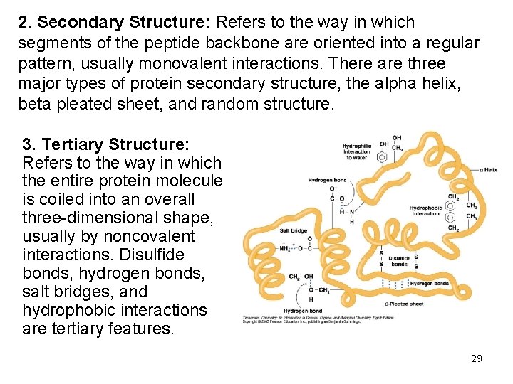
2. Secondary Structure: Refers to the way in which segments of the peptide backbone are oriented into a regular pattern, usually monovalent interactions. There are three major types of protein secondary structure, the alpha helix, beta pleated sheet, and random structure. 3. Tertiary Structure: Refers to the way in which the entire protein molecule is coiled into an overall three-dimensional shape, usually by noncovalent interactions. Disulfide bonds, hydrogen bonds, salt bridges, and hydrophobic interactions are tertiary features. 29
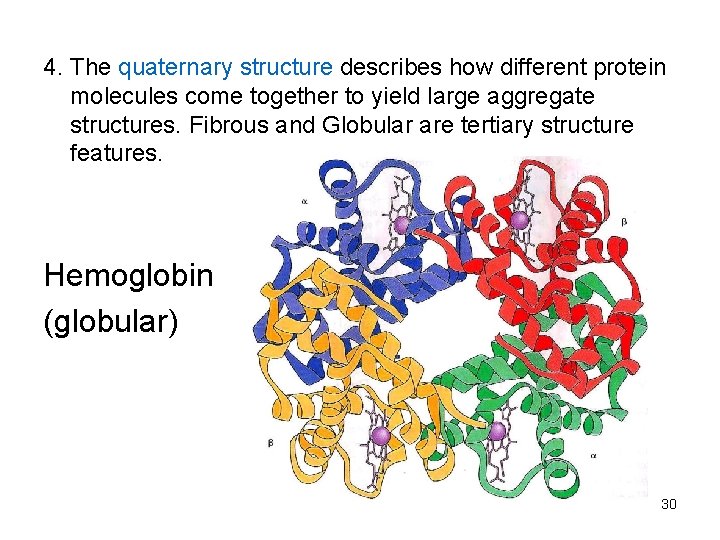
4. The quaternary structure describes how different protein molecules come together to yield large aggregate structures. Fibrous and Globular are tertiary structure features. Hemoglobin (globular) 30
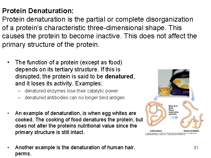
Protein Denaturation: Protein denaturation is the partial or complete disorganization of a protein’s characteristic three-dimensional shape. This causes the protein to become inactive. This does not affect the primary structure of the protein. • The function of a protein (except as food) depends on its tertiary structure. If this is disrupted, the protein is said to be denatured, and it loses its activity. Examples: – denatured enzymes lose their catalytic power – denatured antibodies can no longer bind antigen • An example of denaturation, is when egg whites are cooked. The cooking of food denatures the protein, but does not alter the proteins nutritional value since the primary structure is still intact. • Another example is the denaturation of human hair, perms. 31
YOU ARE NOW FINISHED WITH CHEM 102 Lectures!! 32