Section 3 2 Composite Cell Reem Heba sarah

Section 3. 2: Composite Cell Reem, Heba, sarah, and yasmeen 2 nd hour

Objectives Content Objectives: Students will show understanding of the cell, its organelles and their functions. Language Objectives: Students will show understanding of the cell, its organelles and their functions by taking notes during a powerpoint presentation and completing a four question quiz after.

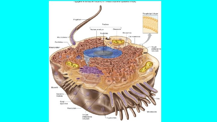
Cells Describing a typical cell is not possible because cells are different in size, shape, function, content, and structure. Three basic parts of a cell ★Cell Membrane that encloses the cell, allows certain substances to enter and leave. ★Nucleus that houses the genetic material and controls cellular activity. ★Cytoplasm that fills out the cell. ● In the cytoplasm are structures called organelles.
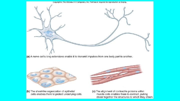

Cell Membrane is also known as the plasma membrane. Controls movement of substances that are coming in or leaving the cell. Some of the cell’s actions that help it survive and interact with other cells use a molecular communication process called signal transduction. Cell membrane also helps cells stick to other cells, which helps them form tissues. General Characteristics ★Extremely thin, flexible, and elastic. ★Selectively permeable which means that certain substances can leave and enter the cell.

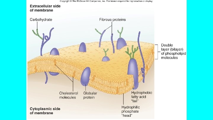
Cell Membrane Structure Cell membrane is mainly made up of lipids, proteins, and some carbohydrates. Basic framework is a double layer, or bilayer, of phospholipid molecules. Each phospholipid molecule has a phosphate group and two fatty acids that come together and make a glycerol molecule. Water soluble phosphate heads form the surfaces of the membrane, and the water- insoluble fatty acids “tails” make up the interior of the membrane. Lipid molecules can move sideways within the plane of the membrane. Two layers form a soft, flexible, but stable, fluid film. Membranes inside is oily because it is made up of mainly fatty acids portions and

Cell Membrane structure: Continued Bilayer is impermeable to water soluble molecules, like amino acids, sugars, proteins, nucleic acids, and various ions. Cholesterol molecules in cell membrane help make the membrane less permeable to water- soluble substances. Includes a few types of lipid molecules, but many kinds of proteins, that have special functions. Membrane proteins are classified according to their positions. Membrane spanning (transmembrane) proteins extend through the lipid bilayer and may protrude from one or both faces. Peripheral membrane proteins associate mostly with one side of the bilayer.

Cell Membrane structure: continued Membrane proteins have many different functions. Some form receptors on the cell surface that bind incoming hormones or growth factors, starting signal transduction. Other proteins transport ions or molecules across the cell membrane. Membrane proteins form selective channels that allow only certain ions to enter and leave. Proteins that extend inward from the inner face of the cell membrane anchor it to the protein rods and tubules that support the cell from within. Proteins that extend from the outer surface of the cell membrane mark the cell as part of a particular tissue or a organ in a particular person.

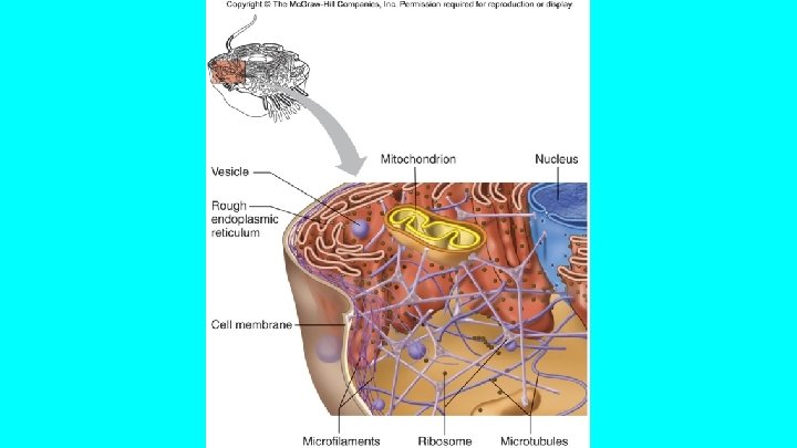
Cytoplasm is the gel like material in which organelles are suspended, it makes up most of the cell’s volume. Contains networks of membranes and organelles that suspend in the liquid cytosol. Cytoplasm also includes abundant protein rods and tubules that form a cytoskeletal, meaning “cell skeleton” Cell activities occur mainly in the cytoplasm, where nutrients are received, processed, and used.

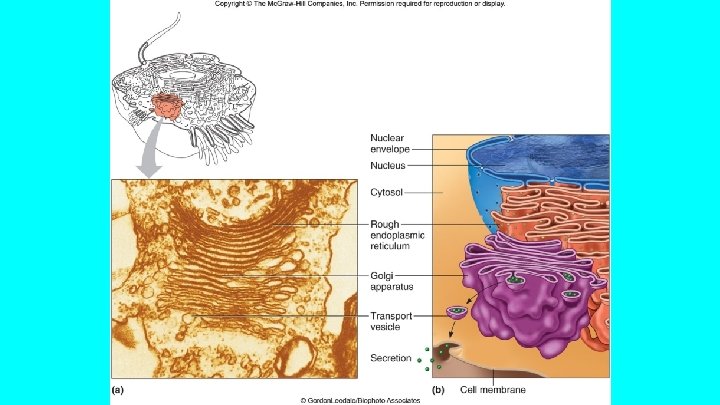
Organelles 1. Endoplasmic Reticulum- a complex organelle composed of membrane bounded flattened sacs, elongated canals, and fluid filled, bubble like sacs called vesicles. Provides a vast tubular network that transports molecules from one cell part to another. Participates in the synthesis of protein and lipid molecules. These molecules can be used to produce new ER or cell membrane as the cell grows. ER’s outer membrane is studded with many tiny, round structures called ribosomes, which give the ER a textured appearance. Rough ER
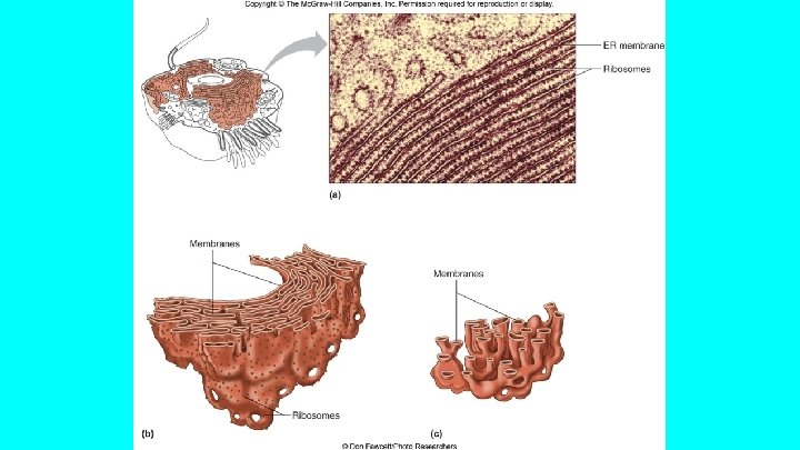

2. Ribosomes is where protein synthesis occurs, they are attached to ER membranes or are scattered throughout the cytoplasm. Clusters of Ribosomes in the cytoplasm called polysomes, enable a cell to quickly manufacture proteins required in large amounts. All ribosomes are composed of protein and RNA molecules. . 3. Golgi Apparatus Stack of about six flattened, membranous sacs. It refines, packages, and transports proteins synthesized on ribosomes associated with the ER Proteins arrive at the golgi apparatus enclosed in vesicles composed of the ER membrane.

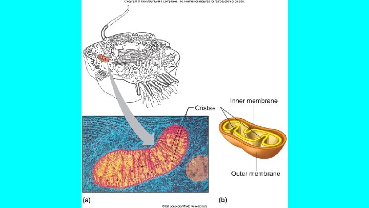
4. Mitochondria Are elongated, fluid filled sacs that vary in size and shape. Can move slowly through the cytoplasm and reproduce by dividing. Has an inner and outer layer. Inner layer is folded into partitions called cristae. Enzymes are connected to the cristae that control some chemical reactions that release energy from certain nutrients molecules. Mitochondria are the major sites of chemical reactions that capture and store this energy within the chemical bonds of adenosine triphosphate (ATP), a formula a cell can use.

5. Lysosomes The “Garbage disposals of the cell”, are tiny membranous sacs. Contain powerful enzymes that break down nutrient molecules of particles. Also destroy worn cellular stores. 6. Peroxisomes Membranous sacs that are abundant in liver and kidney cells. House enzymes that catalyze (speed) a variety of biochemical reactions, including synthesis of bile acids (used to digest fats); detoxification of hydrogen peroxide, a byproduct of metabolism; breakdown of certain lipids and rare biochemicals; and detoxification of alcohol.

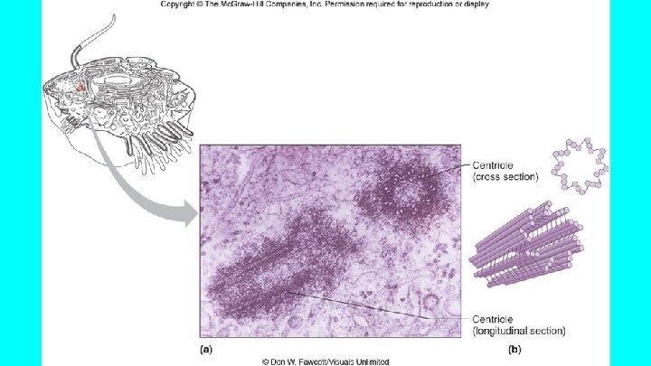
7. Microfilaments and Microtubules Are two types of thin, threadlike strands within the cytoplasm. Form the cytoskeleton and are also part of certain structures that have specialized activities. Microfilaments are tiny rods of actin protein that form meshworks or bundles. Provide cell movement. Microtubules are long, slender tubes with diameters two or three times those of microfilaments. Are composed of molecules of a globular protein called tubulin, attached in a spiral to form a long tube. 8. Centrosome is a structure near the Golgi apparatus and nucleus. Nonmembranous and consists of two hollow cylinders, called centrioles, that are composed of microtubules and organized in nine groups of three. Centrioles lie at the right angles to each other. During mitosis, the centrioles distribute

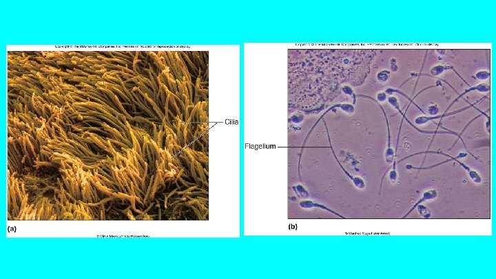
9. Cilia and Flagella Motile structures that extend from the surfaces of certain cells. Composed of microtubules in a “ 9+2” array. Are similar structures but differ in length and abundance. Cilia fringe the free surfaces of some lining cells. Cilia form in precise patterns. Move in a to and fro manner, so that rows of them beat in succession, producing a wave of motion. This wave moves fluids, like mucus, over the surface of tissues. Flagella are longer than cilia. Cell usually only has one.

10. Vesicles Are membranous sacs formed by part of the cell membrane folding inward and pinching off. As a result a tiny bubble like vesicle, containing a solid or liquid outside the cell appears in the cytoplasm. Golgi apparatus and ER also form vesicles that play a role in secretion.
, which directs all cell activities. It")
Cell Nucleus The nucleus houses the genetic material(DNA), which directs all cell activities. It is a large roughly spherical structure enclosed in a double layered nuclear envelope. Nuclear envelope consists of an inner and outer lipid bilayer membranes. Protein lined channels are known as nuclear pores. they allow certain molecules to exit the nucleus. A nuclear pore is a very complex opening formed from 100 or so types of proteins. Chromatin consist of loosely coiled fibers of DNA and protein that condense to form structures called chromosomes. The DNA contains info. for protein synthesis.

Nucleus contains a fluid called nucleoplasm in the nucleolus and chromatin. 1. Nucleolus is a small, dense body composed largely of RNA and protein. It has no surrounding membrane and forms in specialized regions of certain chromosomes. Ribosomes form here then go to the cytoplasm. 2. Chromatin consist of loosely coiled fibers of DNA and protein that condense to form structures called chromosomes. The DNA contains information for protein synthesis.

Quiz 1. What are three basic parts of a cell and their functions? 2. What is the difference between a microfilament and a microtubule? 3. What is the Endoplasmic Reticulum? 4. What is the difference between a Cilia and Flagella?
- Slides: 27