SECOND AND THIRD TRIMESTER BLEEDING Vaginal bleeding is

SECOND AND THIRD TRIMESTER BLEEDING Vaginal bleeding is less common in the second trimester (14+0 to 27+6 weeks) and third trimester (28+0 weeks to delivery). The major causes of bleeding at these times are: Bloody show associated with labor (by definition, labor occurs after 20 weeks) or, less commonly, cervical insufficiency Placenta previa Abruptio placentae Uterine rupture (rare) Vasa previa (rare) Cervical, vaginal, or uterine pathology (eg, polyps, inflammation/infection, trophoblastic disease).

Bleeding after 20 weeks of gestation Antepartum hemorrhage typically refers to vaginal bleeding after 20 weeks of gestation that is unrelated to labor and delivery. Antepartum bleeding complicates 4 to 5 percent of pregnancies. The major causes are: Placenta previa (20 percent) Abruptio placentae (30 percent) Uterine Vasa rupture (rare) previa (rare) In the remaining cases, the exact etiology of the antepartum bleeding cannot be determined and is frequently attributed to marginal separation of the placenta.
 as a placenta developing within")
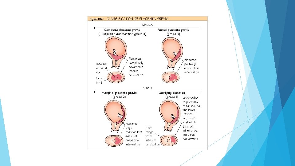
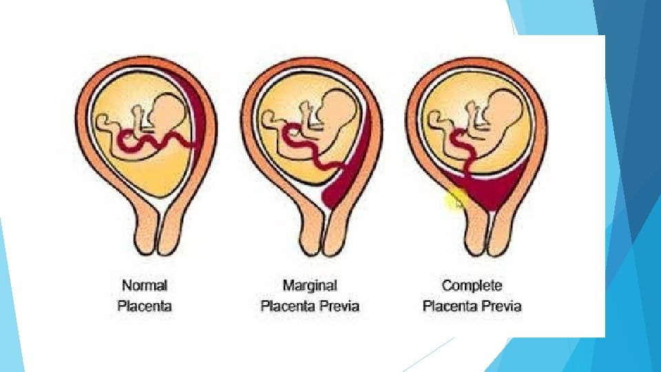
Placenta praevia was originally defined using transabdominal scan(TAS) as a placenta developing within the lower uterine segment and graded according to the relationship and/or the distance between the lower placental edge and the internal os of the uterine cervix. For pregnancies at more than 16 weeks of gestation the term low-lying placenta should be used when the placental edge is less than 20 mm from the internal os on transabdominal or transvaginal scanning (TVS). Grade I or minor praevia is defined as a lower edge inside the lower uterine segment grade II or marginal praevia as a lower edge reaching the internal os; grade III or partial praevia when the placenta partially covers the cervix; and grade IV or complete praevia when the placenta completely covers the cervix. Grades I and II are ‘minor’ placenta praevia whereas grades III and IV are referred to as ‘major’ placenta praevia. For pregnancies greater than 16 weeks of gestation, the placenta should be reported as ‘low lying’ when the placental edge is less than 20 mm from the internal os, and as normal when the placental edge is 20 mm or more from the internal os on TAS or TVS. The estimated incidence of placenta praevia at term is 1 in 200 pregnancies. The rising incidence of the main risk factors, i. e. prior caesarean delivery and pregnancies resulting from ART.

Management depends on the numerous factors gestational age cause of bleeding the severity of bleeding fetal status.

Placenta previa: Epidemiology, clinical features, diagnosis, morbidity and mortality Prevalence prevalence is about 4 per 1000 births, but varies worldwide The prevalence is several-fold higher around 20 weeks of gestation (as high as 2 percent), but most previas identified early in pregnancy resolve before delivery. Risk factors ●Previous placenta previa – Placenta previa recurs in 4 to 8 percent of subsequent pregnancies ●Previous cesarean delivery – previous cesarean delivery was found to increase the risk for placenta previa by 47%. The risk increases with an increasing number of cesarean deliveries ●Multiple gestation – The prevalence of placenta previa was 40 % higher among twin births than among singleton births (3. 9 and 2. 8 per 1000 births, respectively) in one study In another study, dichorionic twin pregnancies were more likely to have a placenta previa than monochorionic twin pregnancies

CLINICAL PRESENTATION AND COURSE Natural history : The lower uterine segment lengthens from 0. 5 cm at 20 weeks of gestation to more than 5 cm at term The development of the lower uterine segment relocates the stationary lower edge of the placenta away from the internal os. Progressive unidirectional growth of trophoblastic tissue toward the fundus results in upward migration of the placenta away from the cervix. Cervical length measurement may help facilitate management decisions in asymptomatic women with placenta praevia. A short cervical length on TVS before 34 weeks of gestation increases the risk of preterm emergency delivery and massive hemorrhage at caesarean section.

P. P antenatal care hospitalisation, . distance between home and hospital and availability of trans-portation, previous bleeding episodes, haematology laboratory results, and acceptance of receiving donor blood products Where hospital admission has been decided, an assessment of risk factors for venous thromboembolism in pregnancy should be performed as outlined in the RCOG This will need to balance the risk of developing a venous thromboembolism against the risk of bleeding from a placenta praevia or low lying placenta. It should be made clear to any woman being treated at home in the third trimester that she should attend the hospital immediately if she experiences any bleeding, including spotting, contractions or pain (including vague suprapubic period-like aches A single course of antenatal corticosteroid therapy is recommended between 34+0 and 35+6 weeks of gestation for pregnant women with a low-lying placenta or placenta praevia andis appropriate prior to 34+0 weeks of gestation in women at higher risk of preterm birth. Tocolysis for women presenting with symptomatic placenta praevia or a low-lying placenta maybe considered for 48 hours to facilitate administration of antenatal corticosteroids. If delivery is indicated based on maternal or fetal concerns, tocolysis should not be used in an attempt to prolong gestation.
 delivery should be considered for")
Late preterm (34+0 to 36+6 weeks of gestation) delivery should be considered for women presenting with placenta praevia or a low-lying placenta and a history of vaginal bleeding or other associated risk factors for preterm delivery. The major risk factors for placenta accreta spectrum are history of accreta in a previous pregnancy, previous caesarean delivery and other uterine surgery, including repeated endometrial curettage. This risk rises as the number of prior caesarean sections increases. Women requesting elective caesarean delivery for non-medical indications should be informed of the risk of placenta accreta spectrum and its consequences for subsequent pregnancies Unit with on-site blood transfusion services

epidemiological studies of the last 2 decades have shown a direct association between the increase in caesarean deliveries and the incidence of placenta accreta spectrum (abnormally adherent and invasive placenta) in subsequent pregnancies worldwide. the risk of invasive placentation increases seven-fold after one prior caesarean section. Placent praevia is another important risk factor for placenta accreta spectrum Other additional risk factors include maternal age and ART, in particular in vitro fertilisation. Advanced maternal age (35 years or more) in women

CLINAL PICTURE ASYMPTOMATIC Bleeding the most common symptom of placenta previa is relatively painless vaginal bleeding, which occurs in up to 90 percent of persistent cases 10 to 20 %of women present with uterine contractions, pain, and bleeding, similar to the presentation of abruptio placenta one-third of pregnancies with persistent previa, the initial bleeding episode occurs prior to 30 weeks of gestation; this group is more likely to require blood transfusions and is at greater risk of preterm delivery and perinatal mortality than women whose bleeding begins later in gestation About 10 percent of women reach term without bleeding. Anterior placenta previas are more likely to be associated with antepartum bleeding than posterior or lateral previas they are also associated with greater bleeding at cesarean Antepartum bleeding from any cause is a risk factor for preterm labor and preterm premature rupture of membranes.

DIAGNOSIS Diagnostic criteria The diagnosis of placenta previa is based on identification of placental tissue extending over the internal cervical os on a second or third trimester imaging study preferably transvaginal ultrasound Of note, when the placental edge is <2 cm from, but not over, the internal os, the placenta is labeled "low-lying“ digital vaginal examination, should be avoided Ultrasonography Transabdominal —can be considered a screening test for placenta previa. Transvaginal sonography The overall false positive rate of transabdominal ultrasound for diagnosis of placenta previa is up to 25 percent. Magnetic resonance imaging

Characteristics that appear to be predictive of antepartum bleeding include: Extension over the internal os rather than lying proximate to it Cervical length ≤ 3 cm Decrease in cervical length in the third trimester RECURRENCE Placenta previa recurs in 4 to 8 percent of subsequent pregnancies

ASSOCIATED FINDINGS Placenta previa-accrete First cesarean birth, 3 percent Second Third cesarean births, 40 percent Fourth Fifth cesarean birth, 11 percent cesarean births, 61 percent or greater cesarean birth, 67 percent ●Malpresentation ●Vasa previa and velamentous umbilical cord ●Intrauterine growth restriction –remains controversial ●Congenital anomalies but no single anomaly or syndrome was associated with the disorder

MORBIDITY AND MORTALITY Maternal — primarily related to antepartum and/or postpartum hemorrhage 52 percent of women with placenta previa had antepartum bleeding and 22 percent had postpartum hemorrhage receive blood transfusions more likely to undergo postpartum hysterectomy hemodynamic instability, decreased oxygen delivery, decreased tissue perfusion, cellular hypoxia, organ damage, and death. the maternal mortality rate associated with placenta previa is less than 1 percent Severe maternal morbidity and maternal mortality can also be a consequence of amniotic fluid embolism syndrome. Neonatal The principal cause is preterm delivery a three- to fivefold increase in risk of Neonatal intensive care unit admission Neonatal death /Perinatal death Neonatal anemia

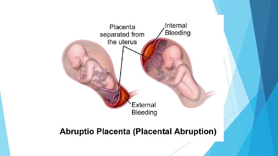
placental abruption: Pathophysiology, clinical features, diagnosis, and consequences Placental abruption (also referred to as abruptio placentae) refers to partial or complete placental detachment prior to delivery of the fetus. The diagnosis is typically reserved for pregnancies over 20 weeks of gestation. The major clinical findings are vaginal bleeding and abdominal pain, often accompanied by hypertonic uterine contractions, uterine tenderness, and a non reassuring fetal heart rate (FHR) pattern. Abruption is a significant cause of both maternal morbidity and neonatal morbidity and mortality, particularly when it occurs preterm. INCIDENCE 1 in 100 with two-thirds classified as severe based on associated maternal, fetal, and neonatal morbidity The incidence appears to be increasing

PATHOPHYSIOLOGYT the immediate cause of the premature placental separation is rupture of maternal vessels in the decidua basalis. Rarely, the bleeding originates from the fetal-placental vessels. The accumulating blood splits the decidua, separating off a thin layer of decidua with its placental attachment. This leads to rapid development of potentially life-threatening clinical manifestations of abruption (eg, severe bleeding, maternal disseminated intravascular coagulation, fetal heart rate [FHR] abnormalities). Thrombin plays a key role in the clinical consequences of placental abruption, and may be important in its pathogenesis, as well. It is formed via two pathways: in one pathway, decidual bleeding leads to release of tissue factor (thromboplastin) from decidual cells, which generates thrombin In the other pathway, decidual hypoxia, which then generates thrombin The production of thrombin can lead to the following clinical sequelae: Uterine hypertonus and contractions, as thrombin is a potent, direct uterotonic agent and induced expression of inflammatory cytokines (predominantly interleukin-8), and often leading to initiation of labor and rupture of membranes Triggering of coagulation due to widespread intravascular fibrin deposition, ischemic tissue injury and microangiopathic hemolytic anemia (ie, disseminated intravascular coagulation [DIC]).

RISK FACTORS Clinical — Previous abruption is the strongest risk factor for abruption, with recurrence risks of 10 - to 15 -fold higher Cocaine use and smoking are important, but less common risk factors for abruption. Smoking is one of the few modifiable risk factors for Hypertensive women have a fivefold increased risk of severe abruption compared with normotensive women. Antihypertensive therapy does not appear to reduce this risk in women with chronic hypertension Uterine anomalies (eg, bicornuate uterus), uterine synechiae, and leiomyoma are modest risk factors for abruption trauma Laboratory — Abnormalities of maternal serum biochemical markers used for Down syndrome or neural tube defect screening carry up to a 10 -fold increase in risk of subsequent abruption (increased alpha fetoprotein or human chorionic gonadotropin)

CLINICAL FEATURES Acute abruption Patient presentation — Women with an acute abruption classically present with the abrupt onset of vaginal bleeding (mild to severe and life-threatening) mild to moderate abdominal and/or back pain(Back pain is prominent when the placenta is on the posterior wall of the uterus) uterine contractions. . The uterus is often firm, and may be rigid and tender. Contractions are usually high frequency and low amplitude, but a contraction pattern typical of labor is also possible, and labor may proceed rapidly. The amount of vaginal bleeding correlates poorly with the degree of placental separation and does not serve as a useful marker of impending fetal or maternal risk. In contrast, abdominal pain , hypotension, and fetal heart rate (FHR) abnormalities suggest clinically significant separation that could result in fetal death and severe maternal morbidity When the presenting symptom is abdominal pain, the incidence of preeclampsia, preterm birth, maternal hemorrhage, and neonatal compromise is increased compared with women whose presenting symptom is vaginal bleeding, highlighting the point that the degree of bleeding correlates poorly with outcomes When placental separation exceeds 50 percent, acute disseminated intravascular coagulation and fetal death are common
. DIC occurs in")
Laboratory findings Severe abruption can lead to disseminated intravascular coagulation (DIC). DIC occurs in 10 to 20 percent of severe abruptions with death of the fetus. Diagnosis of DIC in pregnant women is reviewed separately. The Kleihauer-Betke test is positive in a small proportion of abruptions. There is poor correlation between the results of this test and the presence or absence of abruption sensitivity is only 4 percent Imaging ●Ultrasound A retroplacental hematoma is the classic ultrasound finding of placental abruption Retroplacental hematomas have a variable appearance; they can appear solid, complex, and hypo-, hyper-, or iso-echoic compared with the placenta. Hypoechogenicity and sonolucency are features of resolving rather than acute hematomas The sensitivity of ultrasound findings for diagnosis of abruption is only 25 to 60 percent However, whether a hematoma is identified depends on the extent of hemorrhage, chronicity of the bleeding, and extent that blood has escaped through the cervix Although the worst outcomes appear to occur when there is sonographic evidence of a retroplacental hematoma the absence of retroplacental hematoma does not exclude the possibility of severe abruption ●Magnetic resonance imaging ●Computed tomography

Consequences Maternal consequences – Serious maternal consequences of abruption include: • Excessive blood loss and DIC, which generally necessitate blood transfusion and can lead to hypovolemic shock, renal failure, adult respiratory distress syndrome, multiorgan failure, peripartum hysterectomy and, rarely, death • Emergency cesarean delivery for fetal or maternal indications Increased overall mortality Fetal/neonatal consequences – Serious fetal and neonatal consequences of abruption include: Increased perinatal morbidity and mortality related to hypoxemia, asphyxia, low birth weight, and/or preterm delivery Fetal growth restriction (with chronic abruption)]. Deaths in the postnatal period are primarily related to preterm delivery Placental abruption is implicated in up to 10 percent of preterm births Preterm birth may be iatrogenic due to the non reassuring fetal or maternal condition, or it may be related to preterm labor or preterm premature rupture of membranes Of note, perinatal mortality associated with abruption appears to be decreasing Fetal asphyxia, preterm birth, and growth restriction can be associated with short- and long-term sequelae, and abruption appears to compound the risk. Recurrence Women with placental abruption are at several-fold higher risk of abruption and other manifestations of ischemic placental disease in a subsequent pregnancy

DIFFERENTIAL DIAGNOSIS In pregnant women with suspected abruption, the differential diagnosis of vaginal bleeding accompanied by pain and contractions includes labor, placenta previa, uterine rupture, and subchorionic hematoma. Uterine rupture is most common in women with a prior hysterotomy, and usually occurs during labor. Signs of uterine rupture may include the sudden onset fetal heart rate (FHR) abnormalities, vaginal bleeding, constant abdominal pain, cessation of uterine contractions, recession of the presenting part, and maternal hypotension and tachycardia.

Administer corticosteroids Antenatal fetal assessment – Fetal assessment with a nonstress test or biophysical profile is performed at least weekly Hospitalization Delivery For patients managed conservatively and without any further symptoms, we schedule delivery at 37 to 38 weeks because of the increased risk of stillbirth Delivery before 37 weeks is indicated if additional complications arise Placental abruption occurring in the second trimester carries an especially poor prognosis when accompanied by oligohydramnios.

COUVELAIRE UTERUS In severe abruptions, blood may extravasate into the myometrium (called a Couvelaire uterus), and this can be seen at cesarean. The Couvelaire uterus is atonic and prone to postpartum hemorrhage. Aggressive management of atony is needed to prevent disseminated intravascular coagulation and exsanguination; however, atony in this setting is less likely to respond to standard therapies for postpartum hemorrhage than atony from other causes; thus, these women are at high risk for requiring hysterectomy.

MANAGEMENT OF FUTURE PREGNANCIES Recurrence risk Women with placental abruption are at severalfold higher risk of abruption in a subsequent pregnancy Three to 15 percent of women have a recurrence, compared with a baseline incidence of 0. 4 to 1. 3 percent in the general population In one longitudinal population-based study, the risk of placental abruption in a subsequent pregnancy was approximately 6 percent in women with an abruption in their first pregnancy versus 0. 06 percent in women without an abruption In this study, women with a placental abruption at term were at higher risk for recurrence than those with preterm abruption. After two consecutive abruptions, the risk of a third rises to 20 to 25 percent The risk of recurrence is higher after a severe abruption than after a mild abruption. When the abruption is sufficiently severe to kill the fetus, there is a 7 percent incidence of abruption with fetal demise in a future pregnancy Placental abruption resulting from trauma is not likely to recur in the absence of recurrent trauma, so these women can be reassured.

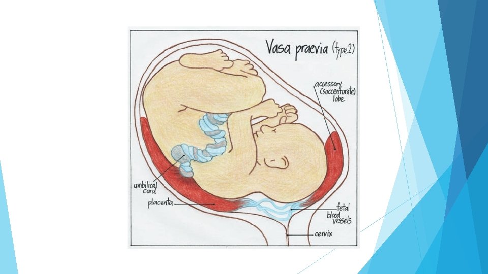
Vasa praevia occurs when the fetal vessels run through the free placental membranes. Unprotected by placental tissue or Wharton's jelly of the umbilical cord, a vasa praevia is likely to rupture in active labour, or when amniotomy is performed to induce or augment labour, in particular when located near or over the cervix, under the fetal presenting part. Vasa praevia is classified as type I when the vessel is connected to a velamentous umbilical cord, and type II when it connects the placenta with a succenturiate or accessory lobe. Vasa praevia may be diagnosed during early labour by vaginal examination, detecting the pulsating fetal vessels inside the internal os, or by the presence of dark‐red vaginal bleeding and acute fetal compromise after spontaneous or artificial rupture of the placental membranes. The fetal mortality rate in this situation is at least 60% despite urgent caesarean delivery. Vasa praevia is uncommon in the general population with a prevalence ranging between 1 in 1200 and 1 in 5000 pregnancies, although the condition may have been under‐r

emergency caesarean delivery and neonatal resuscitation, including the use of blood transfusion if required, are essential in the management of ruptured vasa praevia diagnosed during labour. Placental pathological examination should be performed to confirm the diagnosis of vasa praevia, in particular when stillbirth has occurred or where there has been acute fetal compromise during delivery. A combination of both transabdominal and transvaginal colour Doppler imaging (CDI) ultrasonography provides the best diagnostic accuracy for vasa praevia.

Because of the speed at which fetal exsanguination can occur and the high perinatal mortality rate associated with ruptured vasa praevia, delivery should not be delayed while trying to confirm the diagnosis, particularly if there is evidence that fetal wellbeing is compromised. In the presence of confirmed vasa praevia in the third trimester, elective caesarean section should ideally be carried out prior to the onset of labour. A decision for prophylactic hospitalisation from 30– 32 weeks of gestation in women with confirmed vasa praevia should be individualised and based on a combination of factors, including multiple pregnancy, antenatal bleeding and threatened premature labour. In cases of vasa praevia that develop premature rupture of membranes and/or labour at viable gestational ages, a caesarean section should be performed without delay. To avoid unnecessary anxiety, admissions, prematurity and caesarean section, it is essential to confirm persistence of vasa praevia by ultrasound in the third trimester. Delivery by caesarean section of women with confirmed vasa praevia is intuitive and logical, and not based on RCTs.
- Slides: 31