
Screening for high risk pregnancy History Age Social

- Slides: 63
Screening for high risk pregnancy History • * Age • *Social burden • *Smoking • *Past medical conditions e. g D. M, HTN • *Past Obstetric history
Fetal assessment Aim: Ensure fetal wellbeing ( Identify patients at risk of fetal asphyxia) To prevent prenatal mortality & morbidity
When to start fetal Assessment ** Risk assessed individually ** uncomplicated D. M. : start from 32 weeks onward ***If complicated D. M. : start at 24 weeks onward **For Post date pregnancy start at 40 weeks **For any patient with decrease fetal movement start immediately ** Fetal assessment is done once or twice weekly
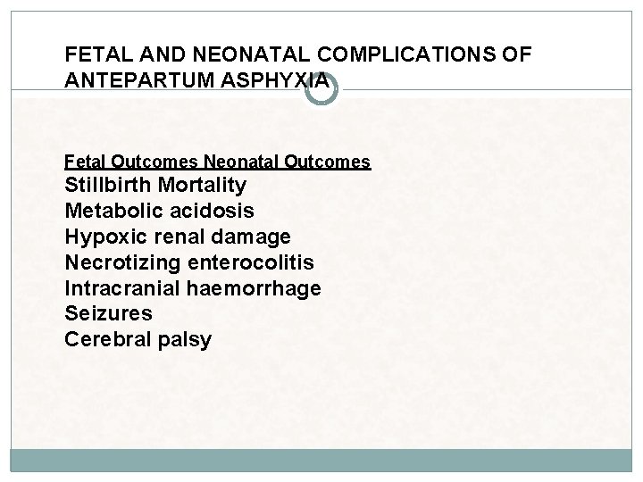
FETAL AND NEONATAL COMPLICATIONS OF ANTEPARTUM ASPHYXIA Fetal Outcomes Neonatal Outcomes Stillbirth Mortality Metabolic acidosis Hypoxic renal damage Necrotizing enterocolitis Intracranial haemorrhage Seizures Cerebral palsy
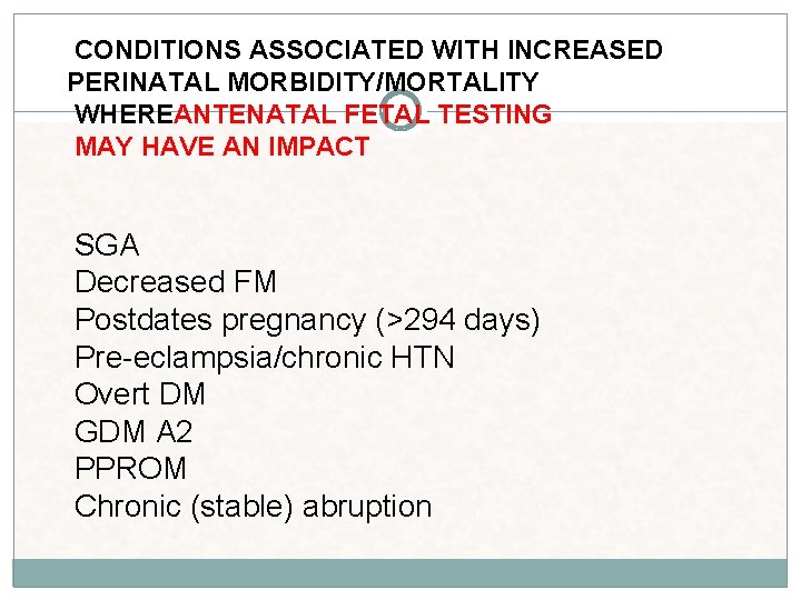
CONDITIONS ASSOCIATED WITH INCREASED PERINATAL MORBIDITY/MORTALITY WHEREANTENATAL FETAL TESTING MAY HAVE AN IMPACT SGA Decreased FM Postdates pregnancy (>294 days) Pre-eclampsia/chronic HTN Overt DM GDM A 2 PPROM Chronic (stable) abruption
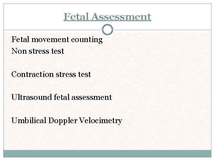
Fetal Assessment Fetal movement counting Non stress test Contraction stress test Ultrasound fetal assessment Umbilical Doppler Velocimetry
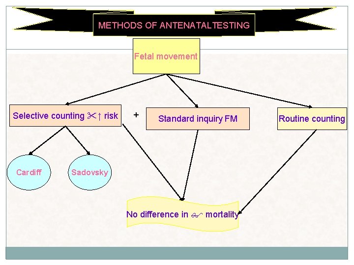
METHODS OF ANTENATALTESTING Fetal movement Selective counting ↑ risk Cardiff + Standard inquiry FM Sadovsky No difference in mortality Routine counting
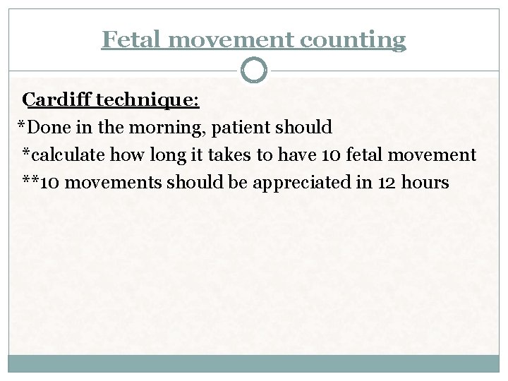
Fetal movement counting Cardiff technique: *Done in the morning, patient should *calculate how long it takes to have 10 fetal movement **10 movements should be appreciated in 12 hours
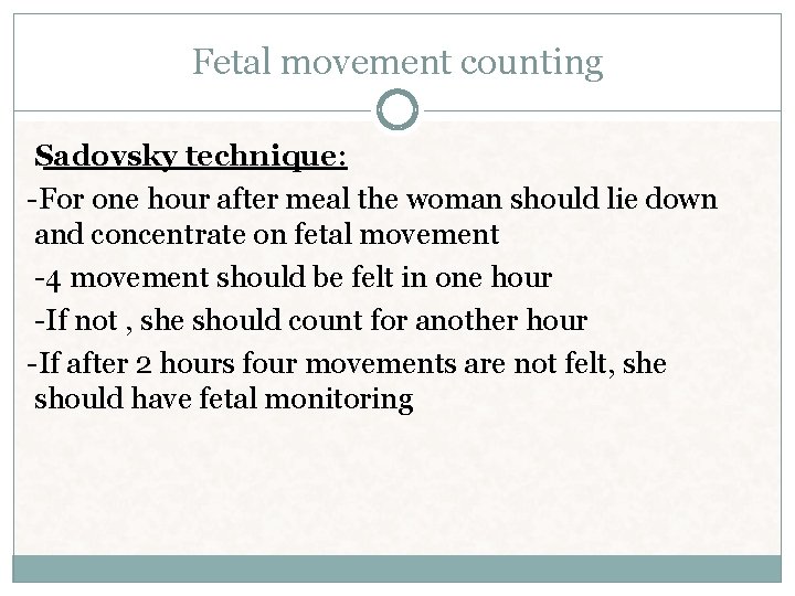
Fetal movement counting Sadovsky technique: -For one hour after meal the woman should lie down and concentrate on fetal movement -4 movement should be felt in one hour -If not , she should count for another hour -If after 2 hours four movements are not felt, she should have fetal monitoring
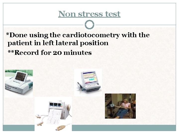
Non stress test *Done using the cardiotocometry with the patient in left lateral position **Record for 20 minutes
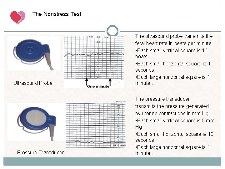
The Nonstress Test Ultrasound Probe Pressure Transducer The ultrasound probe transmits the fetal heart rate in beats per minute. • Each small vertical square is 10 beats. • Each small horizontal square is 10 seconds. • Each large horizontal square is 1 minute. The pressure transducer transmits the pressure generated by uterine contractions in mm Hg. • Each small vertical square is 5 mm Hg • Each small horizontal square is 10 seconds. • Each large horizontal square is 1 minute.
Interpretation of the Fetal Heart Tracing The interpretation of the fetal heart rate tracing should follow a systematic approach with a full qualitative and quantitative description of the following: Baseline fetal heart rate (FHR) variability Presence of accelerations Periodic or episodic decelerations Changes or trends of FHR patterns over time Frequency and intensity of uterine contractions
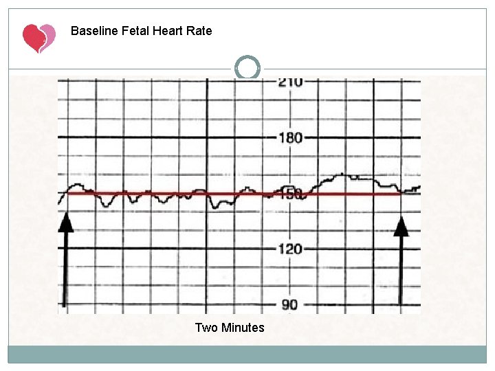
Baseline Fetal Heart Rate The baseline FHR is the heart rate during a 10 minute segment rounded to the nearest 5 beat per minute increment excluding periods of marked FHR variability, periodic or episodic changes, and segments of baseline that differ by more than 25 beats per minute. The minimum baseline duration must be at least 2 minutes. If minimum baseline duration is < 2 minutes then the baseline is indeterminate. (wandering base line)
Baseline Fetal Heart Rate Two Minutes
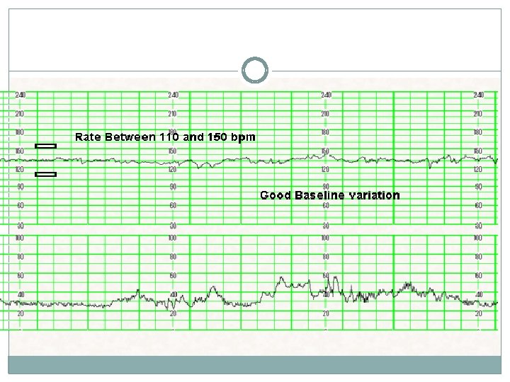
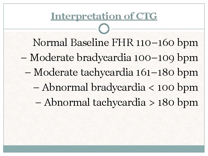
Interpretation of CTG Normal Baseline FHR 110– 160 bpm – Moderate bradycardia 100– 109 bpm – Moderate tachycardia 161– 180 bpm – Abnormal bradycardia < 100 bpm – Abnormal tachycardia > 180 bpm
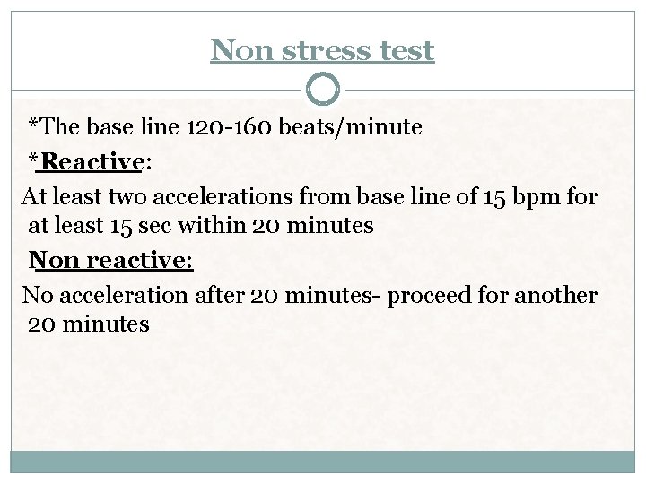
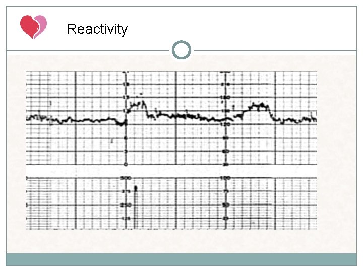
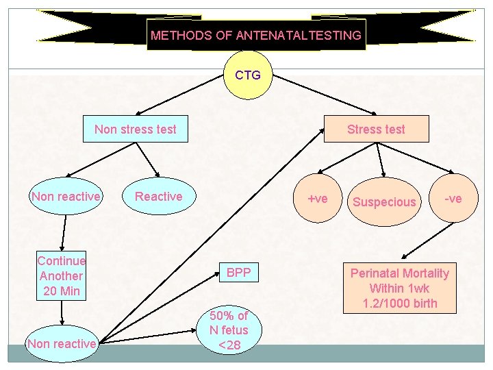
Non stress test *The base line 120 -160 beats/minute *Reactive: At least two accelerations from base line of 15 bpm for at least 15 sec within 20 minutes Non reactive: No acceleration after 20 minutes- proceed for another 20 minutes
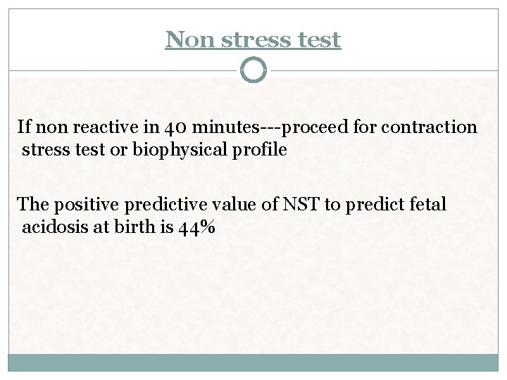
Non stress test If non reactive in 40 minutes---proceed for contraction stress test or biophysical profile The positive predictive value of NST to predict fetal acidosis at birth is 44%
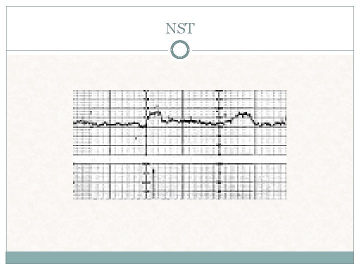
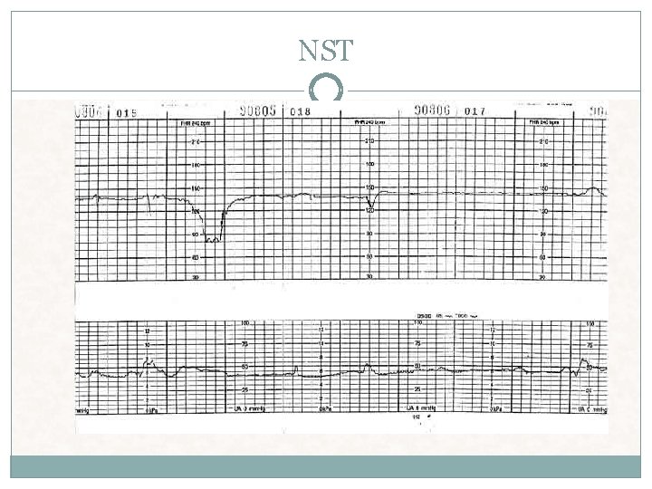
NST
NST
Bradycardia Mean FHR < 110 BPM A rate of 100 -119 BPM in the absence of other non reassuring patterns is not usually a sign of compromise Causes Heart block (little or no variability) Occiput posterior or transverse position Serious fetal compromise. Patients with new onset bradycardia should be transferred to Labor and Delivery for further observation and physician notified
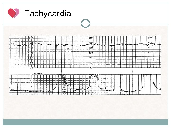
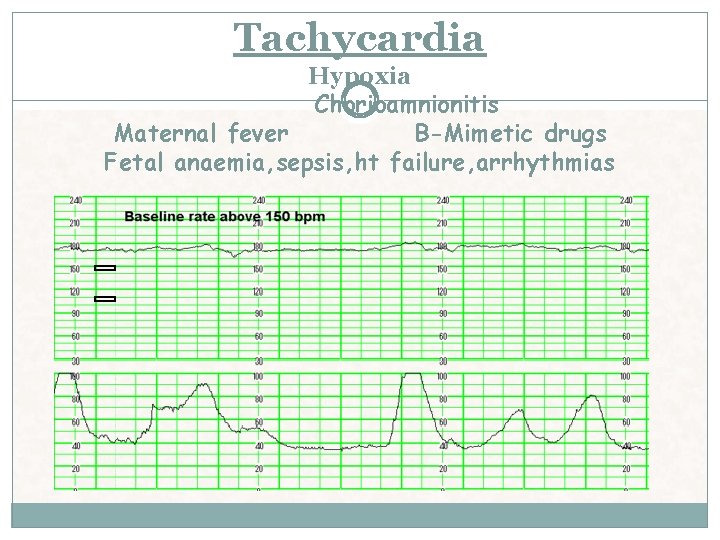
Tachycardia Mean FHR>160 BPM In the presence of good variability tachycardia is not a sign of fetal distress Causes: Maternal fever Fetal hypoxia Fetal anemia Amnionitis Normal variant Fetal tachyarrhythmia (usually > 200 BPM with abrupt onset little to no variability) SVT (200 -240 BPM) Fetal heart failure Drugs Beta sympathomimetics Vistaril Phenothiazines Rebound transient tachycardia following a deceleration accompanied by decreased variability)
Tachycardia
Tachycardia Hypoxia Chorioamnionitis Maternal fever B-Mimetic drugs Fetal anaemia, sepsis, ht failure, arrhythmias
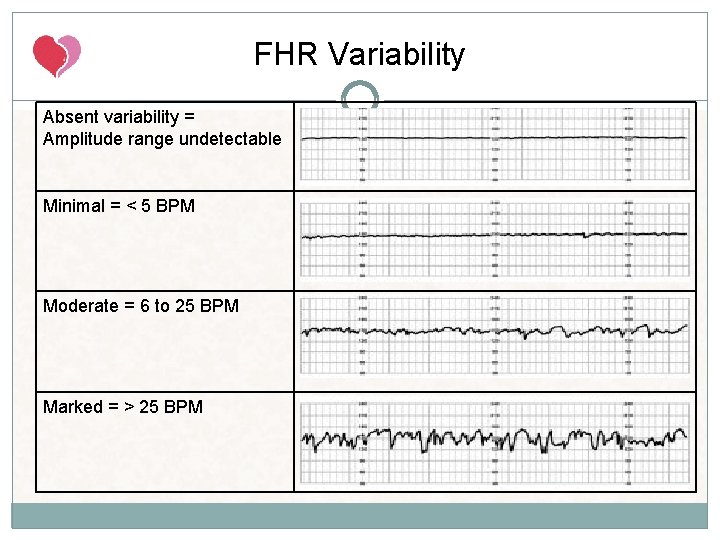
FHR Variability Fluctuation in baseline FHR > 2 cycles per minute. No distinction is made between shortterm variability (or beat-to-beat variability or R-R wave period differences in the electrocardiogram) and long-term variability.
Development of FHR Variability Early in gestation the fetal heart rate is predominately under the control of the sympathetic nervous system and arterial chemoreceptors. As the fetus develops its heart rate decreases in response to parasympathetic (vagal stimulation) nervous system maturation and variability becomes more pronounced.
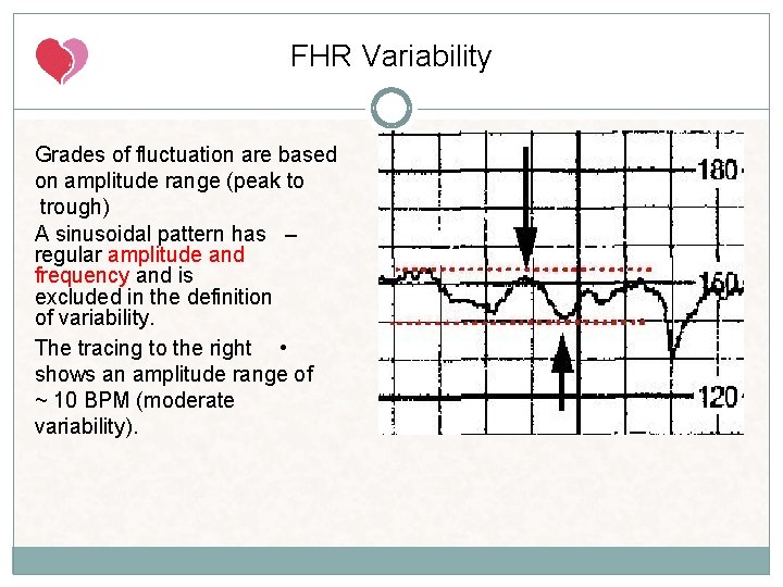
FHR Variability Grades of fluctuation are based on amplitude range (peak to trough) A sinusoidal pattern has – regular amplitude and frequency and is excluded in the definition of variability. The tracing to the right • shows an amplitude range of ~ 10 BPM (moderate variability).
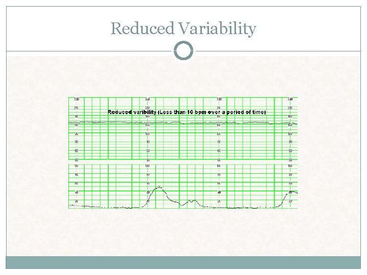
FHR Variability Absent variability = Amplitude range undetectable Minimal = < 5 BPM Moderate = 6 to 25 BPM Marked = > 25 BPM
Reduced Variability
The Presence of Moderate FHR Variability is Reassuring. q. Persistently minimal or absent FHR variability appears to be the most significant intrapartum sign of fetal compromise. q. On the other hand the presence of good FHR variability may not always be predictive of a good outcome (such as may occur with an abruption).
Causes of Decreased Variability • Fetal metabolic • • acidosis CNS depressants Fetal sleep cycles Congenital anomalies Prematurity • Fetal tachycardia • Preexisting neurologic abnormality • Normal variant • Betamethasone
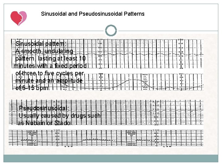
Sinusoidal and Pseudosinusoidal Patterns Sinusoidal pattern: A smooth, undulating pattern, lasting at least 10 minutes with a fixed period of three to five cycles per minute and an amplitude of 5 -15 bpm. Pseudosinusoidal: Usually caused by drugs such as Nubain or Stadol.
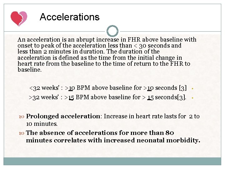
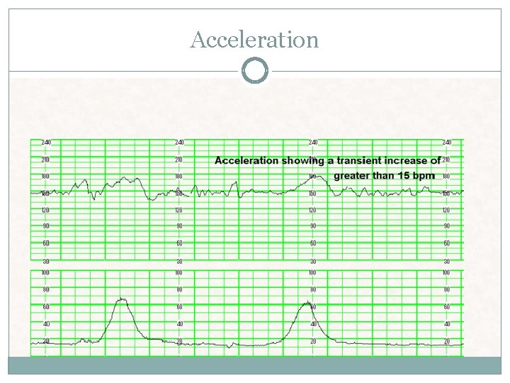
Accelerations An acceleration is an abrupt increase in FHR above baseline with onset to peak of the acceleration less than < 30 seconds and less than 2 minutes in duration. The duration of the acceleration is defined as the time from the initial change in heart rate from the baseline to the time of return to the FHR to baseline. <32 weeks' : >10 BPM above baseline for >10 seconds [3] >32 weeks' : >15 BPM above baseline for > 15 seconds[3]. • • Prolonged acceleration: Increase in heart rate lasts for 2 to 10 minutes. The absence of accelerations for more than 80 minutes correlates with increased neonatal morbidity.
Reactivity A NST is considered reactive when two or more fetal heart rate accelerations peak (but do not necessarily remain) at least 15 beats per minute above the baseline and last 15 seconds from baseline to baseline within a 20 minute period with or without fetal movement discernible by the woman.
Acceleration
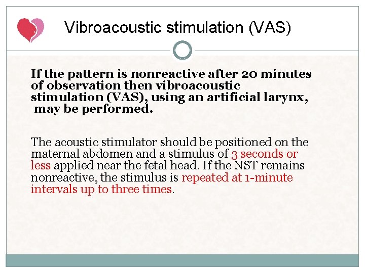
Vibroacoustic stimulation (VAS) If the pattern is nonreactive after 20 minutes of observation then vibroacoustic stimulation (VAS), using an artificial larynx, may be performed. The acoustic stimulator should be positioned on the maternal abdomen and a stimulus of 3 seconds or less applied near the fetal head. If the NST remains nonreactive, the stimulus is repeated at 1 -minute intervals up to three times.
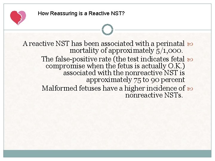
How Reassuring is a Reactive NST? A reactive NST has been associated with a perinatal mortality of approximately 5/1, 000. The false-positive rate (the test indicates fetal compromise when the fetus is actually O. K. ) associated with the nonreactive NST is approximately 75 to 90 percent Malformed fetuses have a higher incidence of nonreactive NSTs.
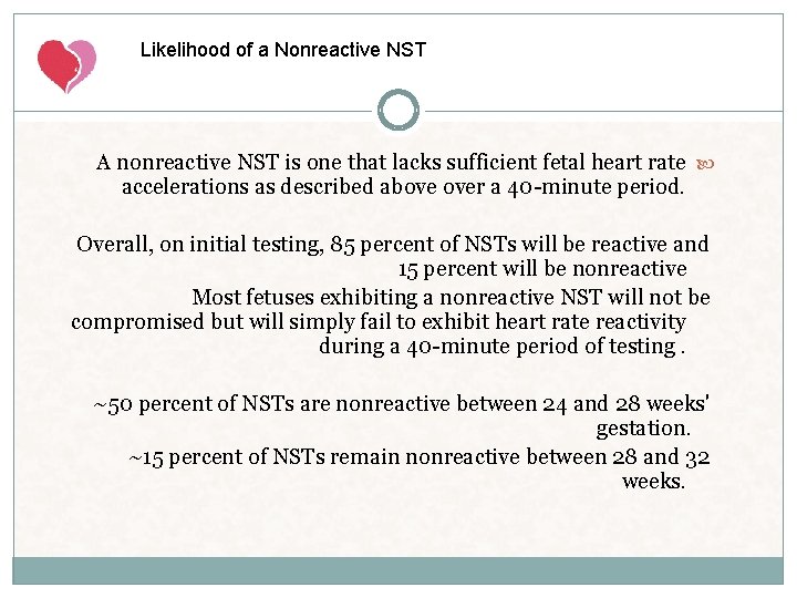
Likelihood of a Nonreactive NST A nonreactive NST is one that lacks sufficient fetal heart rate accelerations as described above over a 40 -minute period. Overall, on initial testing, 85 percent of NSTs will be reactive and 15 percent will be nonreactive Most fetuses exhibiting a nonreactive NST will not be compromised but will simply fail to exhibit heart rate reactivity during a 40 -minute period of testing. ~50 percent of NSTs are nonreactive between 24 and 28 weeks' gestation. ~15 percent of NSTs remain nonreactive between 28 and 32 weeks.
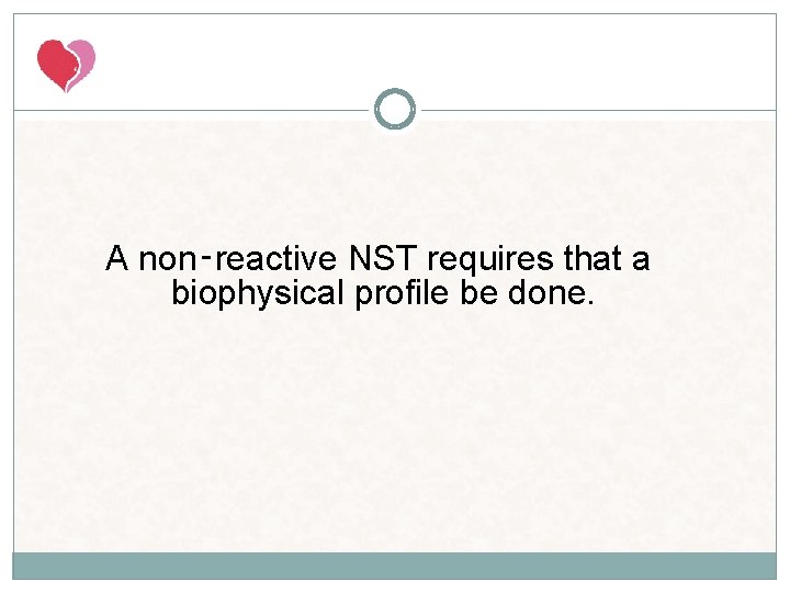
A non‑reactive NST requires that a biophysical profile be done.
METHODS OF ANTENATALTESTING CTG Non stress test Non reactive Continue Another 20 Min Non reactive Stress test Reactive +ve BPP 50% of N fetus <28 Suspecious -ve Perinatal Mortality Within 1 wk 1. 2/1000 birth
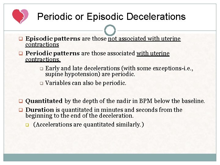
Periodic or Episodic Decelerations q Episodic patterns are those not associated with uterine contractions q Periodic patterns are those associated with uterine contractions. q Early and late decelerations (with some exceptions-i. e. , supine hypotension) are periodic. q Variables can also be periodic. q Quantitated by the depth of the nadir in BPM below the baseline. q Duration is quantitated in minutes and seconds from the beginning to the end of the deceleration. q (Accelerations are quantitated similarly. )
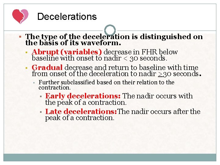
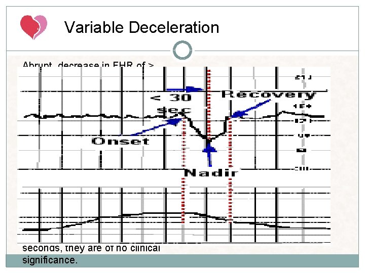
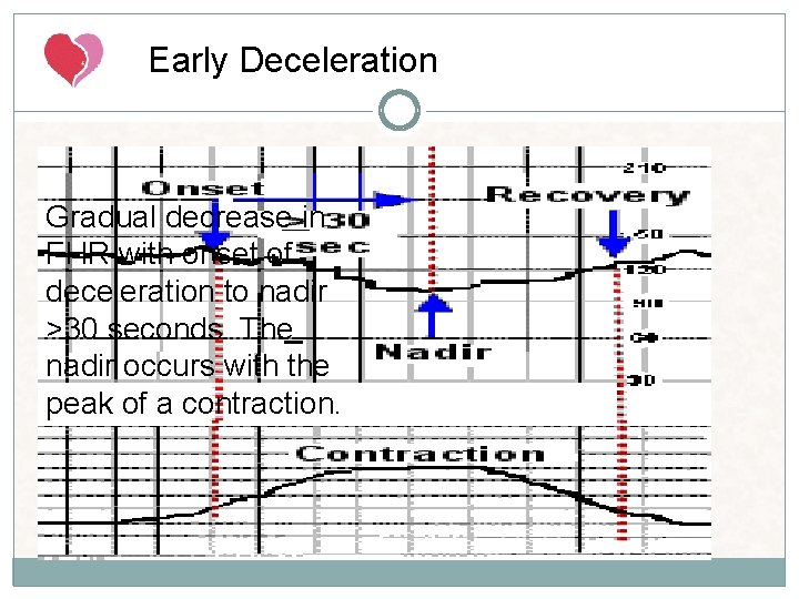
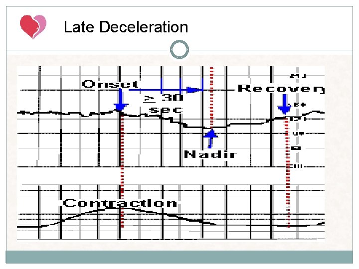
Decelerations § The type of the deceleration is distinguished on the basis of its waveform. § Abrupt (variables) decrease in FHR below baseline with onset to nadir < 30 seconds. § Gradual decrease and return to baseline with time from onset of the deceleration to nadir >30 seconds. § Further subclassified based on their relation to the contraction. § § Early decelerations: The nadir occurs with the peak of a contraction. Late decelerations: The nadir occurs after the peak of a contraction.
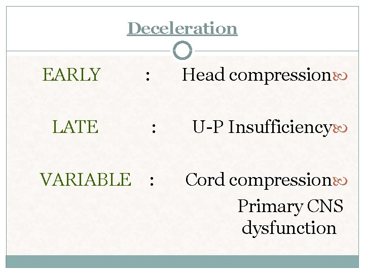
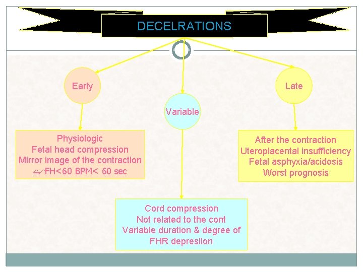
Deceleration EARLY : Head compression LATE : U-P Insufficiency VARIABLE : Cord compression Primary CNS dysfunction
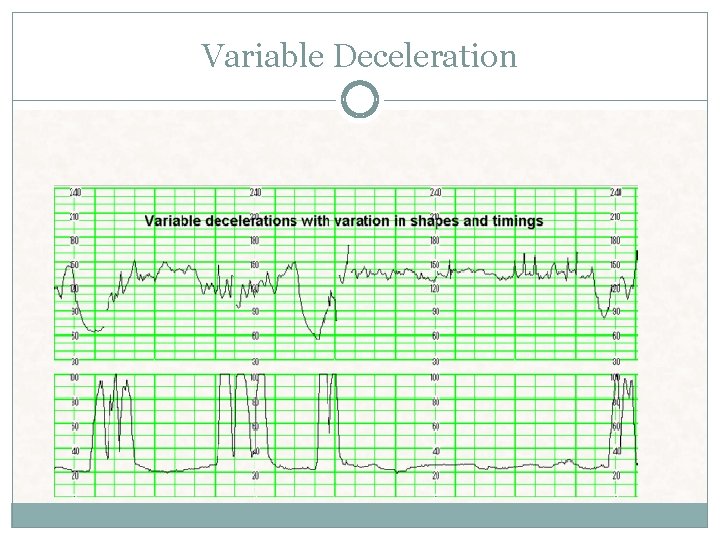
Variable Deceleration Abrupt decrease in FHR of > 15 beats per minute measured from the most recently determined baseline rate. The onset of deceleration to nadir is less than 30 seconds. The deceleration lasts > 15 seconds and less than 2 minutes. A shoulder, if present, is not included as part of the deceleration. Variable decelerations may be observed in up to 50% of NSTs. If nonrecurrent and <30 seconds, they are of no clinical significance.
Variable Deceleration
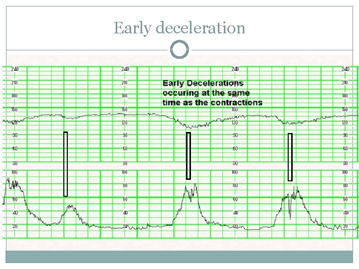
Early Deceleration Gradual decrease in FHR with onset of deceleration to nadir >30 seconds. The nadir occurs with the peak of a contraction.
Early deceleration
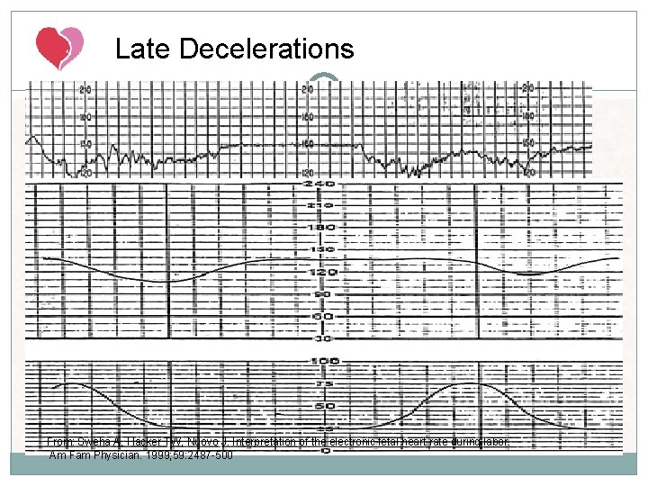
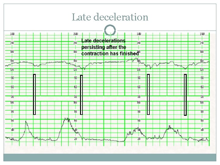
Late Deceleration Gradual decrease in FHR with onset of deceleration to nadir >30 seconds. The nadir of the deceleration occurs after the peak of the contraction
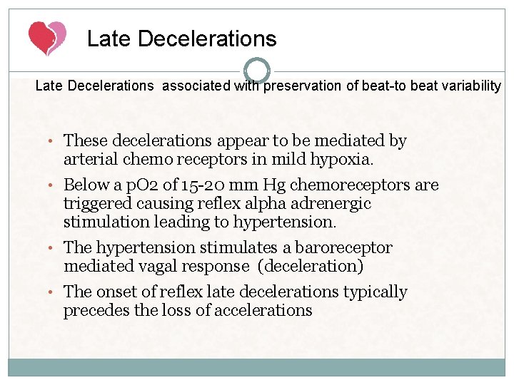
Late Decelerations associated with preservation of beat-to beat variability • These decelerations appear to be mediated by arterial chemo receptors in mild hypoxia. • Below a p. O 2 of 15 -20 mm Hg chemoreceptors are triggered causing reflex alpha adrenergic stimulation leading to hypertension. • The hypertension stimulates a baroreceptor mediated vagal response (deceleration) • The onset of reflex late decelerations typically precedes the loss of accelerations
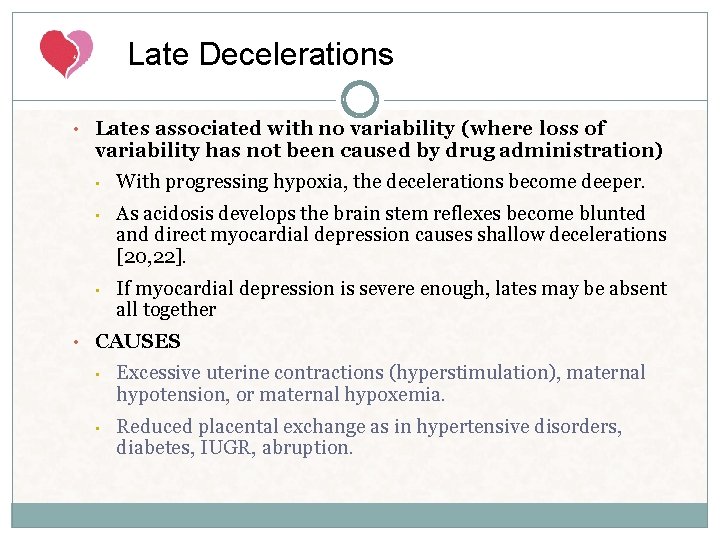
Late Decelerations • Lates associated with no variability (where loss of variability has not been caused by drug administration) • With progressing hypoxia, the decelerations become deeper. • As acidosis develops the brain stem reflexes become blunted and direct myocardial depression causes shallow decelerations [20, 22]. • If myocardial depression is severe enough, lates may be absent all together • CAUSES • Excessive uterine contractions (hyperstimulation), maternal hypotension, or maternal hypoxemia. • Reduced placental exchange as in hypertensive disorders, diabetes, IUGR, abruption.
Late Decelerations From: Sweha A, Hacker TW, Nuovo J. Interpretation of the electronic fetal heart rate during labor. Am Fam Physician. 1999; 59: 2487 -500
Late deceleration
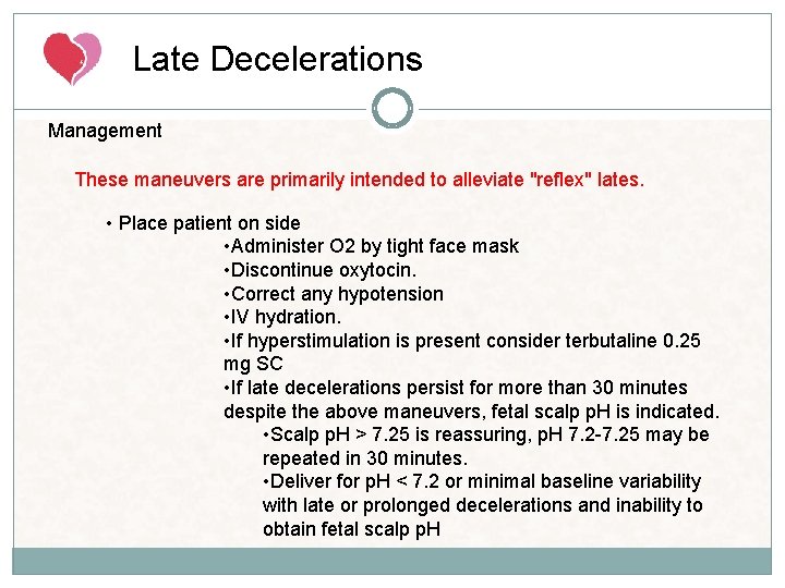
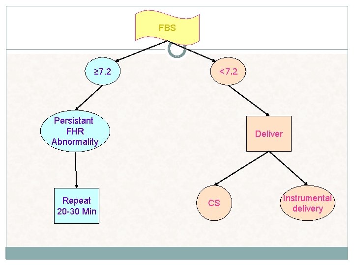
Late Decelerations Management These maneuvers are primarily intended to alleviate "reflex" lates. • Place patient on side • Administer O 2 by tight face mask • Discontinue oxytocin. • Correct any hypotension • IV hydration. • If hyperstimulation is present consider terbutaline 0. 25 mg SC • If late decelerations persist for more than 30 minutes despite the above maneuvers, fetal scalp p. H is indicated. • Scalp p. H > 7. 25 is reassuring, p. H 7. 2 -7. 25 may be repeated in 30 minutes. • Deliver for p. H < 7. 2 or minimal baseline variability with late or prolonged decelerations and inability to obtain fetal scalp p. H
Recurrent Decelerations occur with > 50% of uterine contractions in any 20 minute segment. Recurrent variable decelerations (at least 3 in 20 minutes) may be observed. However, close follow up is recommended because cord accidents with subsequent fetal death may occur even in the presence of normal amounts of amniotic fluid. Recurrent late decelerations should lead to consideration of cesarean delivery unless the abnormal results are believed to be the result of a reversible maternal condition such as diabetic ketoacidosis or pneumonia with hypoxemia.
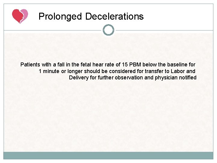
Prolonged Decelerations A decrease in FHR of > 15 beats per minute measured from the most recently determined baseline rate. The deceleration lasts >= 2 minutes but less than 10 minutes. Causes: Maternal hypotension Uterine hyperactivity Cord prolapse Cord compression* Rapid descent of fetal head Abruption • • Artifact (maternal heart rate Maternal seizure • •
Prolonged Decelerations Patients with a fall in the fetal hear rate of 15 PBM below the baseline for 1 minute or longer should be considered for transfer to Labor and Delivery for further observation and physician notified
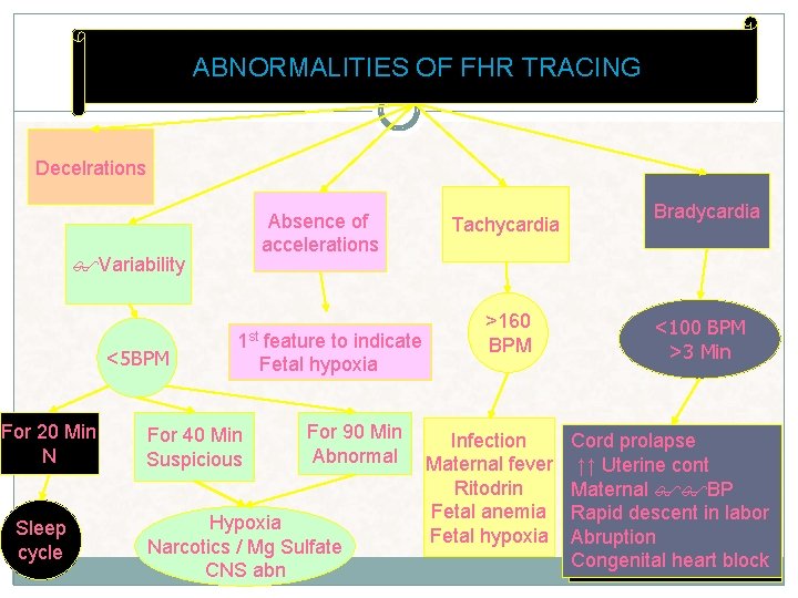
ABNORMALITIES OF FHR TRACING Decelrations Absence of accelerations Variability <5 BPM For 20 Min N Sleep cycle 1 st feature to indicate Fetal hypoxia For 40 Min Suspicious For 90 Min Abnormal Hypoxia Narcotics / Mg Sulfate CNS abn Tachycardia >160 BPM Infection Maternal fever Ritodrin Fetal anemia Fetal hypoxia Bradycardia <100 BPM >3 Min Cord prolapse ↑↑ Uterine cont Maternal BP Rapid descent in labor Abruption Congenital heart block
DECELRATIONS Early Late Variable Physiologic Fetal head compression Mirror image of the contraction FH<60 BPM< 60 sec Cord compression Not related to the cont Variable duration & degree of FHR depresiion After the contraction Uteroplacental insufficiency Fetal asphyxia/acidosis Worst prognosis
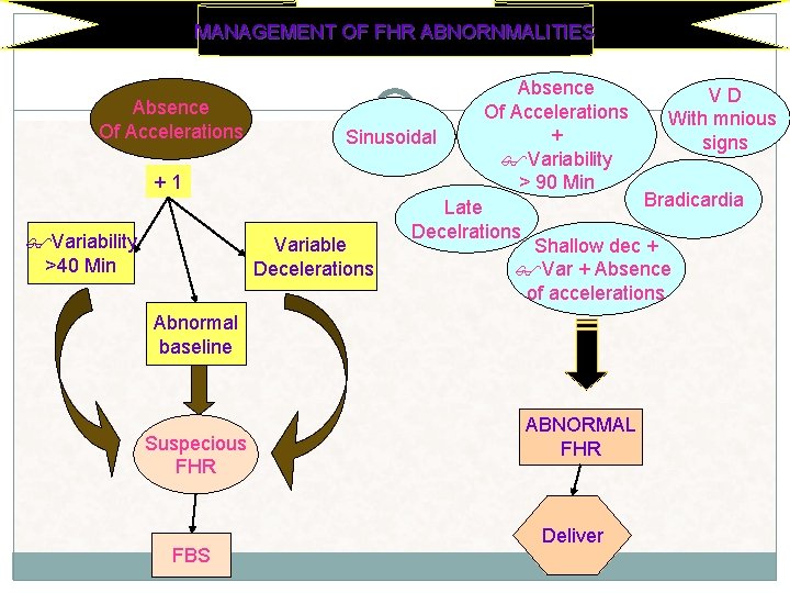
MANAGEMENT OF FHR ABNORNMALITIES Absence V D Absence Of Accelerations With mnious Of Accelerations + Sinusoidal signs Variability + 1 > 90 Min Bradicardia Late Decelrations Variability Variable Shallow dec + >40 Min Decelerations Var + Absence of accelerations Abnormal baseline Suspecious FHR FBS ABNORMAL FHR Deliver
FBS ≥ 7. 2 <7. 2 Persistant FHR Abnormality Repeat 20 -30 Min Deliver CS Instrumental delivery
MBPP The MBPP is an abbreviated • biophysical profile that includes only the nonstress test (NST) and amniotic fluid index (AFI). A normal result is a reactive NST with an AFI > 5. 0 cm.
Thank you