scoliosis outlines 1 normal vertebral curvature 2 Definition

scoliosis

outlines 1 -normal vertebral curvature 2 -Definition 3 -pathology 4 -types 5 -clinical features 6 -investigations 7 - treatment and prognosis

Normal spine -Primary curves are the natural curves in the spine that we are born with (thoracic/pelvic curves) -secondary curves develop after birth in response to learned motor skills Cervical when infant learn to hold his/her head upright Lumbar develop as child learn to walk
 curvature of the spine. Apparent’ because, although")
definition Scoliosis is an apparent lateral (sideways) curvature of the spine. Apparent’ because, although lateral curvature does occur, the commonest form of Scoliosis is actually a triplanar deformity with lateral , anteroposterior and rotational components.

The scoliosis research society has defined scoliosis as a lateral curvature of the spine greater than 10 degrees as measured using the cobb method on standing radiograph *Pts usually present between the age of 10 and 15. *Mostly occur in female
 Usually in the thoracic spine")
dextroscoliosis Describes a spinal curve to the right (dextro=right) Usually in the thoracic spine This is the most common type of curve Forming either c shape or s shape Levoscoliosis Describes a spinal curve to the left (levo=left) Common in lumbar spine

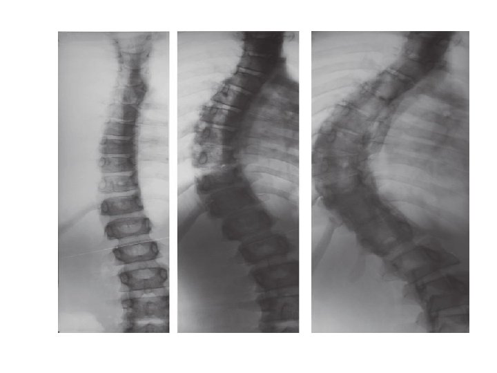
Pathology: - May occur anywhere in the thoracic or lumbar spine. *The vertebrae that make up the curve are always rotated around the vertebral axis. *the bodies point to the convexity. And the spinous processes to the concavity of the curve *coexistence of congenital spine anomalies with congenital cardiac and kidney defects.

types 1 - postural 2 -structural

postural type • • Secondary or compensatory to some condition outside the spine such as short leg, or pelvic tilt , Local muscle spasm associated with a prolapsed lumbar disc may cause a skew back (sciatic scoliosis). It can become structural if it exceed a certain point. 2 dimensional ( AP only ) The deformity disappear when the patient sits or bend forward.

Structural type • non-correctable deformity. • rib hump on bending forward. I. The deformity is liable to increase throughout the growth period. curves greater than 50 degrees may go on increasing by 1 degree per year. II. very severe curves ( angle >70 -80 degree) accompanied with chest deformity and cardiopulmonary dysfunction

Functional type Structural type
 2")
How to differentiate between functional and structural? 1 - bending test (adam’s test) 2 - make the patient sit The scoliosis will disappear on conducting the two tests in functional type
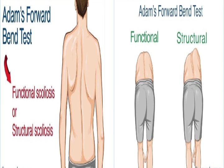

Test for scoliosis Bending test
 2 - Osteopathic scoliosis: due")
Types of structural scoliosis 1 - idiopathic scoliosis (80%) 2 - Osteopathic scoliosis: due to congenital vertebral anomalies 3 - Neuropathic scoliosis: due to asymmetrical muscle weakness (e. g. In poliomyelitis or cerebral palsy). 4 - Myopathic scoliosis: in muscular dystrophies. 5 - Neurofibromatosis.
 idiopathic Structural scoliosis Neuropathic and myopathic Congenital or osteopathic")
Infantile (0 -3 years old) idiopathic Structural scoliosis Neuropathic and myopathic Congenital or osteopathic miscellaneous group of connective-tissue disorders Juvenile(4 -9 years old) Adolescent(10 years to maturity)

1 - Infantile idiopathic scoliosis: *Infantile idiopathic scoliosis is the only type of idiopathic scoliosis whose most common curve pattern is left thoracic. *is the only type of scoliosis that is more common in males. *the only one to resolve spontaneously 90%

2 - Juvenile idiopathic scoliosis *most closely mimics the epidemiology and demographics of the adolescent version of the disease. *It is more common in female. 50% resolve spontaneously *its most common curve pattern is a right thoracic curve. *high rate of progression, and need for surgery, juvenile idiopathic scoliosis might be considered to be a malignant subtype of adolescent idiopathic scoliosis.

3 - Adolescent idiopathic scoliosis: *is the most common type of idiopathic scoliosis and the most common type of scoliosis overall. *Small curves in more mature patients have a substantially lower risk of progression (about 2%) than larger curves in more immature patients, in whom the risk is much higher *Present before puberty and progresses until skeletal growth ceases.

Infantile thoracic Adolescent thoracic Thoracolumbar Lumbar Combined
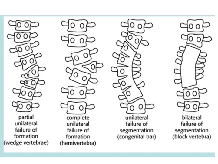
 SCOLIOSIS The commonest bony cause is some type of vertebral anomaly: ü")
OSTEOPATHIC (CONGENITAL) SCOLIOSIS The commonest bony cause is some type of vertebral anomaly: ü Hemivertebra ü wedged vertebra (failure of formation) ü fused vertebrae ü fractures ü bone softening ( rickets or osteogenesis imperfecta) • more aggressive and need early surgery.

NEUROPATHIC AND MYOPATHIC SCOLIOSIS ü Poliomyelitis ü cerebral palsy ü Syringomyelia ü Friedreich’s ataxia *Also aggressive and bracing may fail, need surgery.

SCOLIOSIS AND NEUROFIBROMATOSIS • 1/3 of patients with neurofibromatosis develop spinal deformity. • accompanied by skin lesions, multiple neurofibromata and bony dystrophy affecting the vertebrae and ribs

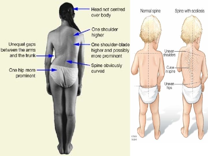
Clinical features *Deformity is the only symptom. *skew back and rib hump *High curves are noticed early, whereas lumber curves may pass virtually unnoticed. *It always looks worse on flexion. *The shoulder is elevated on the side of the convexity, and the hip sticks out on the side of the concavity. *With thoracic scoliosis; the breast are asymmetrical, and the rib angles protrude. *pain is rare complaint ( if present should be alert to possibility of neural tumor and need for MRI) *family history is not uncommon

• Adams test : deviation when the patient bends forward. … used as a screening test in schools along with scoliometer. • Scoliometer : if the angle below 7 normal … above 7 need further investigations

investigations v. Plain X-ray : • Full-length PA and lateral x-rays of the spine and iliac crests must be taken with the patient erect. • Vertebral rotation. • PA x-ray shows asymmetry in vertebra at the apex of the curve
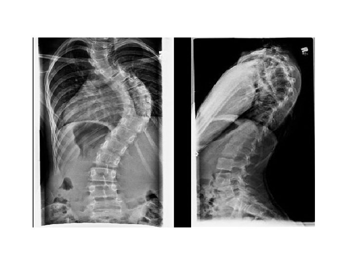

Cobb angle for measuring the spinal curvature. Choose the most tilted verterbrae above & below apex of the curve. Angle b/t intersecting lines drawn perpendicular to the top of the superior vertebrae and bottom of the inferior vertebrae is the Cobb angle
 : <25 degree Primary curve : >25")
• • • Secondary curve (compensatory) : <25 degree Primary curve : >25 degree Treatment : <25 degree : conservative/ follow up 25 -45 degree : bracing and also depend on risser sign.

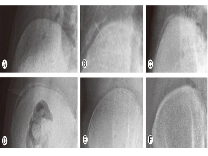
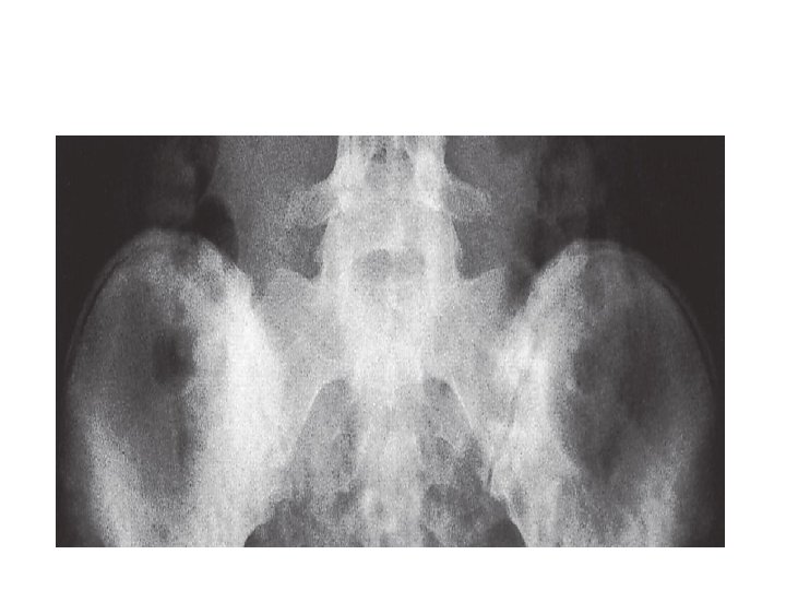
• Risser’s sign : • this is important because the curve often progresses most during the period of rapid skeletal growth and maturation. • iliac apophyses ossification start anteriorly from ASIS & proceeds posteriorly towards PSIS. once the iliac crests are completely ossified ( 14 -16 year) further progression of the scoliosis is minimal. • graded as Risser ( 0 -5 ) • Risser 5 complete fusion of apophysis • Risser 0 (no ossification) • Risser 1 coincide with rapid growth

Ossification of iliac crest starts laterally and proceeds medially toward sacrum Risser staging Based on iliac crest apophysis ossification Type 1= ossification of lateral 25% Type 2=lateral 50% Type 3=lateral 75% Type 4=lateral 100% Type 5=fusion of ilium

v. CT and MRI needed in: • • Pain Abnormal neurological examination Big curves > 50 Rt thoracic curve

Treatment and prognosis • The aim is to prevent severe deformity … usually for cosmetic reasons. • Management differs for the different types of scoliosis. • predictors of bad prognosis are: ü Double curves ü Kyphoscoliosis ü Female ü Rt thoracic ü > 50 degree

Management differs for the different type of scoliosis: 1 - infantile treated by applying serial elongation derotation flexion (EDF) plaster cast until resolve or become big enough for bracing 2 -juvenile treated surgically befor puberty except if child is very young so bracing until age of 10 3 -adolescent treated by : A-non operative = exercise (no effect on curve but maintain muscle tone) And bracing (curve 20 -30 degree)

Types of braces Milwaukee brace Consist of 1 -pelvic corset or girdle 2 -uprights (one anterior and two posterior) 3 -cervical ring (with throat mold and occipital piece 4 - lateral pad (pressure on apical vertebra)

Boston brace Is snug fitting underarm brace Corrective pad is added to device to apply pressure at particular site

Operative Indication of surgery = 1 -curve more than 30 degree 2 -milder deformity that deteriorating rapidly 3 -failure to conservative treatment 4 -balanced double primary curve require operation only if greater than 40 degrees and progress
 3 -cotrel dubousset system")
Methods : 1 -harrington rod 2 -rod and sublaminar wiring(luque) 3 -cotrel dubousset system 4 -anterior instrumentation(dwyer, zielke) 5 -warning

Scoliosis – posterior instrumentation Idiopathic scoliosis treated by posterior double-rod fixation

Scoliosis – anterior instrumentation

osteopathic treatment is difficult because risk of cord injury (surgery =staged resection of curve apex followed by instrumentation and spinal fusion) neuropathic /myopathic Depend on degree of functional disability Mild=not treated Moderate= as idiopathic Sever=fitting suitable sitting support , if not sufficient operation which is stabilization of entire paralytic segment by combined anterior and posterior instrumentation puls fusion

Scoliosis with neurofibromatosis Treatment Mild =as idiopathic Sever =anterior and posterior instrumentation plus fusion

- Slides: 49