Sciatic Nerve Editing File Color Code Important Doctors

 of the sciatic nerve. üList the")


 3 Common Peroneal")
 cutaneous (all sciatic")

 Nerve Ø Course: Course -")


- Slides: 20
Sciatic Nerve Editing File Color Code Important Doctors Notes/Extra explanation
Objectives üDescribe the anatomy (origin, course and distribution) of the sciatic nerve. üList the branches of the sciatic nerve. üDescribe briefly the main motor and sensory manifestations in case of injury of the sciatic nerve or its main branches.
Sciatic nerve • It is the largest branch of the plexus & the largest nerve of the body. • Origin : From the Sacral Plexus , (L 4, L 5, S 1, S 2, S 3). Sacral Plexus Formation: Ventral (anterior)(lower division) rami of a part of L 4 & whole L 5 (lumbosacral trunk) + S 1, 2, 3 and most of S 4. • 4 • Site: On the posterior wall of the pelvis, In front of Piriformis muscle. 5 The nerve in front of the muscle
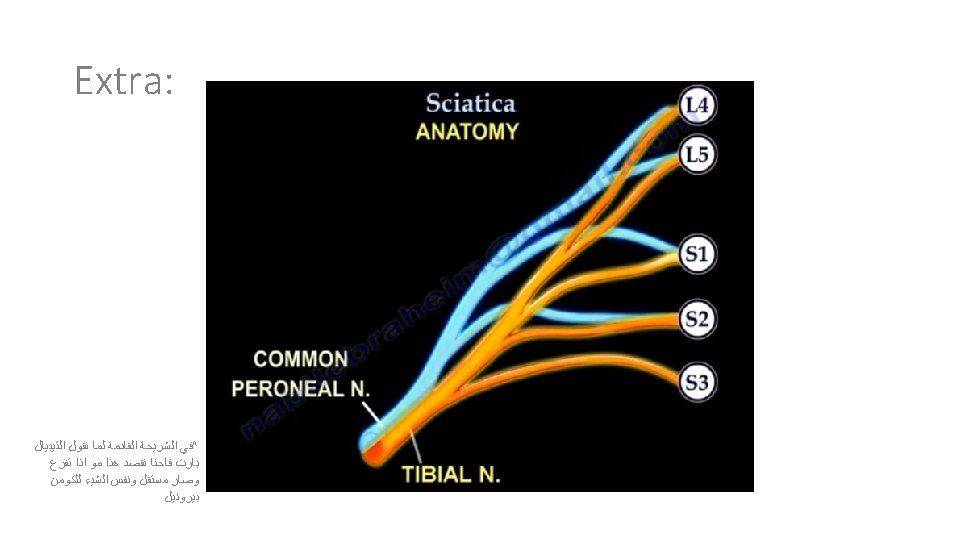
Course & Distribution Ø It leaves the pelvis through greater sciatic foramen, below the piriformis Ø then passes (between 2 bones) in the gluteal region (ischial tuberosity & greater trochanter) Ø then to posterior compartment of thigh. Ø Termination: In the middle of the back of the thigh* It divides into 2 branches: 1. Tibial (medial popliteal) enter the popliteal fossa 2. Common Peroneal or lateral popliteal or (Fibular). Outside popliteal fossa *ﻳﺨﺘﻠﻒ ﻣﻜﺎﻥ ﺍﻟﺘﻔﺮﻉ ﻣﻦ ﺷﺨﺺ ﻵﺨﺮ
Branches of Sciatic Nerve 1 - in thigh (before It divides) 3 Common Peroneal 2 -Tibial
Branches of Sciatic Nerve 1 - in thigh (before It divides) cutaneous (all sciatic branches) To the skin of all leg & foot except Areas supplied by saphenous nerve (branch of femoral nerve ) Muscular To Hamstrings (Flexors of knee & extensors of hip) except The short head of biceps receives its branch from lateral popliteal (common peroneal part) (through tibial part) it gives: 1 - Hamstring(Ischial) part of Adductor Magnus 2 - Long head of Biceps Femoris 3 - Semitendinosus 4 - Semimembranosus : ﻟﻠﺤﻔﻆ Hambi skies on the same sea
Branches of Sciatic Nerve 2 - Tibial nerve Ø Course: Bisect the popliteal fossa. it is the most superficial structure in the popliteal fossa. . ﻋﺸﺎﻥ ﻛﺬﺍ ﻫﻲ ﺃﻜﺜﺮ ﺷﻴﺀ ﻣﻤﻜﻦ ﻳﻨﻘﻄﻊ recall: ^while the deepest structure is the artery Ø Descends through popliteal fossa to posterior compartment of leg, accompanied with posterior tibial vessels. Ø Passes deep to flexor retinaculum (through the tarsal tunnel, tunnel behind medial malleolus) malleolus to reach the sole of foot where it divides into 2 terminal branches (Medial & Lateral planter nerves). Ø Muscular Branches: (in leg) + Hamstrings in thigh Muscles of posterior compartment of leg: 1 - Planter flexors of ankle 2 - Flexors of toes 3 - ONE Invertor of foot (tibialis posterior). + All Intrinsic muscles of sole. (by medial & lateral branches)
Branches of Sciatic Nerve 3 - Common Peroneal (Fibular) Nerve Ø Course: Course - Leaves the lateral angle of popliteal fossa & turns around the lateral aspect of neck of fibula, (Dangerous Position) ﻷﻦ ﻫﺬﺍ ﺍﻟﻤﻜﺎﻥ ﻏﻴﺮ ! ﻣﺘﻐﻄﻲ ﺑﻌﻀﻼﺕ - Then divides into: 1 - Superficial peroneal or (Musculocutaneous) : to supply the Lateral compartment of the leg. 2 - Deep peroneal or (Anterior Tibial): to supply the Anterior compartment of the leg. Ø Muscular Branches: (+ short head of biceps) Muscles of anterior & lateral compartments of leg: 1 - Dorsi flexors of ankle, 2 - Extensors of toes, 3 - Evertors of foot.
Sciatic Nerve Injury -Causes The sciatic nerve is frequently injured by: 1 - Badly placed intramuscular injections in the gluteal region. To avoid this, injections should be done into the gluteus maximus or medius. (into the upper outer (lateral) quadrant of the buttock) *Most nerve lesions are incomplete, in 90% of injuries, the common peroneal (part of the nerve) is the most affected. Because: The common peroneal nerve fibers lie superficial in the sciatic nerve. 2 -- Posterior dislocation of the hip joint ﺑﺴﺒﺐ ﺣﺎﺩﺙ ﺳﻴﺎﺭﺓ ﻣﺜﻼ Posterior = Head of femur ﻳﺮﺟﻊ ﻟﻠﺨﻠﻒ ﻓﻴﻀﻐﻂ ﻋﻠﻰ ﺍﻟﺸﻴﺀ ﺍﻟﻠﻲ ﻣﻮﺟﻮﺩ ﺧﻠﻔﻪ ﺍﻟﻠﻲ ﻫﻮ sciatic nerve !
Sciatic Nerve Injury -Effects NS: ﻟﻤﺎ Dorsiflexion & plantarflexion ﻧﻤﺸﻲ ﺍﻟﺤﺮﻛﺎﺕ ﺍﻟﻠﻲ ﻧﺴﻮﻳﻬﺎ ﻫﻲ Sartorius : Femoral nerve sciatic nerve ﻭﺍﻟﻌﻀﻼﺕ ﺍﻟﻤﺴﺆﻮﻟﺔ ﻋﻦ ﻫﺬﻩ ﺍﻟﺤﺮﻛﺘﻴﻦ ﺗﻐﺬﻳﻬﺎ ﺗﻔﺮﻋﺎﺕ ﻣﻦ ﻓﻠﻤﺎ ﻳﺼﻴﺮ ﻟﻬﺎ ﺍﻧﺠﺮﻱ ﻣﺎ ﺗﺼﻴﺮ ﻭﻻ ﺣﺮﻛﺔ ﻣﻦ ﺍﻟﺜﻨﻴﺘﻦ ﻓﺈﻳﺶ ﺑﺴﺒﺐ ﺍﻟﺠﺎﺫﺑﻴﺔ drop ﻳﺼﻴﺮ ﺑﺎﻟﺮﺟﻞ؟ ﻳﺼﻴﺮ ﻟﻬﺎ Gracilis : Obturator nerve gluteus maximus : Inferior gluteal nerve Motor : Sensory : Weak flexion of the knee (sartorius & gracilis are intact). • Sensation is lost below the knee, except for a narrow area down the medial side of the lower part of the leg (purple) and along the medial border of the foot as far as the ball of the big toe, which is supplied by the saphenous nerve (branch of femoral nerve). • Weak extension of hip (gluteus maximus is intact). • • Marked wasting(atrophy) of the muscles below the knee. • All the muscles below the knee are paralyzed, and the weight of the foot causes it to assume the plantar-flexed position OR Foot Drop. recall • Stamping gait. (high steppage gait) ﻣﺜﻞ ﺗﻠﺰﻳﻖ ﺍﻟﻄﻮﺍﺑﻊ ﻳﻜﻮﻥ ﺑﺴﺮﻋﺔ Stamping gait https: //www. youtube. com/watch? v=SWv. EU 8 FYMFc + Foot Drop: https: //www. youtube. com/watch? v=J 7 -L 9 MFRXD 8
Sciatica ﺍﻟ ﺍ ﻋﺮﻕ • Sciatica describes the condition in which patients have pain along the sensory distribution of the sciatic nerve. ﻓﻘﻂ ﺃﻠﻢ ﺍﻟﻌﻀﻼﺕ ﺗﺸﺘﻐﻞ ﺗﻤﺎﻡ • Thus the pain is experienced in: 1 -the posterior aspect of the thigh 2 -the posterior and lateral sides of the leg 3 -and the lateral part of the foot. Vitamin B 12 also promotes the regeneration and growth of nerve cells. Neuropathy, such as sciatic nerve pain, numbness or tingling, in some cases has been found to be caused and made worse by deficiencies of vitamin B 12 in the body Causes of Sciatica: • Prolapse of an intervertebral disc, with pressure on one or roots of the lower lumbar and sacral spinal nerves • Pressure on the sacral plexus or sciatic nerve by an intrapelvic tumor • Inflammation of the sciatic nerve or its terminal branches. Treatment is according to the Cause. Sciatica - Everything You Need To Know: https: //www. youtube. com/watch? v=XS 2 BTLYsn 5 w
Common Peroneal Nerve Injury: Causes ﻟﻺﺻﺎﺑﺔ ﻣﻦ ﺍﻟﺘﻴﺒﻴﻞ ﺃﻜﺜﺮ ﻋﺮﺿﺔ • The common peroneal nerve is in an exposed position as it leaves the popliteal fossa it winds around neck of the fibula to enter peroneus longus muscle, (Dangerous Position). • The common peroneal nerve is commonly injured 1 - In Fractures of the neck of the fibula and 2 - By pressure from casts or splints. ﺟﺒﺲ ﺃﻮ ﺟﺒﻴﺮﺓ
Common Peroneal Nerve Injury: Manifestations 1 - Motor: Dorsiflexion ankle joint & subtalar joints eversion • The muscles of the anterior and lateral compartments of the leg are paralyzed, Equinovarus. • As a result, the opposing muscles (in the posterior compartment of the leg) , the plantar flexors of the ankle joint & the invertors of the subtalar joints, cause the foot to be Plantar Flexed (Foot Drop) and Inverted, an attitude referred to as Equinovarus. if it is from birth it called Talipes* Equinovarus ( &)ﺗﺒﻘﻰ ﻛﺬﺍ ﻟﻸﺒﺪ if it is from injury it called Paralytic equinovarus( )ﺗﺄﺨﺬ ﻭﻗﺖ ﻋﻠﻰ ﻣﺎ ﺗﺘﺼﻠﺢ “ ﻭﻻﻥ ﺍﻟﻌﻀﻼﺕ ﺍﻟﺨﻠﻔﻴﻪ ﻣﺎﺗﻀﺮﺭﺕ anterior and lateral compartment” ﺍﻹﻋﺎﻗﺔ ﺍﻟﻠﻲ ﺗﺘﺒﻊ ﺍﻹﺻﺎﺑﺔ ﻟﻬﺬﺍ ﺍﻟﻌﺼﺐ ﺗﻜﻮﻥ ﺑﺴﺒﺐ ﺿﻌﻒ ﺍﻟﻐﻀﻼﺕ ﺍﻟﻠﻲ ﻳﻐﺬﻳﻬﺎ “ ﻭﺗﺸﺘﻐﻞ ﻷﻦ ﻳﺘﻢ ﺗﻐﺬﻳﺘﻬﺎ ﻣﻦ Tibial nerve” 2 - Sensory : ﺇﺫﺍ ﻛﻞ ﺍﻻﺛﻨﻴﻦ ﻓﻜﻞ ﺍﻷﺮﺑﻌﺔ ﺍﻟﺘﺎﻟﻴﺔ , ﻋﻠﻰ ﺣﺴﺐ ﺍﻟﺒﺮﺍﻧﺶ ﺍﻟﻠﻲ ﺗﻜﻮﻥ ﻓﻴﻪ ﺍﻹﺻﺎﺑﺔ • Sensation is lost between the first and second toes. (deep peroneal) Superficial peroneal Musculocutaneous • Dorsum of the foot and toes. (Superficial peroneal) deep peroneal Anterior Tibial • Medial side of the big toe. (Superficial peroneal) • Lateral side of the leg. (Superficial peroneal) *Talipes = ﻗﺪﻡ ﻣﺸﻮﻫﺔ ﺧﻠﻘﺔ
Tibial Nerve Injury • Because of its deep and protected position, the tibial nerve is rarely injured. • Complete division results in the following clinical features: 1 -Motor: All the muscles in the back of the leg and the sole of the foot are paralyzed. The opposing muscles Dorsiflex the foot at the ankle joint and Evert the foot at the subtalar joint, an attitude referred to as Calcaneovalgus. If congenital : Taleps Calcaneovalgus If acquired : paralytic Calcaneovalgus Note: it is the opposite of foot drop 2 -Sensory: • Sensation is lost in the sole & on the Lateral side of the leg and foot • Trophic ulcers in the sole. ﻷﻨﻪ ﻳﻤﺸﻲ ﻭﻣﺎ ﻳﺤﺲ ﺑﺎﻟﻠﻲ ﺗﺤﺖ ﻓﻤﻤﻜﻦ ﺗﺼﻴﺮ ﻟﻪ ﺇﺻﺎﺑﺎﺕ ﻣﺎ ﻳﺪﺭﻱ ﻋﻨﻬﺎ (also seen in case of Sciatic nerve injury )
Summary Ø Origin of SCIATIC NERVE: from the sacral plexus (L 4, L 5, S 1, S 2, S 3). ( Paralysis of : 1 - Hamstrings 2 - All muscles of Leg & Foot Movements affected : Flexion of knee Extension of hip All movements of the leg& Foot SENSORY EFFECT MOTOR EFFECT Ø Effect of sciatic nerve injury: Loss of sensation of the areas supplied by sciatic nerve (below knee). EXCEPT area supplied by the (Saphenous nerve).
Common Peroneal Nerve Injury -The muscles of the anterior and lateral compartments of the leg are paralyzed plantar flexors of the ankle joint & the invertors of the subtalar joints Calcaneovalgus Equinovarus Summary Tibial Nerve Injury The muscles of the posterior compartments of the leg and the sole are paralyzed Dorsiflex the foot at the ankle joint & Evert the foot at the subtalar joint If congenital : Taleps If acquired : paralytic
Quiz 1: Which of the following nerve is the largest nerve of the body? A)Radial nerve. B)Ulnar nerve. C)Sciatic nerve. D)Peroneal nerve. 2: The site of sacral plexus: nerve? A)Long head of biceps. B)Short head of biceps. C)Hamstring. D)Semitendinous. 4: The most frequent injuries of the sciatic nerve is: A)Badly placed intramuscular injections in the gluteal region. A)On the anterior wall of the pelvis, in front of piriformis muscle. B)Posterior dislocation of hip joint. B)On the posterior wall of the pelvis, in the back of piriformis C)Both a and b. muscle. D)None of the above C)On the posterior wall of the pelvis, in front of piriformis muscle. D)On the anterior wall of the pelvis, in the back of piriformis muscle. 3: Which one of theses muscles is supplied by common peroneal 1)C 2)C 3)B 4)C
Quiz 5: When all muscles below the knee are paralyzed, the weight of 7: Muscles of posterior compartment of the leg: planter flexors the foot causes it to assume the: of ankle, flexors of toes and one invertor of foot: A)Plantar position. A)Tibialis posterior. B)Foot drop. B)Peroneus teritus. C)Stamping gait. C)Plantaris D)All of the above. D)Calf muscle. 6: In the tibial nerve’s course, it descends through popliteal fossa 8: Which muscle of these is not one of the anterior and lateral to the: compartments of leg? A)Anterior compartment of the leg. A)Dorsi flexors of ankle. B)Posterior compartment of the leg. B)Planter flexors of ankle. C)Posterior compartment of thigh. C)Evertors of foot. D)Anterior compartment of the thigh. D)Extensors of toes. 5)D 6)B 7)A 8)B