Scene from the 1918 influenza pandemic Scene from


")


, a recently emerging viral disease (a) Young ballet")


- Slides: 33
Scene from the 1918 influenza pandemic.
Scene from the 2003 SARS Scare
Comparing the size of a virus, a bacterium, and an animal cell Virus Bacterium Animal cell nucleus 0. 25 μm
T 4 bacteriophage infecting an E. coli cell 0. 5 m 4
Infection by tobacco mosaic virus (TMV)
Viral infection of plants
Viral structure Capsomere of capsid RNA Capsomere Membranous envelope DNA Head Capsid Tail sheath RNA DNA Tail fiber Glycoprotein 18 × 250 mm 20 nm (a) Tobacco mosaic virus Glycoprotein 70– 90 nm (diameter) 80– 200 nm (diameter) 50 nm (b) Adenoviruses (c) Influenza viruses 80 × 225 nm 50 nm (d) Bacteriophage T 4
Classes of Animal Viruses
Entry into cell and uncoating of DNA VIRUS Capsid Transcription Replication HOST CELL Viral DNA A simplified viral reproductive cycle m. RNA Viral DNA Capsid proteins Self-assembly of new virus particles and thei exit from cell
The lytic cycle of phage T 4, a virulent phage 1 Attachment. The T 4 phage uses 2 its tail fibers to bind to specific receptor sites on the outer surface of an E. coli cell. 5 Release. The phage directs production of an enzyme that damages the bacterial cell wall, allowing fluid to enter. The cell swells and finally bursts, releasing 100 to 200 phage particles. Entry of phage DNA and degradation of host DNA. The sheath of the tail contracts, injecting the phage DNA into the cell and leaving an empty capsid outside. The cell’s DNA is hydrolyzed. Phage assembly 4 Head Tails Tail fibers Assembly. Three separate sets of proteins self-assemble to form phage heads, tails, and tail fibers. The phage genome is packaged inside the capsid as the head forms. 3 Synthesis of viral genomes and proteins. The phage DNA directs production of phage proteins and copies of the phage genome by host enzymes, using components within the cell.
The lytic and lysogenic cycles of phage λ , a temperate phage Phage DNA The phage attaches to a host cell and injects its DNA. Phage DNA circularizes Phage Occasionally, a prophage exits the bacterial chromosome, initiating a lytic cycle. Bacterial chromosome Lytic cycle Many cell divisions produce a large population of bacteria infected with the prophage. Lysogenic cycle Certain factors determine whether The cell lyses, releasing phages. Lytic cycle is induced New phage DNA and proteins are synthesized and assembled into phages. or Lysogenic cycle is entered Prophage The bacterium reproduces normally, copying the prophage and transmitting it to daughter cells. Phage DNA integrates into the bacterial chromosome, becoming a prophage.
Bacterial Defense • What defense do bacteria have against phage infection? • Restriction enzymes • What do restirciton enzymes do? • They cut up DNA The bacterial DNA is modified to protect it from the restriction endonucleases. 12
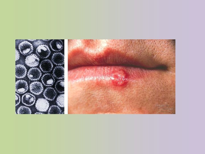
Herpesvirus • Consists of double stranded DNA • Envelope derived from host cell nuclear envelope not from plasma membrane • It, therefore, reproduces within the nucleus • May integrate its DNA as a provirus • Tends to recur throughout lifetime of infected individual. • Why do you think your immune system never removes this virus from your body? 14
SARS (severe acute respiratory syndrome), a recently emerging viral disease (a) Young ballet students in Hong Kong wear face masks to protect themselves from the virus causing SARS. (b) The SARS-causing agent is a coronavirus like this one (colorized TEM), so named for the “corona” of glycoprotein spikes protruding from the envelope.
The reproductive cycle of an enveloped RNA virus 1 Glycoproteins on the viral envelope bind to specific receptor molecules (not shown) on the host cell, promoting viral entry into the cell. Capsid RNA Envelope (with glycoproteins) 2 Capsid and viral genome enter cell HOST CELL 3 The viral genome (red) Viral genome (RNA) Template 5 Complementary RNA strands also function as m. RNA, which is translated into both capsid proteins (in the cytosol) and glycoproteins for the viral envelope (in the ER). functions as a template for synthesis of complementary RNA strands (pink) by a viral enzyme. m. RNA Capsid proteins ER Glycoproteins 4 New copies of viral Copy of genome (RNA) genome RNA are made using complementary RNA strands as templates. 6 Vesicles transport envelope glycoproteins to the plasma membrane. 8 New virus 7 A capsid assembles around each viral genome molecule.
RNA Viruses • Different classes of RNA viruses: single stranded range from class IV to class VI • Class IV: invades as m. RNA, is ready for translation • Clas V: RNA serves as template for m. RNA synthesis • RNA is transcribed and serves as template for m. RNA and genomic RNA • Viral replicase enzyme is packages within viral capsid. 19
Class VI: Retroviruses 20
The structure of HIV, the retrovirus that causes AIDS Glycoprotein Viral envelope Capsid Why are RNA Reverse Viruses transcriptase More deadly than DNA viruses? RNA (two identical strands)
The reproductive cycle of HIV, a retrovirus HIV Membrane of white blood cell The virus fuses with the 1 cell’s plasma membrane. The capsid proteins are removed, releasing the viral proteins and RNA. 2 Reverse transcriptase catalyzes the synthesis of a DNA strand complementary to the viral RNA. HOST CELL 3 Reverse transcriptase catalyzes the synthesis of a second DNA strand complementary to the first. Viral RNA-DNA hybrid 4 The double-stranded DNA is incorporated as a provirus into the cell’s DNA. 0. 25 µm HIV entering a cell DNA NUCLEUS Chromosomal DNA RNA genome for the next viral generation Provirus 5 Proviral genes are transcribed into RNA molecules, which serve as genomes for the next viral generation and as m. RNAs for translation into viral proteins. m. RNA 6 The viral proteins include capsid proteins and reverse transcriptase (made in the cytosol) and envelope glycoproteins (made in the ER). 8 9 New viruses bud New HIV leaving a cell off from the host cell. Capsids are assembled around viral genomes and reverse transcriptase molecules. 7 Vesicles transport the glycoproteins from the ER to the cell’s plasma membrane.
Virus videos
Vaccines / Drugs • What are vaccines and how do they work? • Introduce body to harmless or weakened strain of the virus, so that your immune system learns to recognize the virus prior to invasion • Few drugs around to fight viruses, most interfere with DNA, RNA or protein synthesis 24
Viroids and Prions • Viroids are naked circular RNA that infect plants • Prions are proteins that infect cells • Examples of prions seen in scrapies in sheep, mad-cow disease, and Creutzfeldt-Jakob disease in humans • How can a prion spread infection? • Altered versions of proteins that can alter other proteins 25
Model for how prions propagate Prion Original prion Many prions Normal protein New prion
Viruses and Cancer
Viral Defenses
Dengue Invasion Video
Review Questions
1. Viruses are not classified as prokaryotes because: A. B. C. D. E. They contain membrane bound organelles They are multicellular They are unicellular They are acellular They lack genetic material
2. Which of the following can be found in all viruses: A. B. C. D. E. Ribosomes RNA DNA A Protein Coat Cell membrane
3. The virus HIV replicates using reverse transcriptase. Thus it can be inferred that the virus A. Uses only DNA B. Uses only RNA C. Uses RNA as a template for DNA D. Uses DNA as a template for RNA E. Replicates continuously