Scanning Electron Microscope Innovation Center Analysis performed 16910

")


- Slides: 14
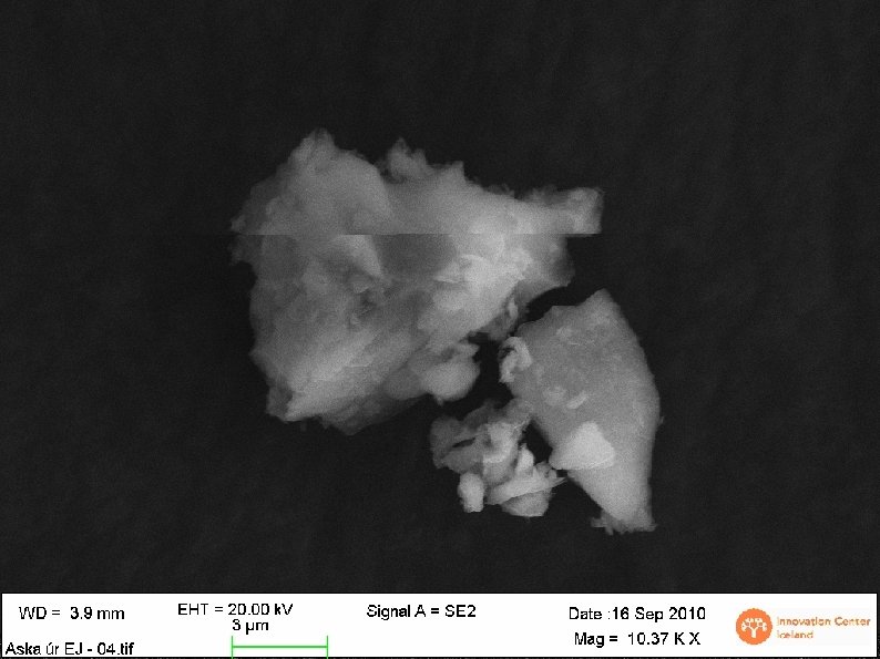
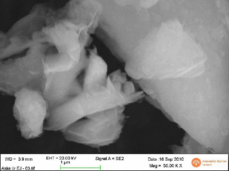
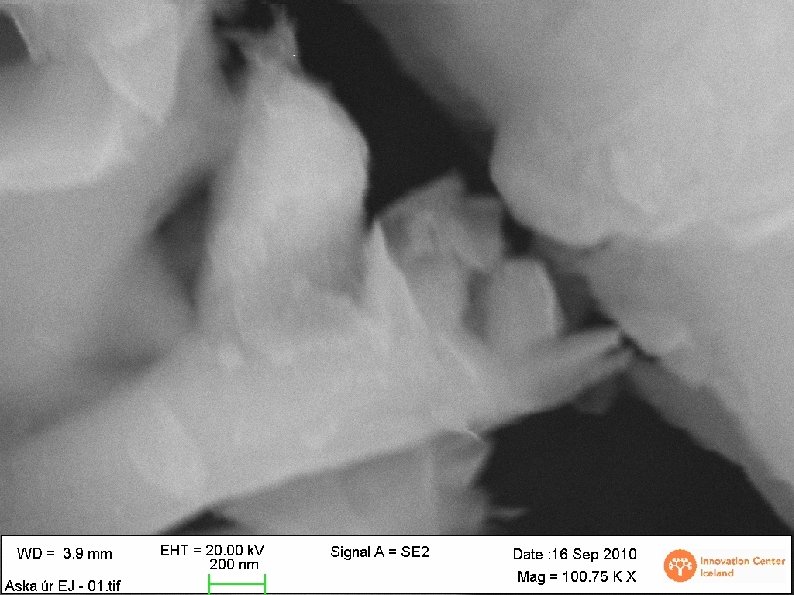
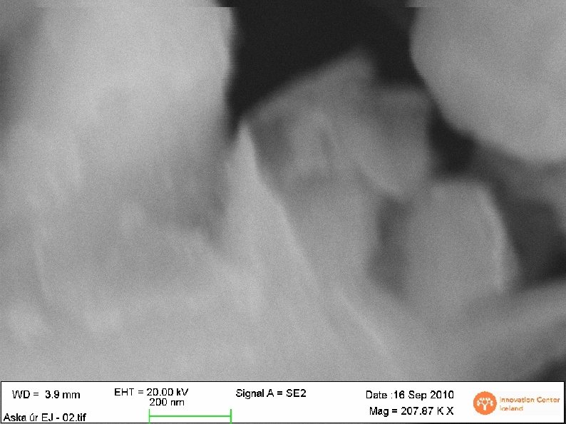
Scanning Electron Microscope Innovation Center, Analysis performed 16/9/10 Supervisor: Birgir Jóhannsson Students: , Frímann, Hafdís, Helgi, Ísak, Þorvaldur
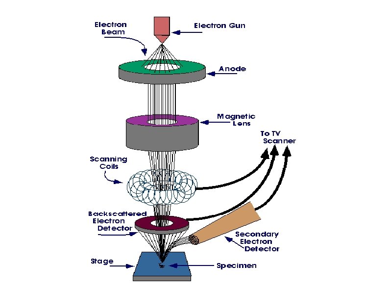
Electron microscope in general Light microscopes can get 2000 x magnification Electron microscopes (EM) can get up to 2. 000 x magnification The resolution is determined by focusing of the electron beam Two most common types, Transmission EM and Scanning EM SEM at the Innovation Center
The electron beam The electrons are generated from a small ceramic tip, radius of the tip about 0. 5μm The acceleration voltage was 20 k. V The electrons are focused to a spot 0. 5 -5 nm in diameter
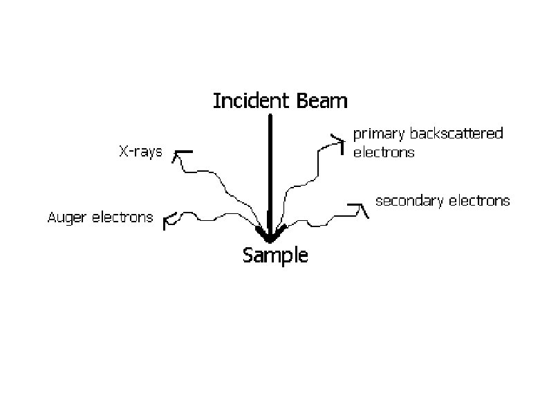
Elemental analysis Energy-dispersive X-ray spectroscopy Electron from an inner shell is excited and ejected from the atom Outer shell electron falls to the gap left by the ejected electron and emits an X-ray Wavelength of the X-ray is characteristic for the atom
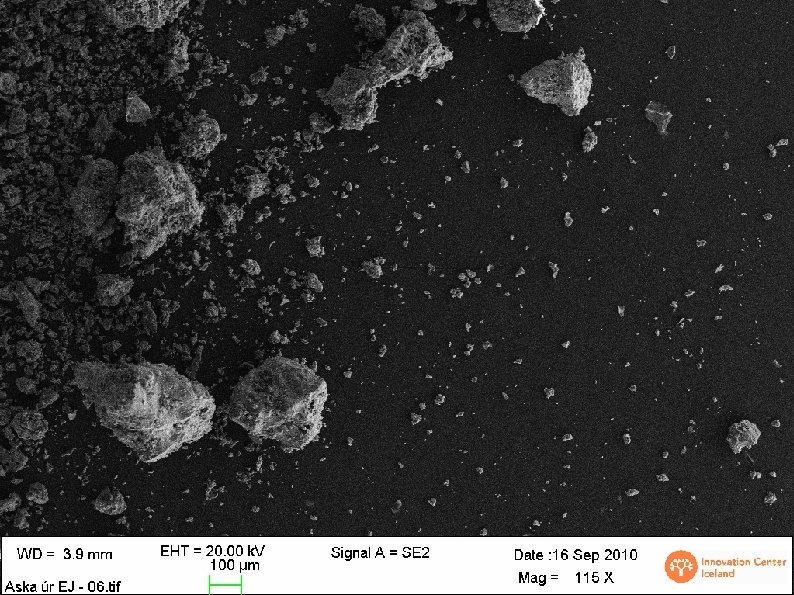
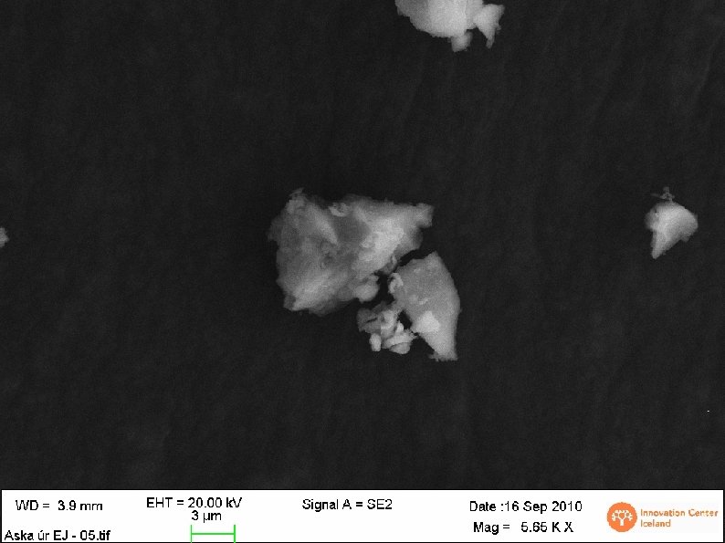
Conclusions Electron microscopes and get an enormous magnification and determine chemical compositions of surfaces with X-ray analysis. EM can also scan over an area and between points. The area around Fjarðarál is polluted. Coins from the early 1980’s differ in composition as opposed to present day.
References http: //en. wikipedia. org/wiki/Scanning_electron_mi croscope http: //www. purdue. edu/rem/rs/sem. htm