Scanning electron micrograph of a preovulatory follicle Scanning


 2 Secondary")

- Slides: 24
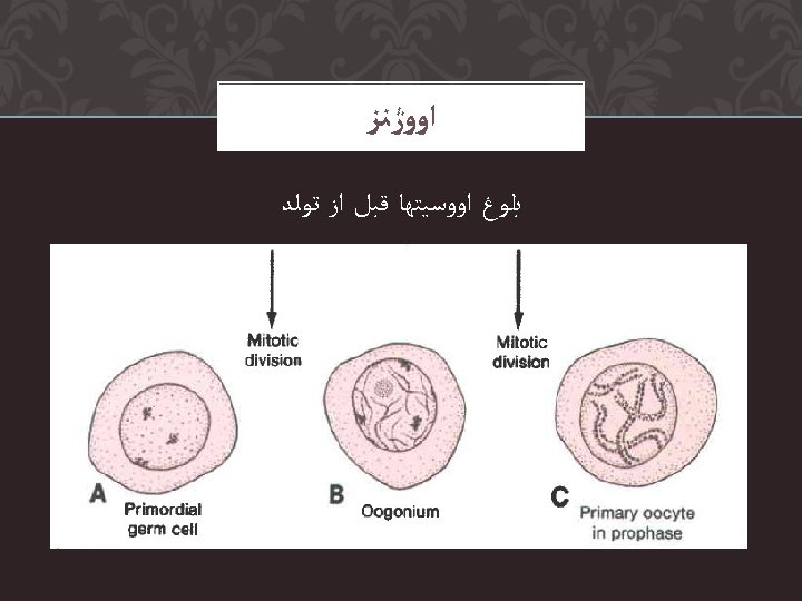
ﺍﻭﻭژﻨﺰ Scanning electron micrograph of a preovulatory follicle
ﺍﻭﻭژﻨﺰ Scanning electron micrograph of the zona pellucida after removal of the cumulus cells
ﺍﻭﻭژﻨﺰ Scanning electron micrograph of the oocyte surface and cumulus oophorus
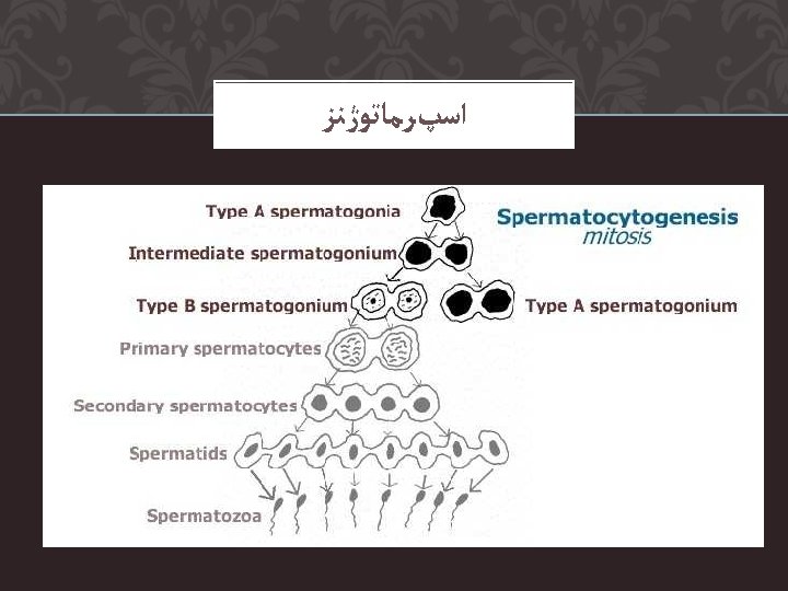
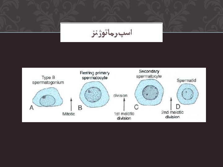
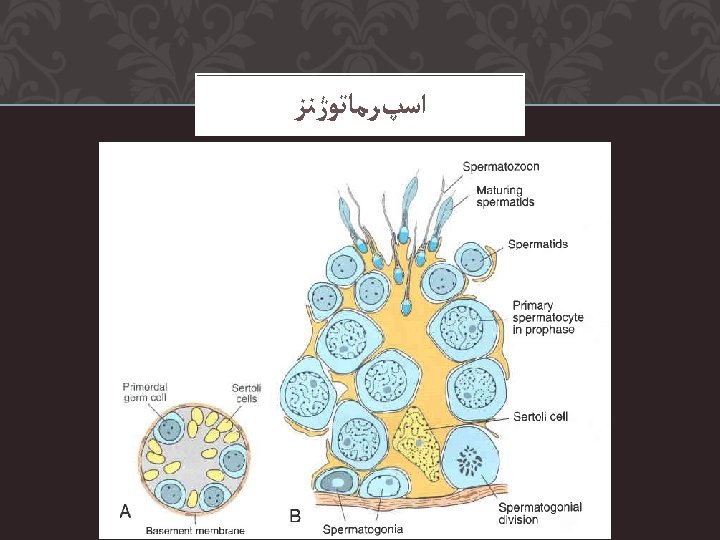
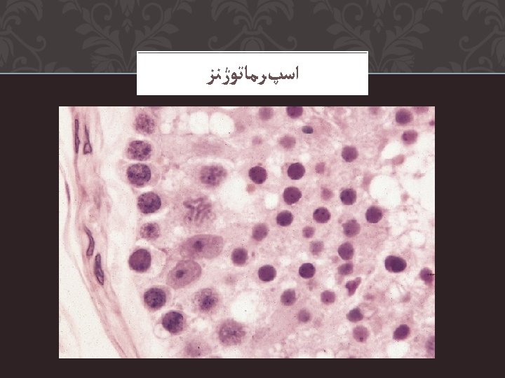
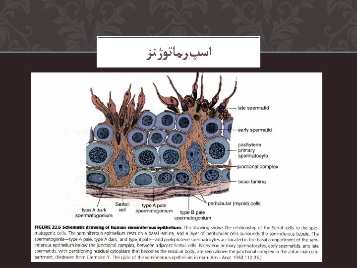
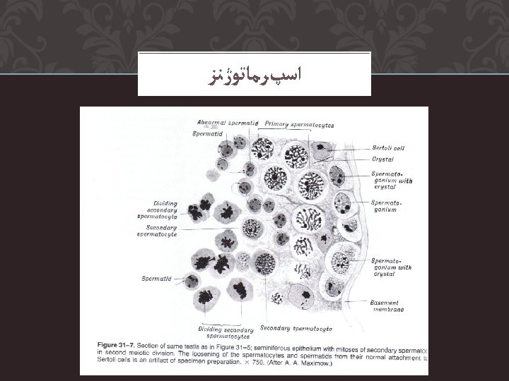
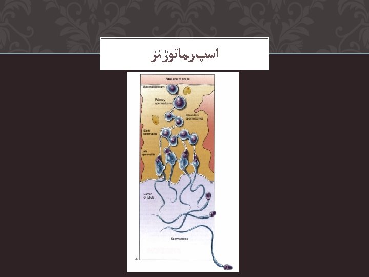
SEMINIFEROUS TUBULES Contain Germ Cells and Sertoli Cells Primary spermatocytes (46 xsomes) 2 Secondary spermatocyte 4 spermatids 4 Sperm Cells
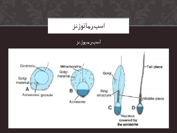
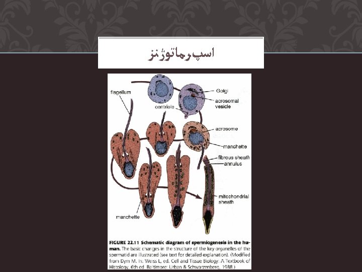
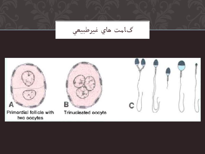
SEMEN ANALYSIS Sperm morphology – morphology types