SCALENE PREVERTEBRAL MUSCLES CERVICAL PLEXUS Dr Mujahid Khan

SCALENE & PREVERTEBRAL MUSCLES CERVICAL PLEXUS Dr. Mujahid Khan

Scalenus Anterior Muscle Ø The scalenus anterior muscle is a key muscle in understanding the root of the neck Ø It is deeply placed Ø It descends almost vertically from the vertebral column to the first rib
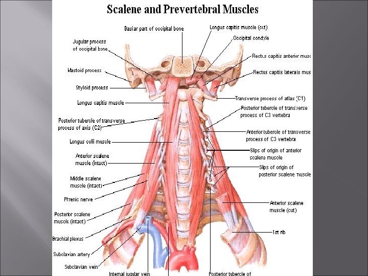

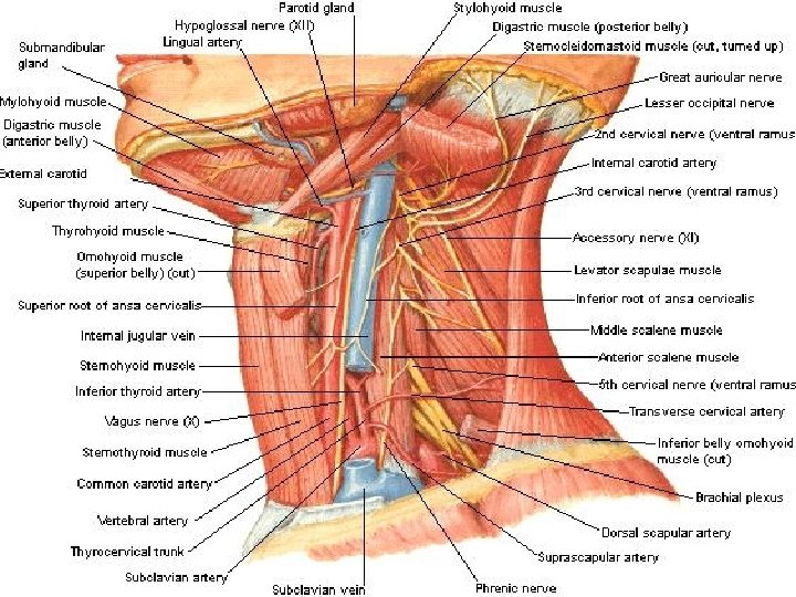
Anterior Relation Ø Related to the carotid arteries, the vagus nerve, the internal jugular vein, and the deep cervical lymph nodes Ø The transverse cervical and suprascapular arteries and the prevertebral layer of deep cervical fascia bind the phrenic nerve to the muscle

Posterior Relation Ø Related to the pleura, the origin of the brachial plexus, and the second part of the subclavian artery Ø The scalenus medius muscle lies behind the scalenus anterior muscle

Medially Ø Related to the vertebral artery and vein and the sympathetic trunk Ø On the left side, the medial border is related to the thoracic duct

Laterally Ø Related to the emerging branches of the cervical plexus, the roots of the brachial plexus, and the third part of the subclavian artery


Scalenus Anterior Muscle Ø Ø Ø Origin: Transverse processes of third, fourth, fifth, and sixth cervical vertebrae Insertion: First rib Action: Elevates first rib, laterally flexes and rotates cervical part of vertebral column

Scalenus Medius Ø It lies behind the scalenus anterior Ø It extends from the transverse process of the atlas and the transverse processes of the next five cervical vertebrae Ø Inserted into the upper surface of the first rib behind the groove for the subclavian artery Ø The muscle lies behind the roots of the brachial plexus and the subclavian artery


Scalenus Posterior Ø Ø Ø Origin: Transverse processes of lower cervical vertebrae Insertion: Second rib Action: Elevates second rib, laterally flexes and rotates cervical part of vertebral column

Longus Colli Muscle Ø Origin: Anterior tubercle of C 1, bodies of C 1 to C 3 and transverse processes of C 3 to C 6 vertebrae Ø Insertion: Bodies of C 5 to T 3 vertebrae, transverse processes of C 3 to C 5 vertebrae Ø Action: Flexes neck with rotation to opposite side


Longus Capitis Muscle Ø Ø Ø Origin: Basilar part of occipital bone Insertion: Anterior tubercles of C 3 to C 6 transverse processes Action: Flexes the head


Rectus Capitis Anterior Ø Origin: Base of the skull, just anterior to the occipital condyle Ø Insertion: Anterior surface of lateral mass of atlas Ø Action: Flexes the head

Rectus Capitis Lateralis Ø Origin: Jugular process of occipital bone Ø Insertion: Transverse process of atlas Ø Action: Flexes head and helps stabilize it


Splenius Capitis Ø Origin: Inferior half of nuchal ligament and spinous processes of superior 6 thoracic vertebrae Ø Insertion: Lateral aspect of mastoid process and lateral third of superior nuchal line Ø Action: Laterally flexes and rotates head and neck to same side, extend head and neck

Cervical Plexus Ø The cervical plexus is formed by the anterior rami of the first four cervical nerves Ø The rami are joined by connecting branches, which form loops that lie in front of the origins of the levator scapulae and the scalenus medius muscles
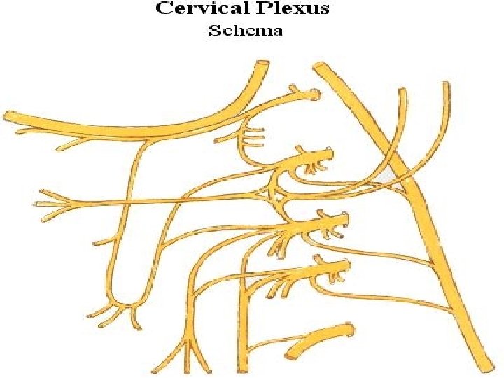
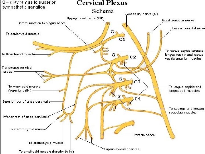

Cervical Plexus Ø The plexus is covered in front by the prevertebral layer of deep cervical fascia Ø Is related to the internal jugular vein within the carotid sheath Ø The cervical plexus supplies the skin and the muscles of the head, the neck, and the shoulders
: Supplies the back")
Cutaneous Branches Ø Ø Ø The lesser occipital nerve (C 2): Supplies the back of the scalp and the auricle The greater auricular nerve (C 2 and 3): Supplies the skin over the angle of the mandible The transverse cervical nerve (C 2 and 3): Supplies the skin over the front of the neck
 Ø The medial, and intermediate,")
Cutaneous Branches The supraclavicular nerves (C 3 and 4) Ø The medial, and intermediate, and lateral branches supply the skin over the shoulder region Ø These nerves are important clinically, because pain may be referred along them from the phrenic nerve (gallbladder disease)
, levator scapulae (C")
Muscular Branches Ø Prevertebral muscles, sternocleidomastoid (proprioceptive, C 2 and 3), levator scapulae (C 3 and 4), and trapezius (proprioceptive, C 3 and 4) Ø A branch from C 1 joins the hypoglossal nerve Ø Some of these C 1 fibers later leave the hypoglossal as the descending branch, which unites with the descending cervical nerve (C 2 and 3), to form the ansa cervicalis

Muscular Branches Ø The first, second, and third cervical nerve fibers within the ansa cervicalis supply the omohyoid, sternohyoid, and sternothyroid muscles Ø Other C 1 fibers within the hypoglossal nerve leave it as the nerve to the thyrohyoid and geniohyoid

Phrenic Nerve Ø It arises in the neck from the third, fourth, and fifth cervical nerves of the cervical plexus Ø It runs vertically downward across the front of the scalenus anterior muscle Ø Enters the thorax by passing in front of the subclavian artery

Phrenic Nerve Ø The phrenic nerve is the only motor nerve supply to the diaphragm Ø It also sends sensory branches to the pericardium, the mediastinal parietal pleura, and the pleura and peritoneum covering the upper and lower surfaces of the central part of the diaphragm
- Slides: 31