
Sarcoidosis Ramesh Kaul MD MS FCCP What is




- Slides: 92
Sarcoidosis Ramesh Kaul, MD, MS, FCCP
What is Sarcoidosis? • • Chronic multi system disorder Unknown cause Most affected organ is the lung Skin, eyes and lymph nodes are frequently involved • Acute or sub acute and self limiting • Waxing and waning over years
Etiology Results from exaggerated cellular immune response (acquired, inherited or both) to a limited class of antigens or self antigens.
Etiology 1. 2. 3. 4. 5. A defect in the immune system An unidentified toxic substance An unknown environmental cause An inherited or genetic cause A viral or bacterial infection
Incidence and Prevalence • In all races and both sexes • Risk greatest in a young black woman • Scandinavian, German, Irish, or Puerto Rican origin • 5/100, 000 whites in the US have sarcoidosis • 40/100, 000 blacks
Incidence and Prevalence • 20 cases/100, 000 in cities on the east coast • 20 to 40 years of age • Black women gets sarcoidosis twice black men • White women equal white men
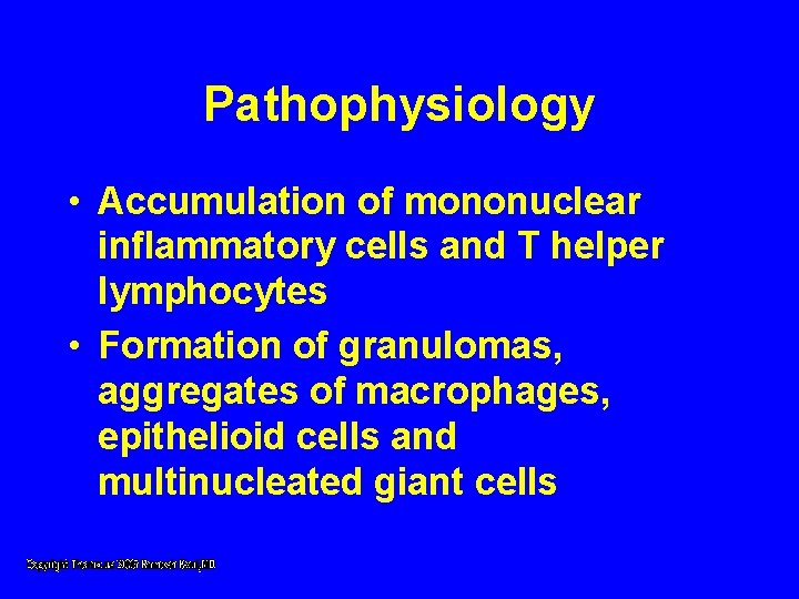
Pathophysiology • Accumulation of mononuclear inflammatory cells and T helper lymphocytes • Formation of granulomas, aggregates of macrophages, epithelioid cells and multinucleated giant cells
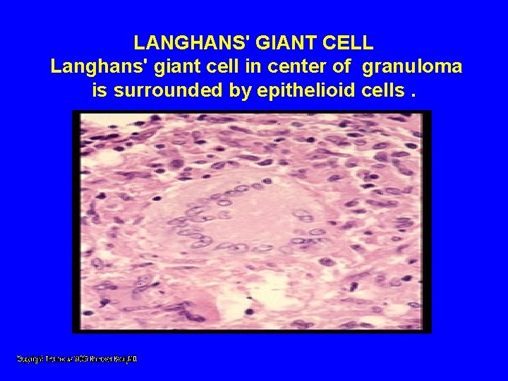
LANGHANS' GIANT CELL Langhans' giant cell in center of granuloma is surrounded by epithelioid cells.
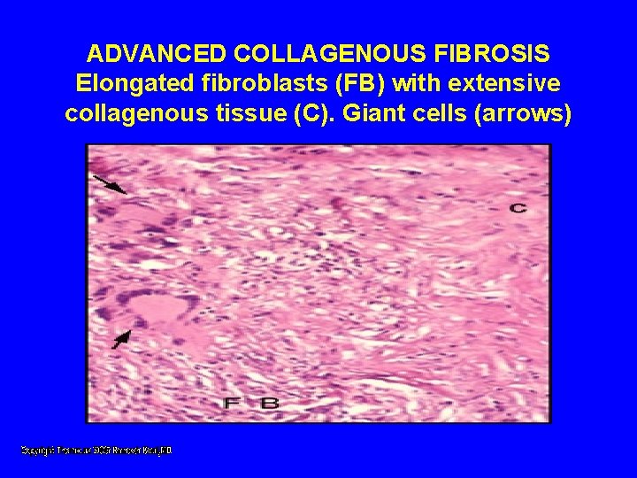
ADVANCED COLLAGENOUS FIBROSIS Elongated fibroblasts (FB) with extensive collagenous tissue (C). Giant cells (arrows)
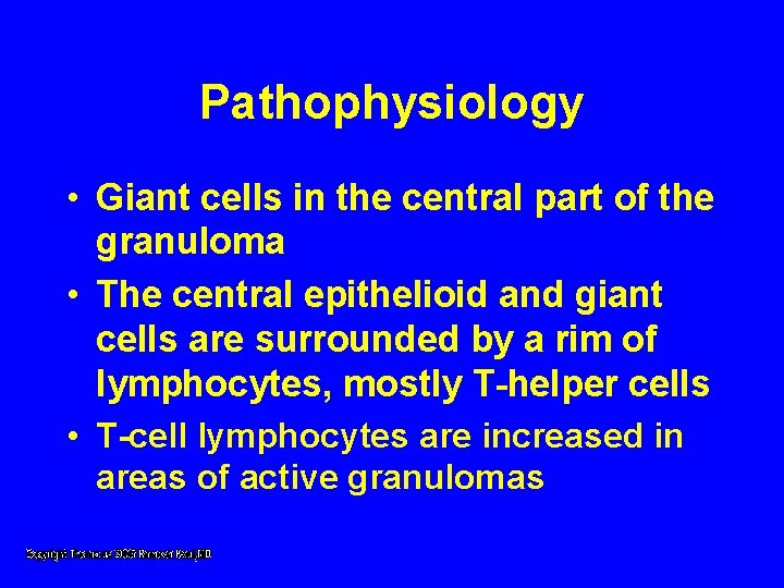
Pathophysiology • Giant cells in the central part of the granuloma • The central epithelioid and giant cells are surrounded by a rim of lymphocytes, mostly T-helper cells • T-cell lymphocytes are increased in areas of active granulomas
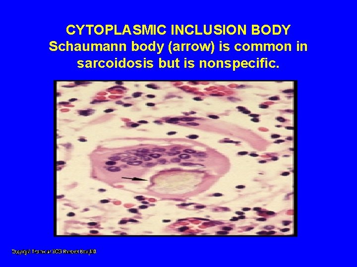
CYTOPLASMIC INCLUSION BODY Schaumann body (arrow) is common in sarcoidosis but is nonspecific.
Pathophysiology • T-helper cells to T-suppressor cells ratio is increased • Exaggerated T-cell activity indicates an altered immune response • Hyper globulinemia • Mass affect of granulomas damages the tissues
Clinical Manifestations • 50% patients are asymptomatic • Abnormal "routine" chest radiograph • Symptomatic patients, with wide variety of symptoms • Onset is usually insidious but can be acute
Clinical Manifestations • Respiratory symptoms are most common • Cough, chest discomfort, and dyspnea • Symptoms reflect the specific organs involved by the granulomas
Lungs • First site involved • Begins with alveolitis involving small bronchi and small blood vessels • Alveolitis either clears up spontaneously or leads to granuloma • Fibrosis
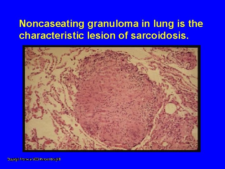
Noncaseating granuloma in lung is the characteristic lesion of sarcoidosis.
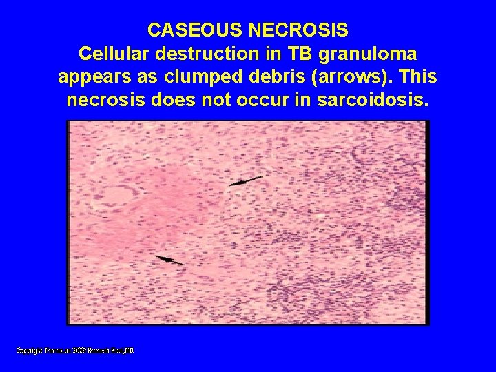
CASEOUS NECROSIS Cellular destruction in TB granuloma appears as clumped debris (arrows). This necrosis does not occur in sarcoidosis.
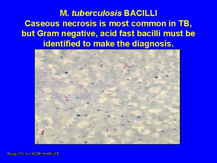
M. tuberculosis BACILLI Caseous necrosis is most common in TB, but Gram negative, acid fast bacilli must be identified to make the diagnosis.
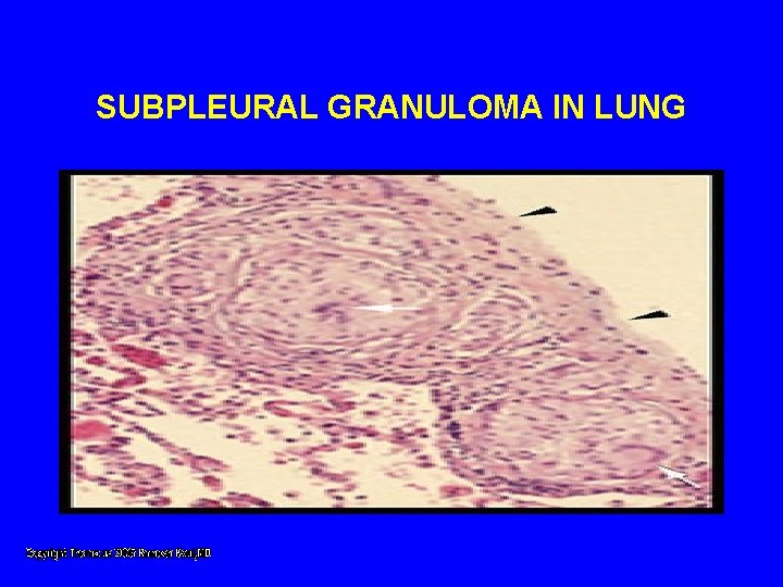
SUBPLEURAL GRANULOMA IN LUNG
Eyes • 25% have eye lesions • Blurred vision, pain, photophobia and dry eyes • Chronic uveitis leads to glaucoma, cataracts and blindness • Keratoconjunctivitis sicca • Papilledema
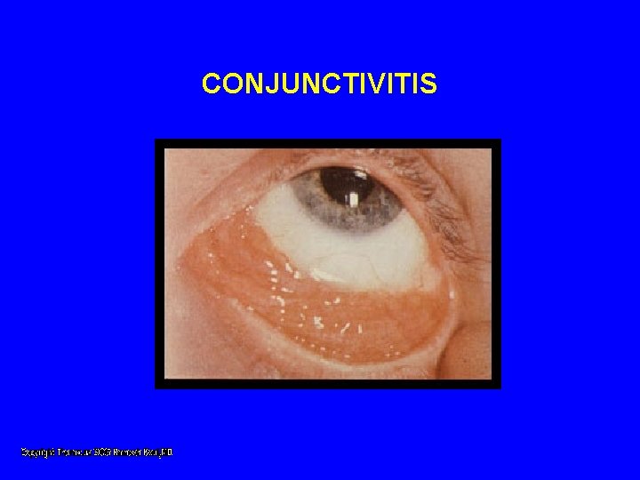
CONJUNCTIVITIS
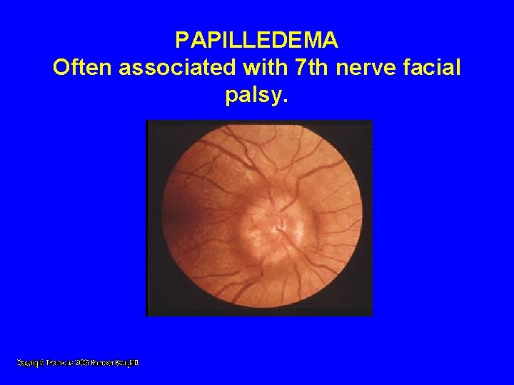
PAPILLEDEMA Often associated with 7 th nerve facial palsy.
Skin • 33% have skin lesions • Cutaneous anergy is common. • LOFGREN'S SYNDROME; acute triad of erythema nodosum, joint pains, and bilateral hilar adenopathy
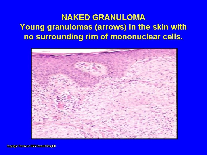
NAKED GRANULOMA Young granulomas (arrows) in the skin with no surrounding rim of mononuclear cells.
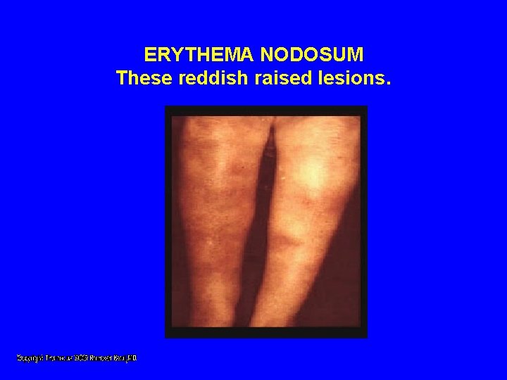
ERYTHEMA NODOSUM These reddish raised lesions.
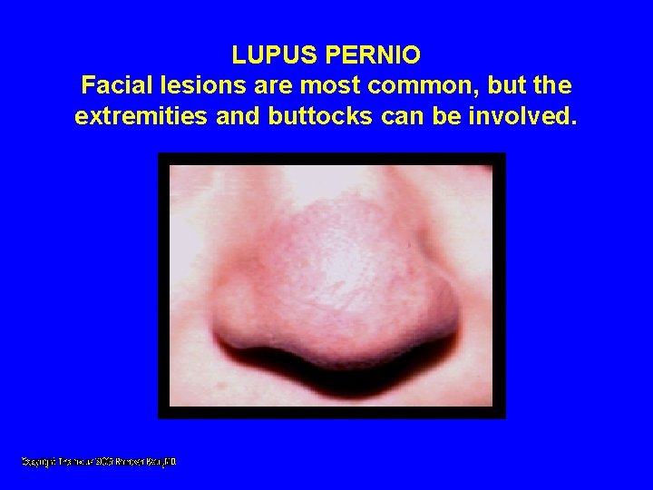
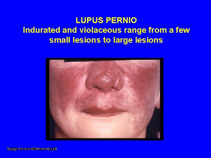
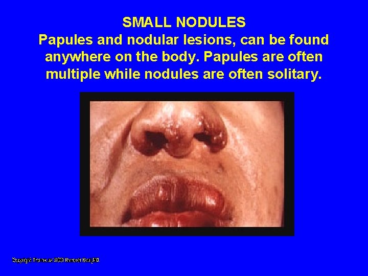
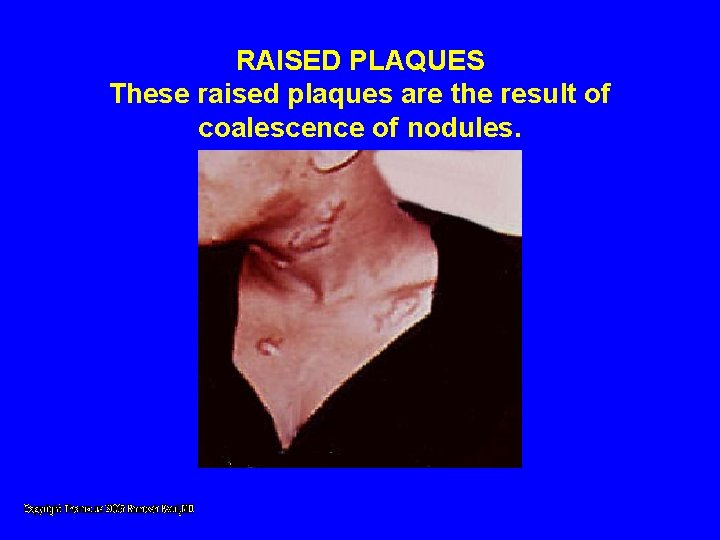
Skin • Lupus pernio- indurated blue purple swollen shiny lesions on nose, cheeks, lips, ears and fingers. • Papules, nodules, and plaques • Psoriatic like lesions • Lesions in scars and tattoos
LUPUS PERNIO Facial lesions are most common, but the extremities and buttocks can be involved.
LUPUS PERNIO Indurated and violaceous range from a few small lesions to large lesions
SMALL NODULES Papules and nodular lesions, can be found anywhere on the body. Papules are often multiple while nodules are often solitary.
RAISED PLAQUES These raised plaques are the result of coalescence of nodules.
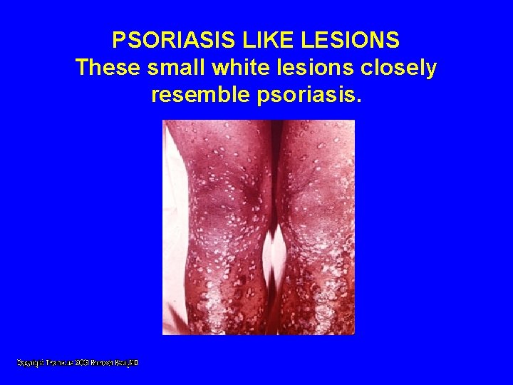
PSORIASIS LIKE LESIONS These small white lesions closely resemble psoriasis.
Liver • 33% have hepatomegaly or biochemical evidence of disease • Symptoms usually absent • Cholestasis, fibrosis, cirrhosis, portal hypertension, and the Budd-Chiari syndrome have been seen
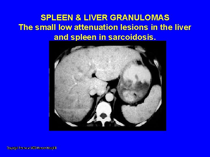
SPLEEN & LIVER GRANULOMAS The small low attenuation lesions in the liver and spleen in sarcoidosis.
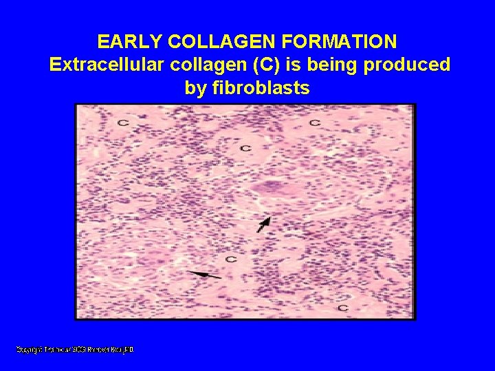
EARLY COLLAGEN FORMATION Extracellular collagen (C) is being produced by fibroblasts
Musculoskeletal • Acute polyarthritis with fever is common • Arthritis is self limited • Chronic destructive bone disease with deformity is rare • Polymyositis and chronic myopathy • Muscle disease is rare
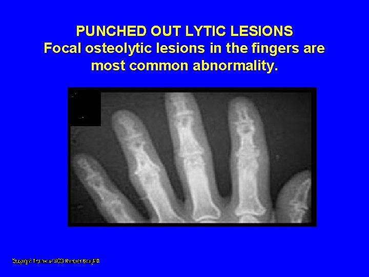
PUNCHED OUT LYTIC LESIONS Focal osteolytic lesions in the fingers are most common abnormality.
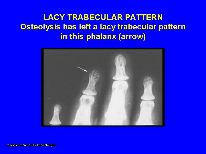
LACY TRABECULAR PATTERN Osteolysis has left a lacy trabecular pattern in this phalanx (arrow)
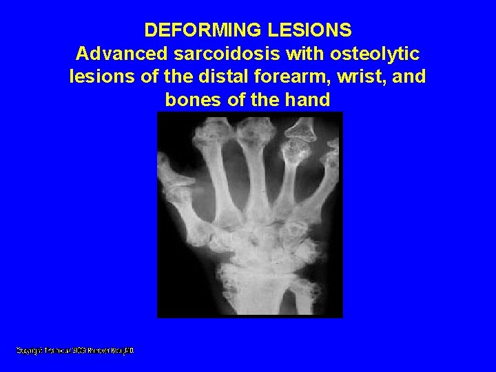
DEFORMING LESIONS Advanced sarcoidosis with osteolytic lesions of the distal forearm, wrist, and bones of the hand
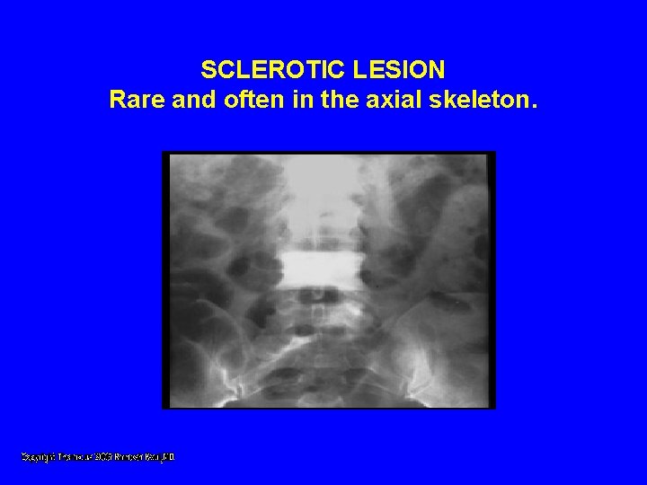
SCLEROTIC LESION Rare and often in the axial skeleton.
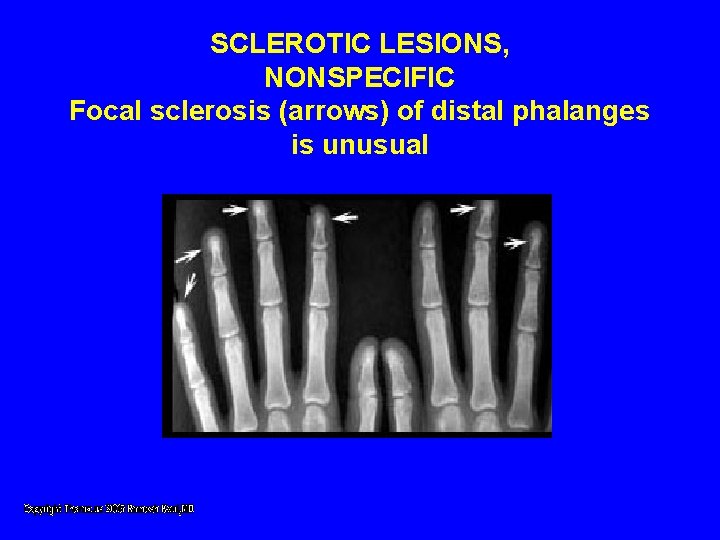
SCLEROTIC LESIONS, NONSPECIFIC Focal sclerosis (arrows) of distal phalanges is unusual
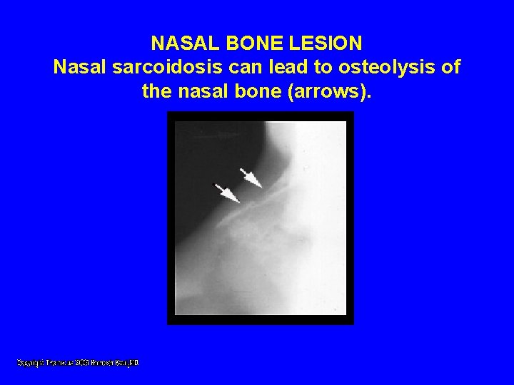
NASAL BONE LESION Nasal sarcoidosis can lead to osteolysis of the nasal bone (arrows).
Heart • • • 5% have heart involvement Conduction abnormalities Cardiomyopathy Chest pain Intractable arrythmias Sudden death
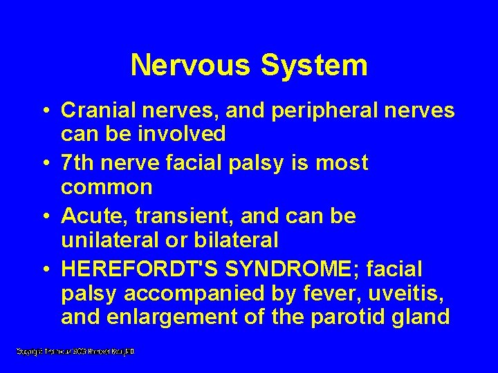
Nervous System • Cranial nerves, and peripheral nerves can be involved • 7 th nerve facial palsy is most common • Acute, transient, and can be unilateral or bilateral • HEREFORDT'S SYNDROME; facial palsy accompanied by fever, uveitis, and enlargement of the parotid gland
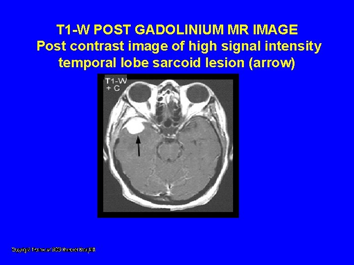
T 1 -W POST GADOLINIUM MR IMAGE Post contrast image of high signal intensity temporal lobe sarcoid lesion (arrow)
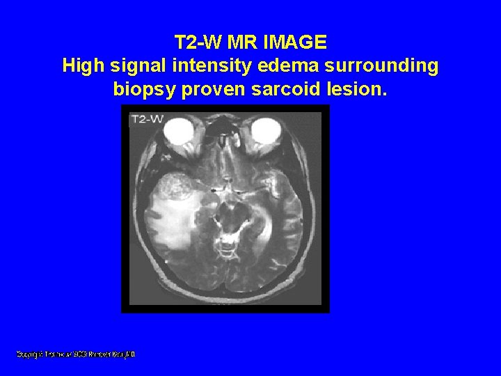
T 2 -W MR IMAGE High signal intensity edema surrounding biopsy proven sarcoid lesion.
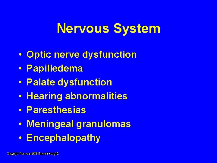
Nervous System • • Optic nerve dysfunction Papilledema Palate dysfunction Hearing abnormalities Paresthesias Meningeal granulomas Encephalopathy
Kidney • Granulomatous interstitial nephritis produces renal failure • Develops over a period of weeks to months • Rapid response to steroid therapy • Kidney stones (nephrolithiasis) and nephrocalcinosis are very unusual secondary to hypercalcemia and hypercalciuria
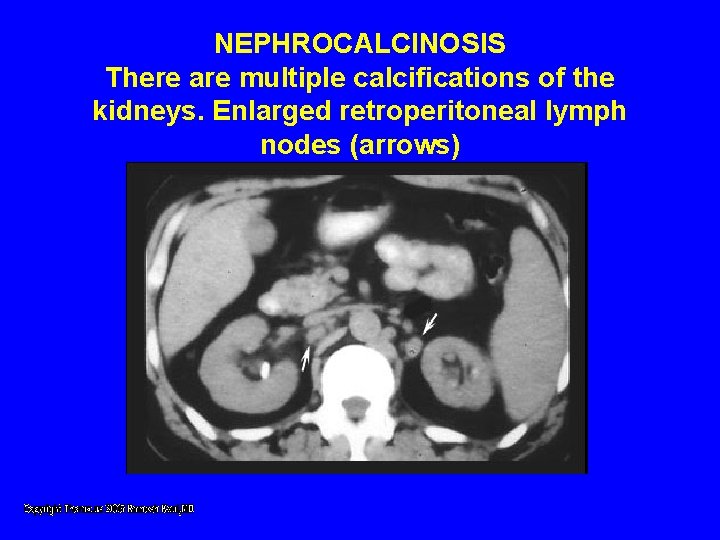
NEPHROCALCINOSIS There are multiple calcifications of the kidneys. Enlarged retroperitoneal lymph nodes (arrows)
Kidney • Increased calcium absorption in the gut • Related to high levels of circulating 1, 25 -dihydroxy vitamin D produced by mononuclear phagocytes in granulomas
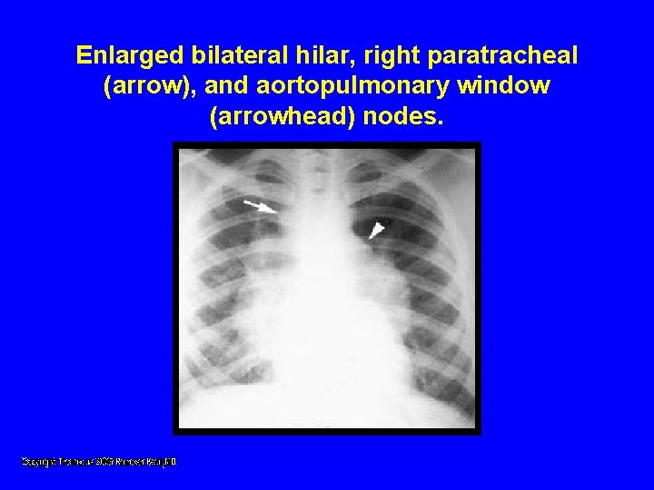
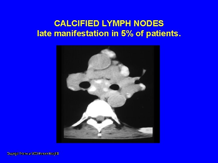
Lymph Nodes • Lymphadenopathy • Intrathoracic nodes enlarged in 7590% patients including hilar nodes and paratracheal nodes. • Peripheral lymphadenopathy
Enlarged bilateral hilar, right paratracheal (arrow), and aortopulmonary window (arrowhead) nodes.
CALCIFIED LYMPH NODES late manifestation in 5% of patients.
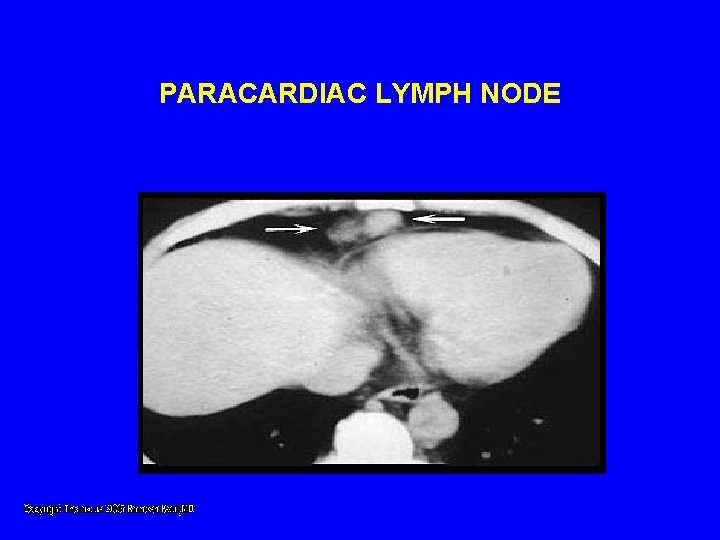
PARACARDIAC LYMPH NODE
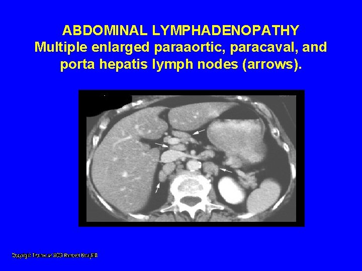
ABDOMINAL LYMPHADENOPATHY Multiple enlarged paraaortic, paracaval, and porta hepatis lymph nodes (arrows).
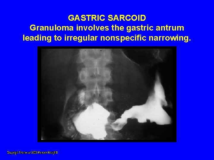
GASTRIC SARCOID Granuloma involves the gastric antrum leading to irregular nonspecific narrowing.
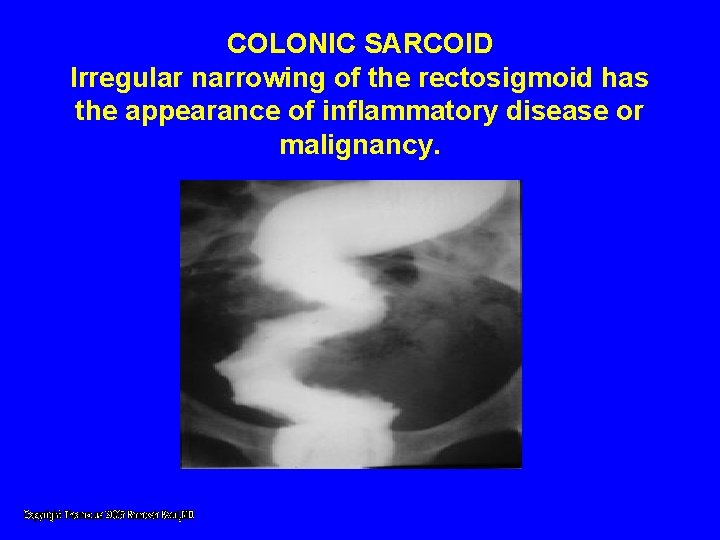
COLONIC SARCOID Irregular narrowing of the rectosigmoid has the appearance of inflammatory disease or malignancy.
Lab Abnormalities • • Lymphocytopenia Mild eosinphilia Increased E. S. R Hyperglobulenemia
Lab Abnormalities • Elevated level of angiotensin converting enzyme • Gallium 67 lung scan showing a pattern of diffused uptake. • Bronchiole alveolar lavage shows increased lymphocytes
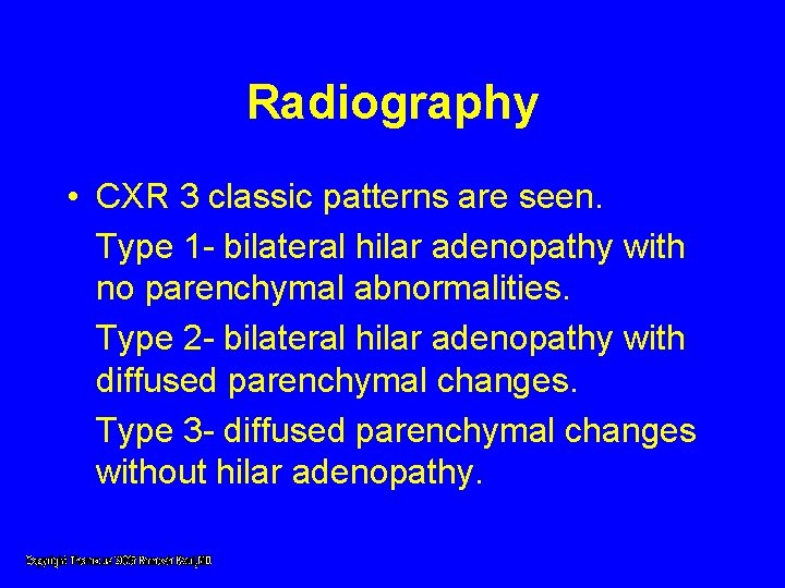
Radiography • CXR 3 classic patterns are seen. Type 1 - bilateral hilar adenopathy with no parenchymal abnormalities. Type 2 - bilateral hilar adenopathy with diffused parenchymal changes. Type 3 - diffused parenchymal changes without hilar adenopathy.
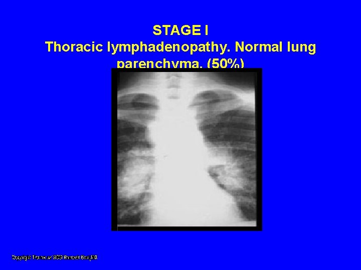
STAGE I Thoracic lymphadenopathy. Normal lung parenchyma. (50%)
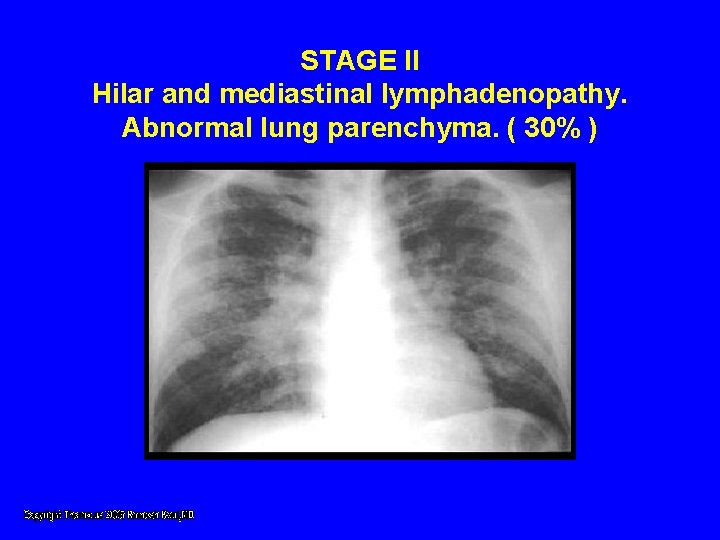
STAGE II Hilar and mediastinal lymphadenopathy. Abnormal lung parenchyma. ( 30% )
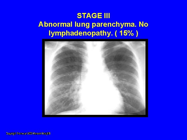
STAGE III Abnormal lung parenchyma. No lymphadenopathy. ( 15% )
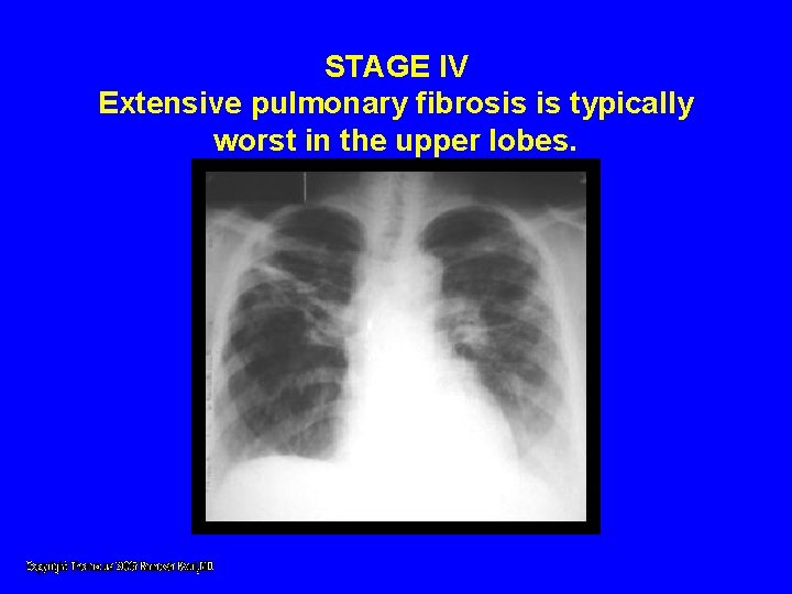
STAGE IV Extensive pulmonary fibrosis is typically worst in the upper lobes.
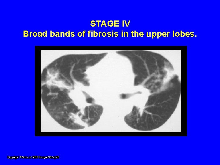
STAGE IV Broad bands of fibrosis in the upper lobes.
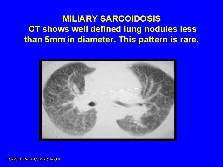
MILIARY SARCOIDOSIS CT shows well defined lung nodules less than 5 mm in diameter. This pattern is rare.
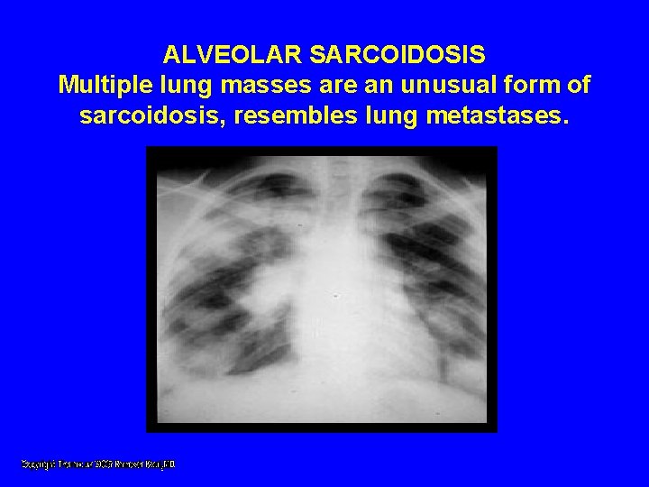
ALVEOLAR SARCOIDOSIS Multiple lung masses are an unusual form of sarcoidosis, resembles lung metastases.
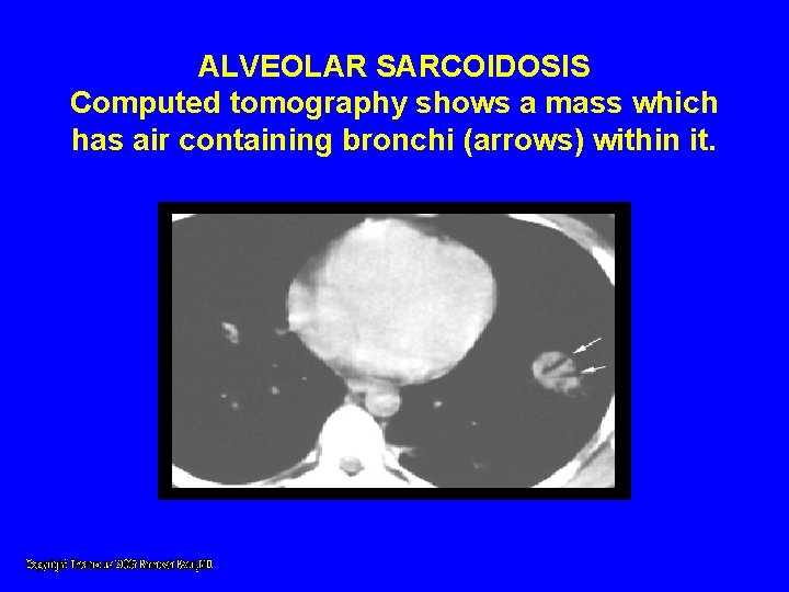
ALVEOLAR SARCOIDOSIS Computed tomography shows a mass which has air containing bronchi (arrows) within it.
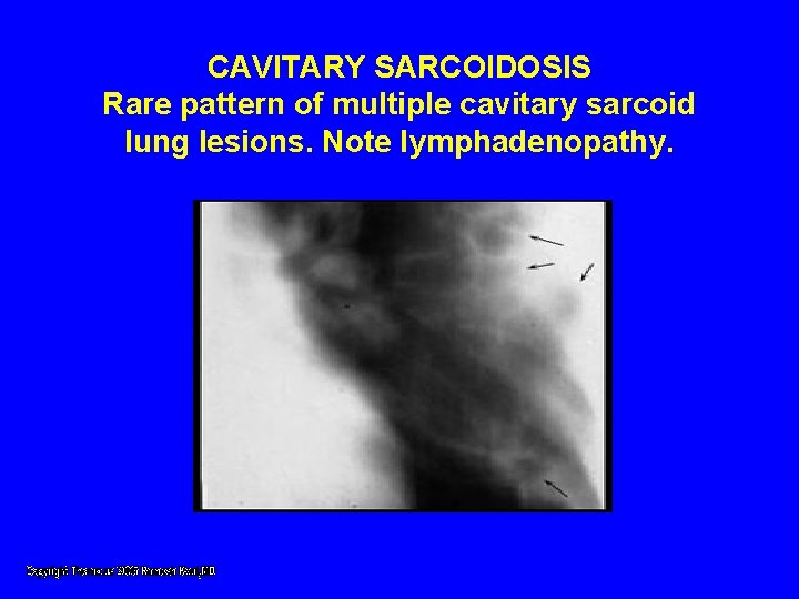
CAVITARY SARCOIDOSIS Rare pattern of multiple cavitary sarcoid lung lesions. Note lymphadenopathy.
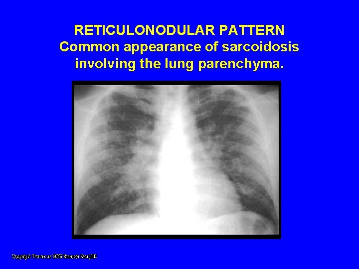
RETICULONODULAR PATTERN Common appearance of sarcoidosis involving the lung parenchyma.
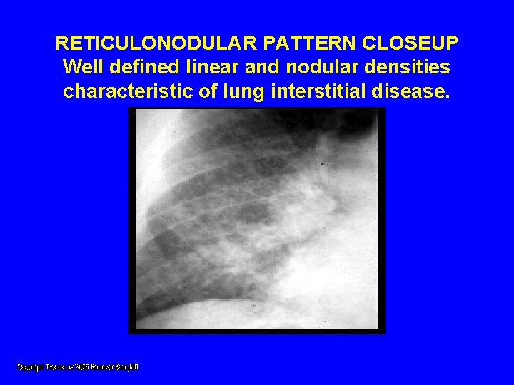
RETICULONODULAR PATTERN CLOSEUP Well defined linear and nodular densities characteristic of lung interstitial disease.
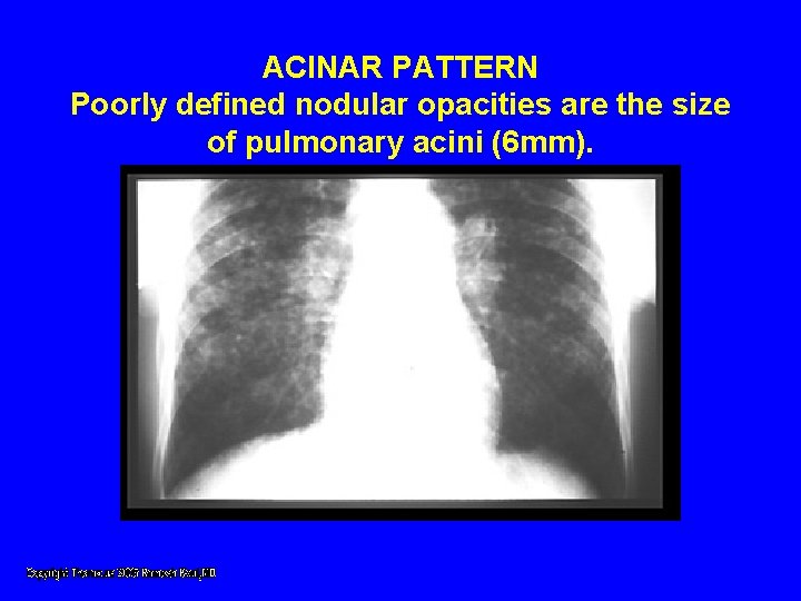
ACINAR PATTERN Poorly defined nodular opacities are the size of pulmonary acini (6 mm).
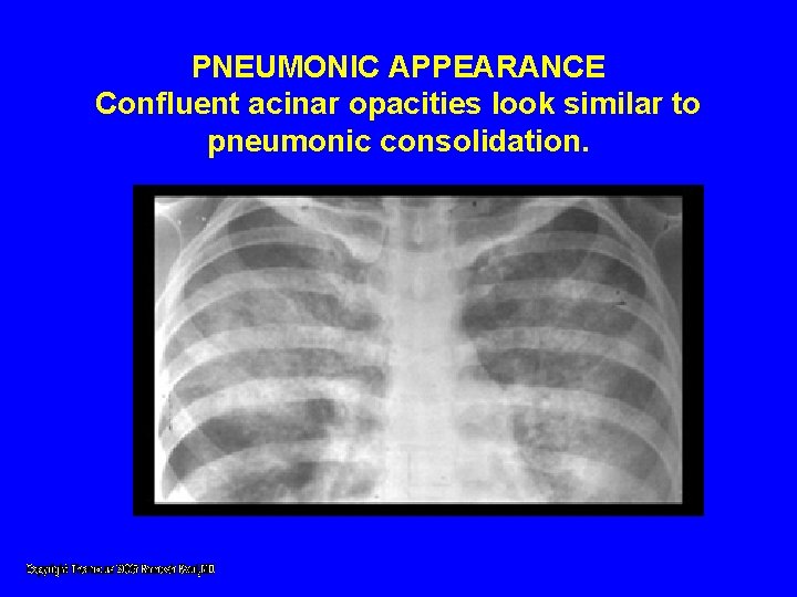
PNEUMONIC APPEARANCE Confluent acinar opacities look similar to pneumonic consolidation.
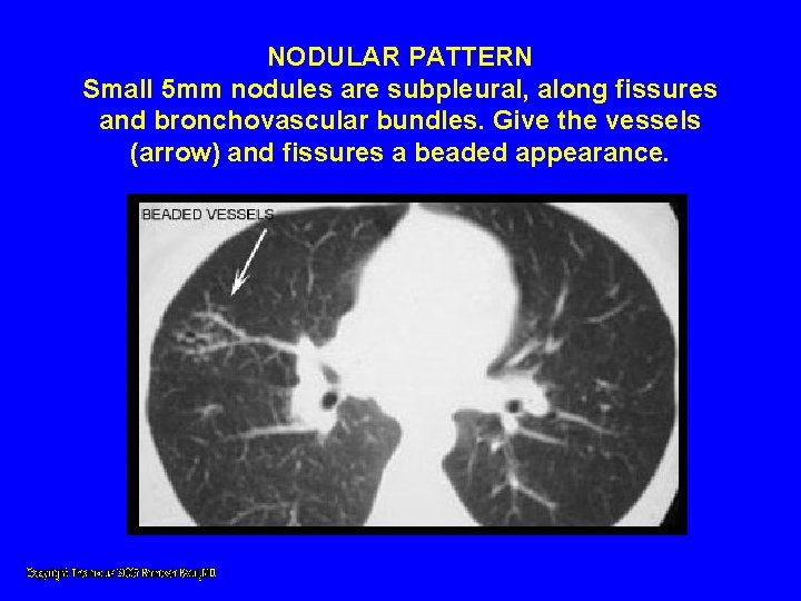
NODULAR PATTERN Small 5 mm nodules are subpleural, along fissures and bronchovascular bundles. Give the vessels (arrow) and fissures a beaded appearance.
Lung Function Test • Lung function abnormalities for interstitial lung disease with decreased lung volumes and diffusing capacities
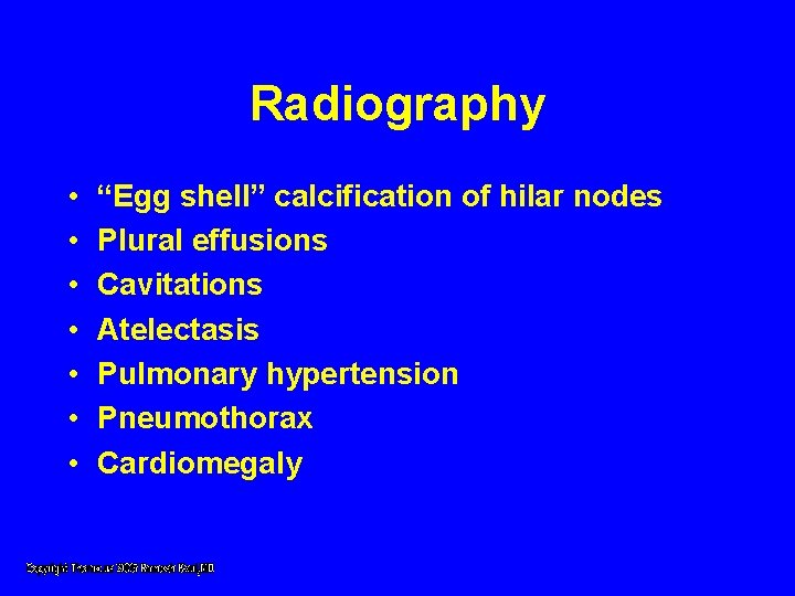
Radiography • • “Egg shell” calcification of hilar nodes Plural effusions Cavitations Atelectasis Pulmonary hypertension Pneumothorax Cardiomegaly
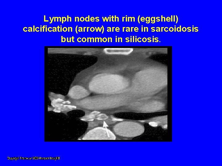
Lymph nodes with rim (eggshell) calcification (arrow) are rare in sarcoidosis but common in silicosis.
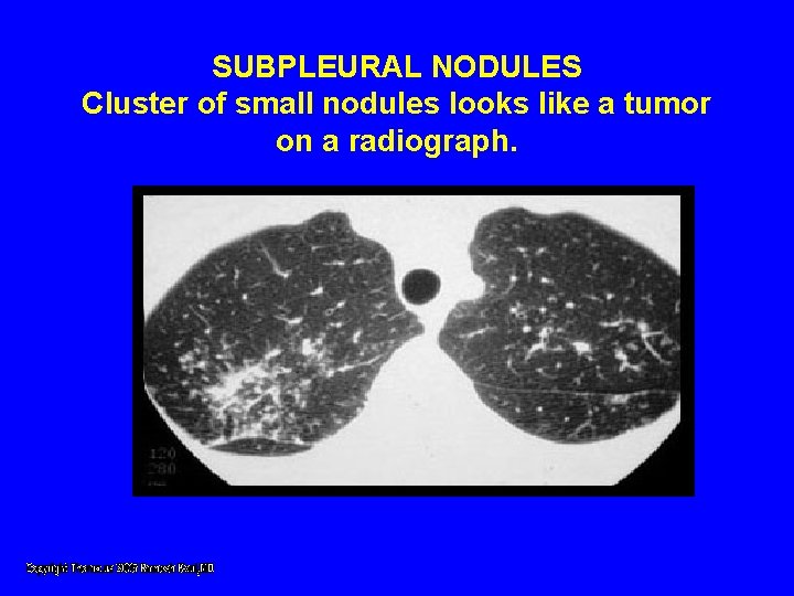
SUBPLEURAL NODULES Cluster of small nodules looks like a tumor on a radiograph.
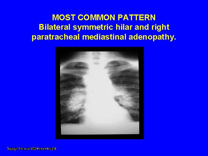
MOST COMMON PATTERN Bilateral symmetric hilar and right paratracheal mediastinal adenopathy.
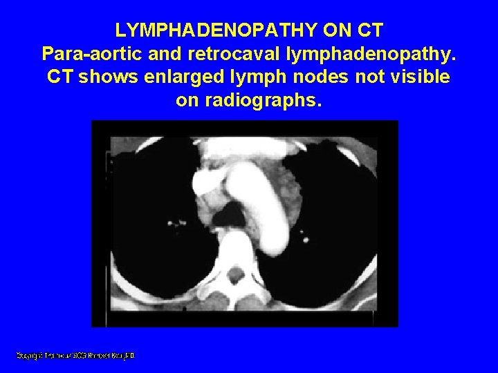
LYMPHADENOPATHY ON CT Para-aortic and retrocaval lymphadenopathy. CT shows enlarged lymph nodes not visible on radiographs.
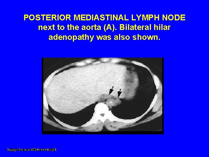
POSTERIOR MEDIASTINAL LYMPH NODE next to the aorta (A). Bilateral hilar adenopathy was also shown.
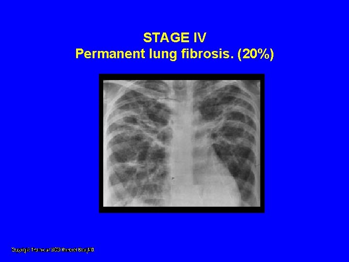
STAGE IV Permanent lung fibrosis. (20%)
Diagnosis • Difficult to differentiate from chronic infections, fungal diseases, T. B. and lymphoma. • Based on combined clinical, radiologic and histologic findings. • Laboratory tests seldom important • Asymptomatic
Diagnosis • Identify noncaseating granulomas • Variety of infections • Transbronchial biopsies positive in 65 -95%, even if no lung parenchymal abnormalities imaged. • Tissue from mediastinoscopy positive in 95% • Scalene node biopsy positive in 80%
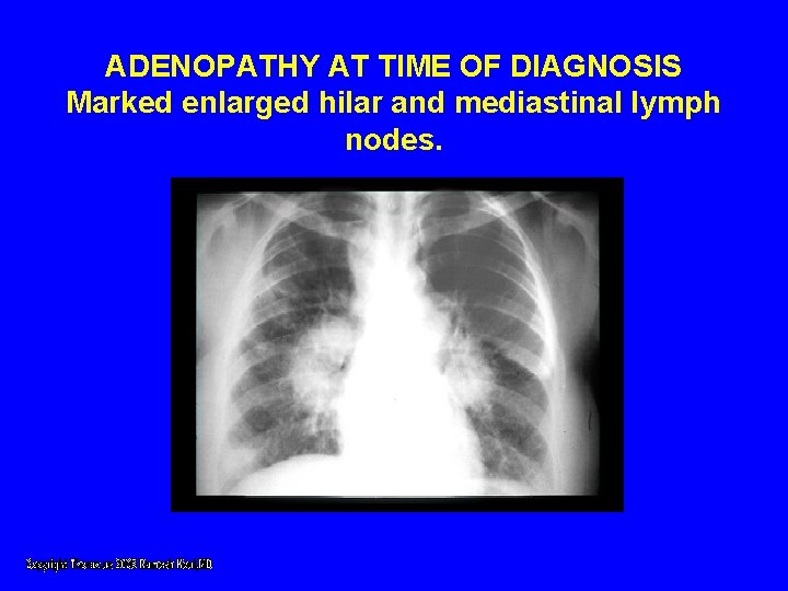
ADENOPATHY AT TIME OF DIAGNOSIS Marked enlarged hilar and mediastinal lymph nodes.
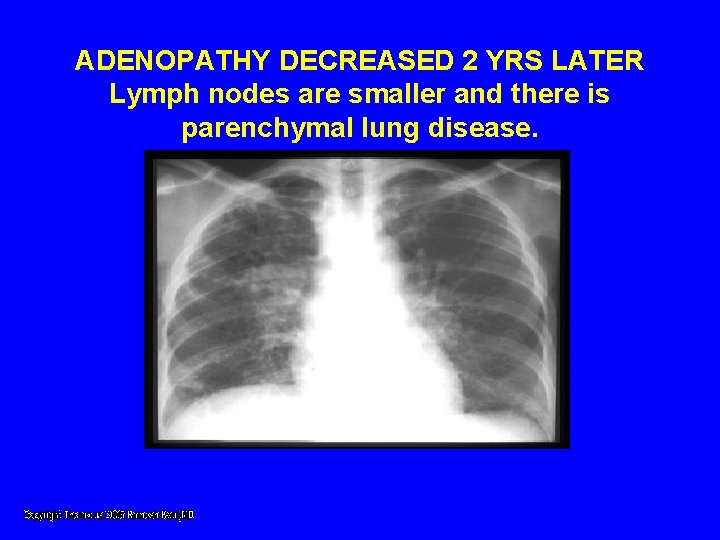
ADENOPATHY DECREASED 2 YRS LATER Lymph nodes are smaller and there is parenchymal lung disease.
Diagnosis • KVEIM TEST • Involves injecting standardized preparation of sarcoid tissue material into the skin. • Unique lump formed at the point of injection is considered positive for sarcoidosis.
Diagnosis • Test not always positive • Not used often in US • Test material not approved for sale by FDA.
Prognosis • Good • 50% have some permanent organ dysfunction • In 15 -20% remains active or recurs intermittently.
Treatment • No known cure • Corticosteroids, primary treatment for inflammation and granuloma formation. • Prednisone, 1 mg/kg for 4 -6 weeks followed by slow taper over 2 -3 months.
Treatment • • • Chloroquine Effectiveness ? D-penicillamine. Effectiveness ? Chlorambucil Azathioprine Methotrexate might suppress alveolitis Cyclophosphamide may suppress alveolitis
Treatment • Risk of drugs high, in pregnant women. • Cyclosporine, evaluated in one controlled trial was found unsuccessful.
God Bless America