Sarcoid in a Teenager 1062010 HPI 17 yo


- Slides: 18
Sarcoid in a Teenager 10_6_2010
HPI • • • 17 yo girl 2 -4 weeks of progressive fatigue and weight loss 12 pound weight loss over the last month Intermittent dry cough since age 11!!! Decreased exercise tolerance
• ROS: – No fever/chills – No headache/change in vision – No abdominal pain/diarrhea/constipation/vomiting – + intermittent erythematous rash of eyelids and extensor surfaces of extremities – No joint/muscle pains • PMH: chronic cough, normal CXR 2 years ago • SH: lives in Nantucket, senior in high school, plays lacrosse
P/E and labs • Normal vitals • General: NAD, thin, tired appearing • Otherwise unremarkable • • • CBC: 9. 4>13. 2/38. 6<293 BUN/Cr : 21/1. 4 Ca/Mg/PO 4: 14. 4/2. 0/3. 5 LFTs: normal CRP/ESR: <0. 1/13
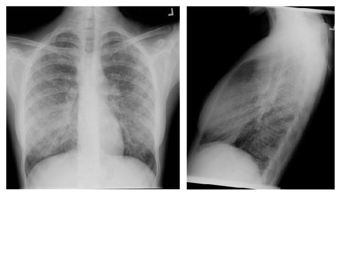
ØDiffuse interstitial markings ØHilar adenopathy ØNo evidence of volume loss or fibrosis ØConsistent with stage II sarcoidosis
Hypercalcemia and Interstitial Lung Disease • Infections • Mycobacteria • M. tuberculosis • Nontuberculous mycobacteria • Fungi • Candidiasis • Histoplasma • Cryptococcus • Coccidioides • Blastomyces • Pneumocystis • Parasites • Dirofilaria • Cat Scratch Disease • Noninfectious diseases – Sarcoidosis – Langerhans-cell histiocytosis – Berylliosis – Silicone-induced granulomas – Hot tub lung – Wegener’s granulomatosis – Churg-Strauss syndrome – Crohn’s Disease
Further Evaluation • ACE: 210 • FH: mom carries diagnosis of sarcoid based on CT scan. Has never had a biopsy and has not required treatment.
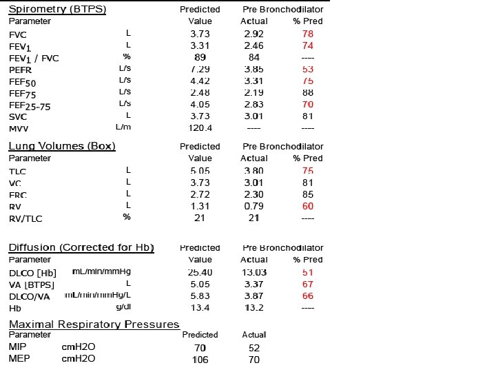
ØRestrictive pattern with decreased diffusion capacity
ØMediastinal and hilar adenopathy Ødiffuse lung disease including diffuse nodular and confluent ground glass opacities
Bronchoscopy • BAL: 73% macrophages, 25% columnar cells, 2% neutrophils • Mucus • A few iron-laden macrophages are present • Fat-laden macrophages are present. (Lipid Index: 32)
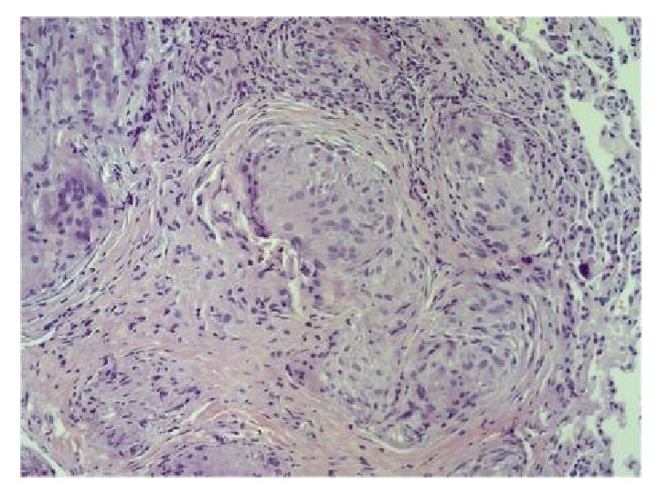
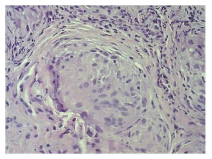
Pathology: transbronchial biopsy ØNoncaseating granulomas
Treatment • Spontaneous remission: – 60 -80% of pts with radiographic Stage I – 50 -60% of pts with Stage II – <30% in Stage III • Indications of treatment: – Worsening pulmonary symptoms – Deteriorating lung function – Progressive radiographic changes – Ocular, neurologic, myocardial or renal involvement – Hypercalcemia
Treatment • Spontaneous remission: – 60 -80% of pts with radiographic Stage I – 50 -60% of pts with Stage II – <30% in Stage III • Indications of treatment: – Worsening pulmonary symptoms – Deteriorating lung function – Progressive radiographic changes – Ocular, neurologic, myocardial or renal involvement – Hypercalcemia This patient had met at least 3 of the 5 above indications for treatment