
Sandstorm in her Chest Manmadha Rao Talluri Nizams




- Slides: 24
Sandstorm in her Chest ? Manmadha Rao Talluri Nizam’s Institute of Medical Sciences, Hyderabad, India
History • • • 32 -year-old woman, housewife Dry cough, exertional dyspnea – 1 year No wheezing/ chest pain/ hemoptysis No malar rash/ photosensitivity No upper respiratory symptoms
Past History No history of: • Bronchial asthma • Varicella in childhood • Recurrent respiratory tract infections • Rheumatic heart disease • Tuberculosis • Occupational dust exposure • Similar complaints in family history
Examination • P – 100, BP – 130/80, respiratory rate – 18, afebrile • Facial puffiness with acne • Upper respiratory tract – normal • Lungs – bilateral basal fine end-inspiratory crepitations, no rhonchi • Abdomen, CVS, CNS – normal • No clubbing
Dr. Newell
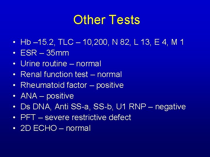
Other Tests • • • Hb – 15. 2, TLC – 10, 200, N 82, L 13, E 4, M 1 ESR – 35 mm Urine routine – normal Renal function test – normal Rheumatoid factor – positive ANA – positive Ds DNA, Anti SS-a, SS-b, U 1 RNP – negative PFT – severe restrictive defect 2 D ECHO – normal
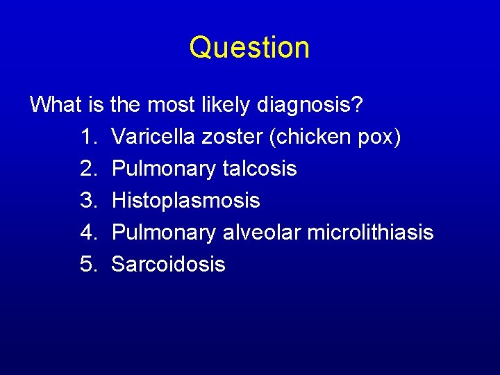
Question What is the most likely diagnosis? 1. Varicella zoster (chicken pox) 2. Pulmonary talcosis 3. Histoplasmosis 4. Pulmonary alveolar microlithiasis 5. Sarcoidosis
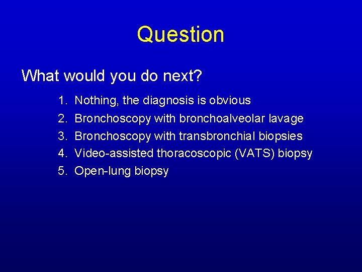
Question What would you do next? 1. Nothing, the diagnosis is obvious 2. 3. 4. 5. Bronchoscopy with bronchoalveolar lavage Bronchoscopy with transbronchial biopsies Video-assisted thoracoscopic (VATS) biopsy Open-lung biopsy
Dr. Heffner
Dr. Leslie
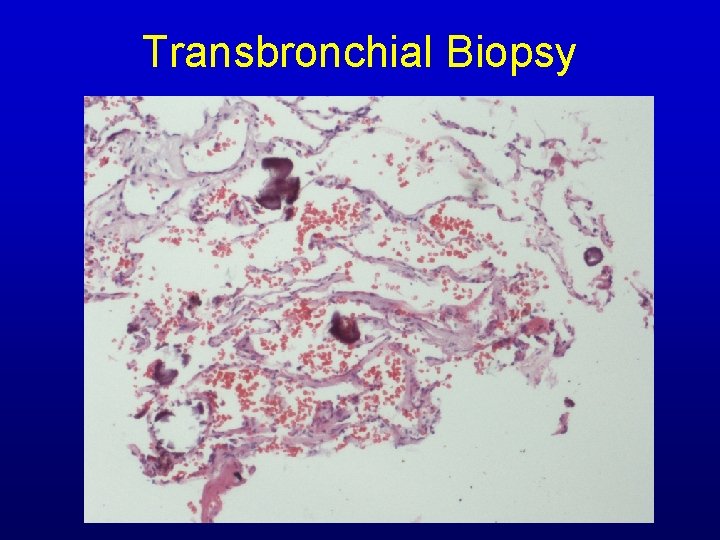
Transbronchial Biopsy
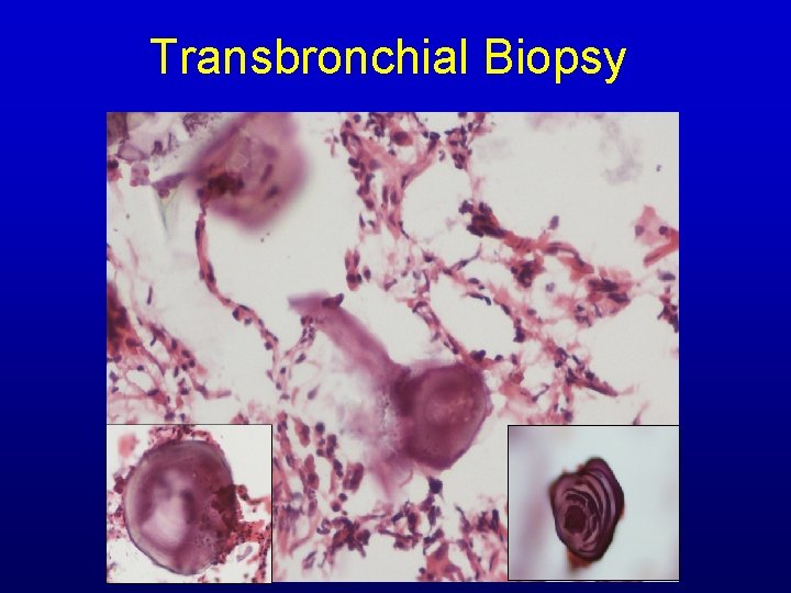
Transbronchial Biopsy
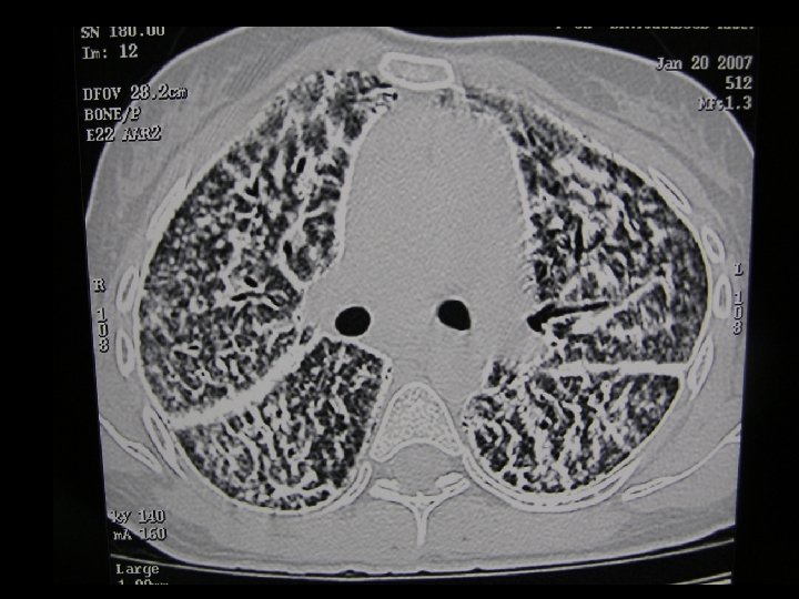
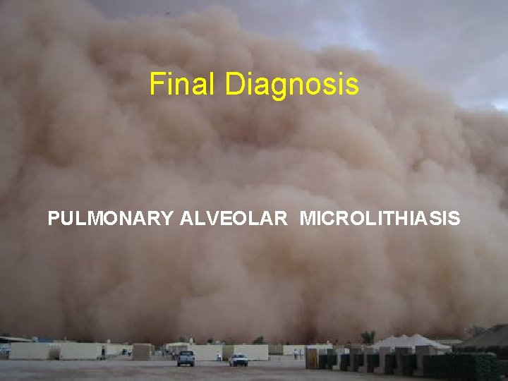
Final Diagnosis PULMONARY ALVEOLAR MICROLITHIASIS
Clinical Course • Given corticosteroids and theophylline for dyspnea • Advised lung transplantation – Not done due to financial constraints • Patient gradually worsened in course of 2 years and succumbed to respiratory failure
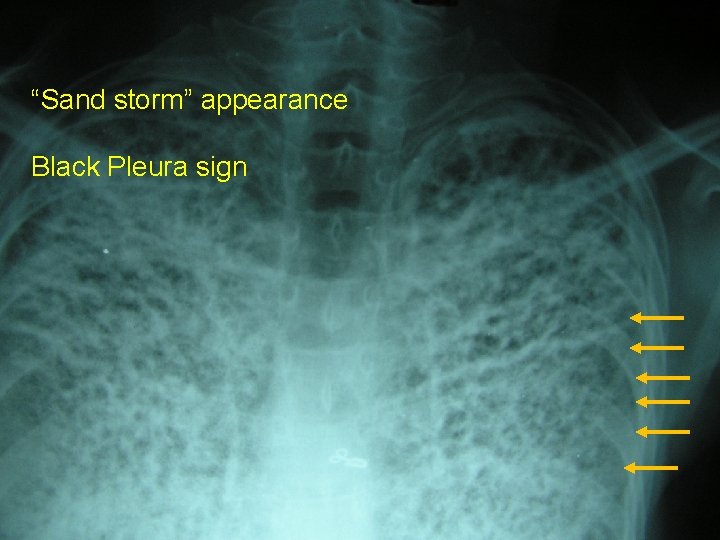
Pulmonary Alveolar Microlithiasis • Rare disease of unknown pathogenesis • Usually sporadic; autosomal recessive form described (Mediterranean countries) • Paucity of symptoms despite widespread involvement • Cough & dyspnea in 3 rd & 4 th decade • Death usually in mid-life due to respiratory failure and cor pulmonale
Pulmonary Alveolar Microlithiasis • Widespread laminated calcispherites in alveolar spaces • Absence of any known disorder of calcium metabolism • Unknown stimulus • Changes in the alveolar lining membrane or secretions result in greater alkalinity, promoting intra-alveolar precipitation of calcium phosphates and carbonates
• Serum surfactant protein – A & D are markedly elevated – Increase as disease progresses – Function as serum markers to monitor disease activity and progression • Mutations in SLC 34 A 2 gene expressed in type II pneumocytes which encode type IIb sodium phosphate co-transporter
• No known therapy • Corticosteroids, chelating agents and BAL have demonstrated no benefit • Role of bisphonates remains to be proven • Bilateral lung transplantation for advanced cases
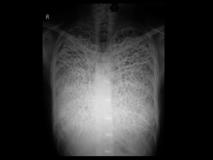
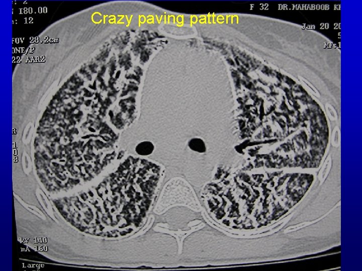
Chest X-ray/ CT “Sand storm” appearance Black Pleura sign
Crazy paving pattern
Take Home Message • In no other condition is the lack of association between roentgenologic and clinical findings so striking as in PAM • PAM should always be considered in the differential diagnosis of calcific micronodular pulmonary lesions
References • • Barbolini G, Rossi G, Bisetti A. Pulmonary alveolar microlithiasis. N Engl J Med 2002; 347: 69– 70. K. Gowrinath and Arun R. Warrier Pulmonary alveolar microlithiasis, Lung India 2006; 23: 42 -44. Gasparetto EL, Tazoniero P, Escuissato DL, et al. Pulmonary alveolar microlithiasis presenting with crazy-paving pattern on high resolution CT. Br. J. Radiol. 2004; 77: 974 -976 Chan Ed, Morales DV, Welsh CH, et al. Calcium deposition with or without bone formation in the lung. Am J Respir Crit Care Med. 2002; 165(12): 1654 -1669 Korn MA, Schurawitzki H, Klepetko W, et al. Pulmonary alveolar microlithiasis: findings on high-resolution CT. AJR Am J Roentgenol. 1992 ; 158(5): 981 -982. Johkoh T, Itoh H, Müller NL, et al. Crazy paving appearance at thinsection CT. Spectrum of disease and pathologic findings. Radiology 1999; 211: 155– 160 Takahashi H, Chiba H, Shiratori M, et al. Elevated serum surfactant protein A and D in pulmonary alveolar microlithiasis. Respirology. 2006; 11(3): 330 -333