Sampling Dr Wafaa Abd ElGhany Poultry Diseases Department

Sampling Dr. Wafaa Abd El-Ghany Poultry Diseases Department, Fac. Vet. Med. , Cairo Univ

Aim of Sampling 1. 2. 3. 4. To monitor poultry flocks from freedom from some diseases. For isolation and identification of the causative agents (Bacteria, Virus, Parasite or Fungi). For examination of the antibody titers either for estimation of the post-vaccinal immune response or due to infection. For determination of mycotoxins level in the ration.
 Flock: A. Number")
It must be a good representative sample for: Flock Problem (complain) Flock: A. Number of birds: The sample must cover the number of the flock. For living birds 0. 5 -1% of the total number. For dead birds all dead ones or the largest number as possible. (Freshly dead, 2 -4 hrs after death acc. to the season). q

B. The sample should be representative to the production and the development of the flock. (body weight, egg production, etc …). C. Odd (culled) birds which have deformities, abnormalities or retarded development are not representative and must be discarded regularly from the flock (1 -2 times/ week).
: A. The samples should be taken from birds showing the")
Problem (Clinical complain): A. The samples should be taken from birds showing the main clinical signs (ailing birds). B. The samples must be represent the chronological sequence (pathogeneses) of the disease. taken at the disease beginning where the birds have mild signs and at the end where the birds are severely ill. E. g: Gumboro disease (Bursa). ND (Resp. & or Nervous signs).

Time of Sampling Sample collection should be done in case of disease threatening poultry performance (body weight, egg production, etc………. ) In case of disease affecting the health status of the flock (appearance of any abnormal sign). It’s preferable to take the sample (tissues) in the acute sage of the disease when the organism present in the organs in large number. Serum samples could be taken in convalescent stage (2 -4 wks after appearance of signs).

Selection of birds for sampling Diseased living birds 1. Blood sampling. 2. Faecal sampling 3. Clinicopathological Exam. 4. Histopathological Exam. 5. Microbiological Exam. 6. Parasitological Exam. 7. Organ-smear Exam. 8. Tissue sections Freshly dead birds 1. Clinicopathological Exam. 2. Histopathological Exam. 3. Microbiological Exam. 4. Organ-smear Exam. 5. Tissue sections

Selection of birds for P/M Exam. Selection of specimens is critical because improper sample choice could lead to: 1. 2. 3. Wrong decision. Economic losses (deaths, decrease in productivity, losses in weights). Increase the cost of treatment.

Samples for Microbiological Exam. Selection of samples for Microbiological Exam. depends mainly the knowledge, care and the attention of the veterinarian. The sample should be taken from the right place at the right time. The right time mean as soon as possible after the onset of clinical signs when the organisms are usually present in maximum amount at this time, then they diminish within few days. Specimens taken after days or weeks of the onset of signs is a waste of effort.
 2) The site for sample collection depends mainly on:")
Samples for Microbiological Exam. 1) 2) The site for sample collection depends mainly on: The clinical findings (signs and lesions). The pathogenesis of the suspected disease.

Samples for Microbiological Exam. The samples should be put in sterile plastic bags, tightly closed and then labeled by writing the followings: The sample code No. The date and time. The name of the taken organ. The flock history, clinical examination and P/M examination report should be sent with the sample to the lab.

Samples for Microbiological Exam. When ambient temperature is moderate and transient time to the laboratory is less than one day, ice or cold bags (<4 C) is usually taken during transportation. When environmental temperature is high and the time longer than a day, Styrofoam boxes containing dry ice should be taken during transportation.

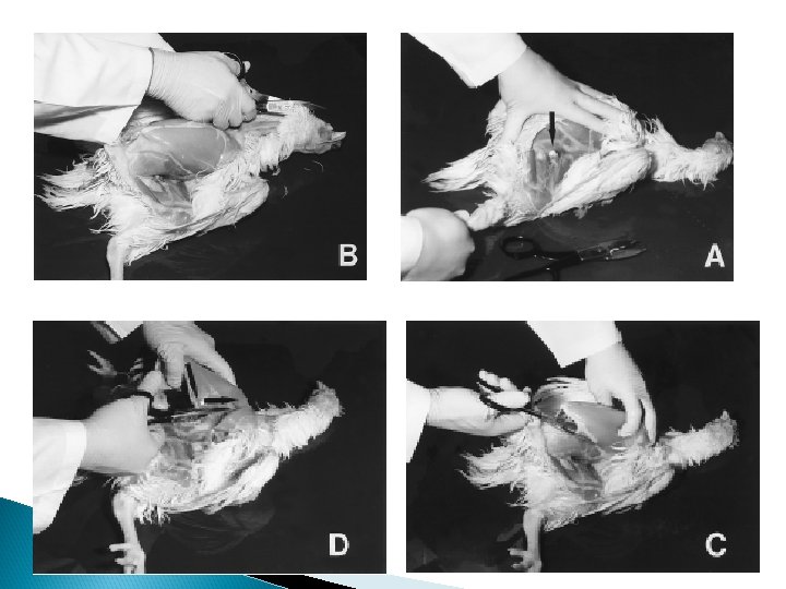
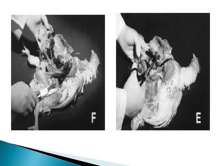
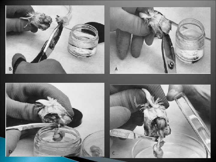
Histopathological sampling § § § The sample should be taken from freshly dead or slaughtered birds. Gentle handling with the sample without hampering. The sample should be approximately 1 cube cm in size. Wide mouth bottle or jar containing 10 -15% neutral formalin solution for samples used. The ratio between the total volume of the sample to the volume of the fixative should range from 1/10 -1/20. Sample immersed in a fixative don’t have to be cooled or stored more than 8 weeks.

Histopathological sampling The jar should be labeled with the full data about the flock and the clinical exam. § The histopathological (microscopical) examn. is important in diagnosis of some important poultry disease like: ? MD (Lymphocytic infiltration in nerves). ? Vit. E Def. (Degenerative changes in the brain). ? AE (Prevascular cuffing & gliosis in the brain and spinal cord). ? ALC (Acc. to the type of the tumor). §

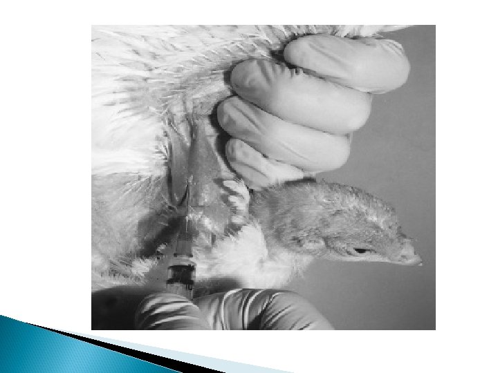
Blood, Serum and Plasma Sampling The blood samples are collected from the birds from the following sites: v The wing vein: ü It is recommended for birds with 2 weeks of age or older. The amount of blood is taken in this method is about 1 -3 ml. v The heart puncture: v Bird slaughtering: It is recommended for birds are 3 weeks of age or older. The amount of blood is taken in this method is about 4 -8 ml. Its recommended in baby chicks (day old). Very large amount of blood could be taken.

Blood, Serum and Plasma Sampling ü Test tubes or Weatherman tubes for collection of blood: With anticoagulant (EDTA, heparin, citrate) For diagnosis of CIA, EDS and ALC. Clinical pathology Without anticoagulant For serum Indirect diagnosis (Antibodies by serology) serum chemistry collection

Blood, Serum and Plasma Sampling Blood film: Considered as the rapid method for diagnosis for the following diseases: • Acute FC The blood film is taken from he wing vien should be thin, then dry it and stain with Giemsa or Leishman’s then examine under oil immersion lens to find bipolar organism (P. multocida in between he nucleated RBCS). • Acute spiroketosis (Spiral shape Borellia anserina organism).

Spiral Borilia anserina Bipolar P. multocida
 specially in hot")
Blood, Serum and Plasma Sampling Blood protozoon parasites: Like plamodium (Malaria) specially in hot countries or haemoproteus infection.

Feed Sampling Samples taken for detection of the level of mycotoxoins (aflatoxin, ochratoxins, T 2 or zeralenone) in the ration should taken from: 1. The mixer of the rations components. 2. From the place for ration preservation. 3. From the feeders in front of the birds and under it. 4. Poultry feed samples for detection and determination of mycotoxins should be representative (quantitative and qualitative) to avoid false positive and negative results.
.")
Feed Sampling ü ü Quantitative represent (not less than 25 kg/ton of tested feed). Qualitative represent (collected from top, middle and bottom of the feed lot). If false positive result is obtained, this constitute a high risk on feed manufactures, while if false negative results leads to risk on the owner. After collection, sample should be thoroughly mixed and blended to become fine and uniformly distributed.

Feed Sampling Detection and determination of mycotoxins: Chemical extraction: ü Mycotoxins are extracted from samples by using non-polar solvent (methanol, chloroform, aceton, etc. . ) as mycotoxins are dissolved in these solvent. Cleaning up procedure: ü Cleaning up of chemical extract to remove or avoid interference materials which could interfere with toxins as pigments.
 Thin layer chromatography (TLC): using silica gel plate")
Feed Sampling Detection and determination: 1) Thin layer chromatography (TLC): using silica gel plate (gel substance adsorb mycotoxins) and Ultra violet light of 365 wave length (330 -350). This depends on that mycotoxins have the ability to fluorescence under UV light as aflatoxins give blue or green fluorescence and ochratoxins give greenish or bluish fluorescence but with different rate in diffusion in silica gel plate. It should be compared with standard mycotoxins to determine the rate of flow. (RF).
 High performance liquid chraomatography (HPLC). (similar to TLC,")
Feed Sampling Detection and determination: 2) High performance liquid chraomatography (HPLC). (similar to TLC, but more accurate). 3) Gas liquid chraomatography (GLC). (similar to TLC, but more accurate). 4) Flourometer: depends on monoclonal antibodies.
- Slides: 28