Same chromophore different absorption maximum Length of the

Same chromophore – different absorption maximum Length of the conjugation Electrostatic interaction with the binding pocket Steric interaction with the protein Protonation / deprotonation
")
Same chromophore – different absorption maximum Length of the conjugation Shorter chain (blue shift) Short wavelength Longer chain (red shift) Longer wavelength

Same chromophore – different absorption maximum Electrostatic interaction with the binding pocket BLUE SHIFT (shorter wavelengths): - Ground state stabilization - Excited state destabilization + S 1 + + S 1 S 0

Same chromophore – different absorption maximum Electrostatic interaction with the binding pocket RED SHIFT (longer wavelengths): - Ground state destabilization - Excited state stabilization + + + S 1 S 0 Very important S 1 S 0

Same chromophore – different absorption maximum Steric interaction with the protein Twisting the bonds in the backbone (blue shift) Straightening the backbone (red shift) Rhodopsin (500 nm) Bacteriorhodopsin (568 nm)

Same chromophore – different absorption maximum Protonation / deprotonation UV vision birds, honeybee Me Me N Me Strong blue shift

Isomerization triggers the biological response Light S 1 S 0 Response Isomerization

Isomerization triggers different cellular responses Light S 1 Ion pump energy S 0 Transducer activation G-protein activation Isomerization signaling

Variety of responses to retinal isomerization bacteriorhodopsin ion pump Rhodopsin G-protein activation Sensory rhodopsin transducer activation

Light Rhodopsin Rod G-protein signaling pathway Cone

Rhodopsin Photocycle Biologically active

Rhodopsin Photocycle

Rhodopsin Architecture
")
Signaling pathway of rhodopsin G-proteins Rhodopsin: Lipid molecules: (effective signaling)

G-proteins animation

G-protein coupled receptors More than 1000 types of receptor specific for: 50% of drugs in the • odorants market act on GPCRs, • Photon which are very difficult to crystallize • Neurotransmitter • Hormones • Calcium • Peptides Importance of • … rhodopsin structure
")
Simulation of rhodopsin in membrane Protein: pdb file, 1 HZX Lipids: POPC bilayer (artificial) Total: ~40, 000 atoms NAMD 2, CHARMm 27, PME 1 ns equilibration, 10 ns relaxation after isomerization
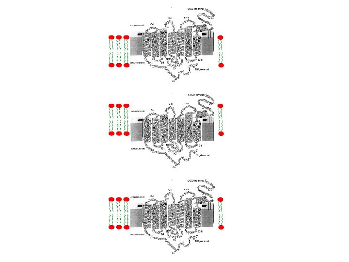

Embedding the protein in membrane 1. Tyrosine residues 2. Palmitoyls connected to Cys 322 and Cys 323

Inducing isomerization 500 nm ~50 kcal/mole

Retinal Charge Distribution QM/MM derived partial atomic charges

In the ground state, the ring of the chromophore blocks the rotation of helix VI

Retinal Isomerization Twist Propagation

In the ground state, the ring of the chromophore blocks the rotation of helix VI

Text book figures not quite right!

Retinal Isomerization Decoupling of Trp 265 and the b-ionone ring

Rhodopsin Isomerization 11 -cis all-trans Bacteriorhodopsin Isomerization: all-trans 13 -cis

Isomerization Barriers and bond selectivity Twisted bonds in Rh Twisted bonds in b. R

Counterion and bond selectivity counterions in Rh counterions in b. R
- Slides: 29