Salmonella Enteric fever Dr Mohit Bhatia Assistant Professor

Salmonella & Enteric fever Dr. Mohit Bhatia Assistant Professor Department of Microbiology AIIMS, Rishikesh

Learning objectives • Describe morphology and antigens of Salmonella spp. • Describe Pathogenesis & Clinical features of Enteric fever • Choose appropriate lab diagnosis and interpret the results • Describe prevention and treatment of Enteric fever

Salmonella • 1885 - Salmon and Smith - discovery of ‘Salmonella’ • Salmonella Typhi, the causative agent of typhoid fever was first observed by Eberth (1880) and Gaffky (1884) Eberth-Gaffky bacillus or Eberthella typhi.

Classification Clinical Classification 1. Typhoidal Salmonella: - Includes serotypes S. Typhi and S. Paratyphi - Restricted to human hosts enteric fever (typhoid/paratyphoid fever) 2. Non-typhoidal Salmonella or NTS: - Colonize intestine of animals - Also infect humans gastroenteritis septicemia &
 Serogroup New Serotype name Vi Ag H Ag* Old Phase")
Antigenic Classification (Kauffmann–White Scheme) Serogroup New Serotype name Vi Ag H Ag* Old Phase 1 Phase 2 2 A S. Paratyphi A 1, 2, 12 - a [1, 5] 4 B S. Paratyphi B 1, 4, [5], 12 - b 1, 2 S. Typhimurium 1, 4, [5], 12 - i 1, 2 S. Agona 1, 4, 12 - f, g, s - S. Heidelberg 1, 4, [5], 12 - r 1, 2 S. Paratyphi C 6, 7 + c 1, 5 S. Choleraesuis 6, 7 - c 1, 5 S. Thompson 6, 7, 14 - k 1, 5 7 C 1
 Serogroup New 8 9 3, 10 Serotype name Vi Ag")
Antigenic Classification (Kauffmann–White Scheme) Serogroup New 8 9 3, 10 Serotype name Vi Ag O Ag* Old C 2 -C 3 S. Muenchen D 1 E 1 H Ag* Phase 1 Phase 2 6, 8 - d 1, 2 S. Newport 6, 8, 20 - e, h 1, 2 S. Typhi 9, 12 + d - S. Enteritidis 1, 9, 12 - g, m [1, 7] S. Gallinarum 1, 9, 12 - - - S. Dublin 1, 9, 12 + g, p - S. Anatum 3, 10, [15], [34] - e, h 1, 6
")
ANTIGENIC STRUCTURE • Three important antigens on their cell wall 1. Somatic antigen (O) 2. Flagellar antigen (H) 3. Surface envelope antigen (Vi)—found in some species - Fimbrial antigens - present in some strains - Nonspecific, widespread among other members of Enterobacteriaceae

Features of O and H Antigen O antigen H antigen Somatic antigen Part of cell wall lipopolysaccharide (LPS) Flagellar antigen Made up of proteins –flagellin confers motility to the bacteria. Heat stable , Alcohol stable Heat labile, Alcohol labile Formaldehyde stable In Widal test- O antigen of S. Typhi is used In Widal test- H antigens of S. Typhi, S. Paratyphi A and B are used O Ag is less immunogenic H Ag is more immunogenic

Features of O and H Antigen O antigen H antigen O antibody appears early, disappears early H antibody appears late, disappears late– indicates recent infection Indicates convalescent stage When O antigen reacts with O antibodyforms compact, granular, chalky clumps Agglutination takes place slowly. Optimum temperature for agglutination is 550 C When H antigen reacts with H antibodyforms large, loose, fluffy clumps. Agglutination takes place rapidly. Optimum temperature for agglutination is 370 C

Features of O and H Antigen O antigen H antigen Serogrouping is based on the O antigen Serogroups are differentiated into serotypes based on H antigen Also called Boivin antigen- extracted from bacterial cell by treatment with trichloracetic acid, first shown by Boivin Flagellar antigens exist in two alternative phases – Phase I and II. Most of them are biphasic except, (monophasic - S. Typhi)

Vi Antigen • Surface polysaccharide envelope or capsular antigen covering the O antigen • Named with belief that Vi antigen is related to virulence • Expressed in only few serotypes - S. Typhi, S. Paratyphi C, S. Dublin and some stains of Citrobacter freundii (Ballerup. Bethesda group) • Renders the bacilli inagglutinable with O antiserum agglutinable after boiling / heating at 100°C for 1 hour, which removes Vi antigen

Vi Antigen • Poorly immunogenic & antibody titres are low Not helpful in diagnosis of cases • Complete absence of Vi antibody poor prognosis • Disappears early in convalescence. If persists carrier state • Phage typing of S. Typhi - using Vi specific bacteriophages • �Vi antigens - used for vaccination
: - Due")
Antigenic Variations Variation in O Antigen • �S-R variation (smooth to rough): - Due to loss of O antigen side chain from LPS, leading to exposure of core polysaccharide portion (or R antigen) of LPS - Smooth colonies - virulent strains carrying the O antigen - � Rough strains - large, rough, and irregular colonies and are avirulent. Autoagglutinable in saline suspensions • �Lysogenic conversion: - S. Anatum + O 15 Ag S. Newington + O 34 Ag S. Minneapolis

Variation in H Antigen v �OH-O variation: - It is associated with the loss of flagella which can be induced by: • Phenol agar: Inhibition of flagella temporarily regained by subculturing on the media without phenol • By mutation: S. Typhi 901 -O strain, employed in the Widal test - Craigie’s tube - U-tube

Variation in H Antigen Phase variation: • Phase 1 antigens - serotype specific Designated as a, b, c, etc • Phase 2 antigens - nonspecific or group antigens. Designated as 1, 2, etc � Serotypes: - Diphasic: Most salmonellae possess antigens of both phases - Monophasic: Some serotypes possess only phase 1 antigens, e. g. S. Typhi, S. Agona, S. Dublin - � Aphasic: No flagellar Ags, S. Gallinarum

Phase Conversion • � A culture in phase 1 can be converted to phase 2 by passing it through a Craigie’s tube containing specific phase 1 antiserum

TYPHOIDAL SALMONELLA • S. Typhi and S. Paratyphi A, B and C which cause enteric fever Pathogenesis • Transmitted by faeco-oral • �Infective dose- Minimum 103– 106 bacilli • �Risk factors that promote transmission: - Stomach acidity - Intestinal integrity
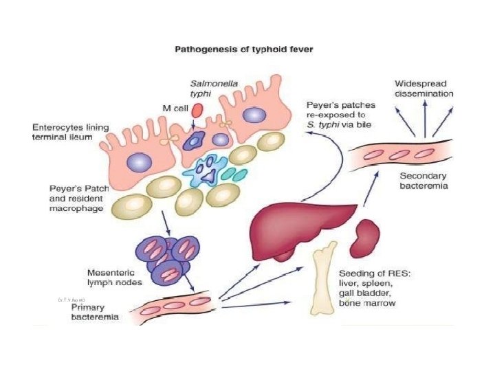

Pathogenesis • Primary bacteraemia • Spread • �Secondary bacteremia occurs from the seeded organs

CLINICAL FEATURES

Epidemiology • Host: Humans the only natural hosts • �Mode of transmission: Ingestion of contaminated water and food • �Prevalence: Worldwide • �Incidence is: - � Highest (>100 cases per 100, 000 population per year) in south central and southeast Asia - Medium (10– 100 cases per 100, 000) in the rest of Asia, Africa, Latin America

Epidemiology �Factors that favour transmission: - � Poor sanitation, � Contaminated water, food and drinks - � Lack of hand washing and toilet access �Typhi vs Paratyphi: • S. Typhi infection is more common than S. Paratyphi A (ratio is 4: 1)

Epidemiology • Carriage: Up to 10% of untreated patients 1. Faecal carriers: Multiply in the gall bladder & excreted in faeces (More common) 2. Urinary carriers: Multiply in kidneys & excreted in urine • - � Duration of shedding: Convalescent carriers 3 weeks to 3 months � Temporary carriers 3 months to 1 year � Chronic carriers for more than 1 year �
: - � Women, infants")
Epidemiology • � Chronic carriers (1– 4% of infected people): - � Women, infants and old age - B �iliary tract abnormalities which leads to increased fecal excretion - A �bnormalities of urinary tract and associated Schistosoma haematobium infection of bladder— leads to increased urinary excretion • Food handlers or cooks who become chronic carriers – dangerous - Mary Mallon (‘Typhoid Mary’) - More than 1300 cases

Laboratory Diagnosis – Sample Collection Duration of illness Specimen used and test done First week Culture of– Blood, Bone marrow aspirate, Duodenal aspirate Second week and Third week Serum For antibody detection by Widal test For antigen detection Stool and urine culture Fourth week Stool and urine culture Carriers Stool and urine culture Serum- for detection of antibodies to Vi antigen Sewage culture- indirect way

Laboratory Diagnosis - Culture and identification Blood Culture • Sensitivity 90% in first week • �Clot culture: Blood is centrifuged serum for Widal test and clot for culture Culture medium: - Conventional: Brain heart infusion broth, Castaneda’s biphasic medium (BHI agar slope and BHI broth) - Automated blood culture systems - BACTEC or Bac. T/ALERT

Media for Blood culture
: Anticoagulant, Counteracts bactericidal action of blood • Incubation:")
Isolation • Sodium polyanethol sulfonate (SPS): Anticoagulant, Counteracts bactericidal action of blood • Incubation: at 37°C for up to 1 week �Repeat subcultures: • Monophasic BHI medium: Periodical subcultures onto blood agar and Mac. Conkey agar • � Biphasic medium BHI is preferred over monophasic - subcultures in the same bottle • � Automated blood culture systems – monitor growth continuously & positive growth flagged subcultures done

Isolation - Enrichment broth - Selenite F broth, tetrathionate broth and gram-negative broth are used • Selective media such as: - Low selective media - Mac. Conkey agar – Colourless colonies
: pale colonies with black")
Isolation • �Highly selective media: - DCA (deoxycholate citrate agar): pale colonies with black center
: red colonies with black center")
Isolation • XLD agar (xylose lysine deoxycholate): red colonies with black center
: colorless with black centers • � Hektoen")
Isolation • SS agar (shigella Salmonella agar): colorless with black centers • � Hektoen enteric agar: Bluegreen colonies with black centers • � Wilson Blair’s brilliant green Bismuth sulfite medium –jet black colored colonies with a metallic sheen due to production of H 2 S
")
Other Specimens • �Bone marrow culture - first week of illness (55– 90% sensitive) when blood culture is negative, especially when patient is on antibiotics • �Duodenal aspirate culture - first week of illness if both blood and bone marrow cultures turn negative • �Combination of blood, bone marrow, and intestinal secretions culture is the best method in the first week, which shows a sensitivity of more than 90%

Identification • Culture Smear and Motility Testing - Gram-negative, non-sporing and non–capsulated bacilli - Motile with peritrichous flagella Biochemical Identification Catalase positive and oxidase negative Indole test—negative Citrate test—positive (except for S. Typhi and S. Paratyphi A, which are citrate negative) - Urease test—negative • -

Biochemical Identification • Biochemical Identification of Salmonellae - TSI shows: Alkaline/acid, Gas present (except for S. Typhi, which is anaerogenic) - Abundant H 2 S present except for: - S. Paratyphi A and S. Choleraesuis: H 2 S not produced �� - S. Typhi: Speck of H 2 S - MR positive and VP negative

Biochemical Identification • Sugar fermentation test: Gluocse, mannitol, arabinose, Maltose, dulcitol and sorbitol are fermented • �Decarboxylation test

Slide Agglutination Test • S. Typhi: Agglutinates with O 9 antisera • �S. Paratyphi A: Agglutinates with O 2 antisera • �S. Paratyphi B: Agglutinates with O 4 antisera Masking effect of Vi antigen: - Tested with Vi antisera - Boiled for 60 minutes at 100 o. C testing with O antisera • Antimicrobial Susceptibility Testing

Demonstration of Serum Antibodies Widal Test • Principle: Tube agglutination test where H and O antibodies against S. Typhi and S. Paratyphi A and B are detected • �Antigens used: Four antigens are used 1. O antigens of S. Typhi (TO) 2. H antigens of S. Typhi (TH) 3. H antigens of S. Paratyphi A (AH) 4. H antigens of S. Paratyphi B (BH)
, with clear")
RESULTS • � O agglutination - compact granular chalky clumps (disk-like pattern), with clear supernatant Fluid � • H agglutination - loose fluffy cotton-woolly clumps, with clear supernatant fluid • � No agglutination - button formation & supernatant fluid remains hazy

Widal Test - Interpretation Widal test result Interpretation Rise of TO and TH antibody Enteric fever due to S. Typhi Rise of TO and AH antibody Enteric fever due to S. Paratyphi A Rise of TO and BH antibody Enteric fever due to S. Paratyphi B Rise of only TO antibody Recent infection -Due to any serotype S. Typhi or S. Paratyphi A or B Rise of only TH antibody ? Convalescent stage/ Anamnestic response Rise of all three TH, AH, BH antibodies- Post TAB vaccination

Widal Test • Titre: The highest dilution of sera, at which agglutination occurs • L� ow titres should be ignored and considered as baseline titres in endemic areas

Widal Test False-positive - Anamnestic response - � Fourfold rise in antibody titre demonstrated by testing paired sera at 1 week interval is more meaningful False-negative O antibodies are serotype nonspecific H antibodies are specific

OTHER TESTS - Typhidot test - IDLTubex test - Ig. M dip stick test and ELISA - Dot blot assay

Other Tests • Demonstration of Salmonella Antigen • Molecular Methods • Other Nonspecific Methods - WBC count: Neutropenia (15– 25%), Leukocytosis in children - Liver function tests moderately deranged - Muscle enzyme levels moderately elevated
 and urine")
Detection of Carriers • �Culture: Stool and bile culture (detects faecal carriers) and urine culture (detects urinary carriers) • �Detection of Vi antibodies: - Tube agglutination test using S. Typhi suspension carrying Vi antigen (Bhatnagar strains) - Titre of 1: 10 is also considered as significant • Isolation of salmonellae from sewage: To trace the carriers in the communities - Sewer–swab technique & Filtration

Treatment of Enteric Fever Drug of choice Alternate drug Empirical treatment This is the treatment given before antimicrobial susceptibility report is available Ceftriaxone Azithromycin Fully Susceptible to all the drugs given for enteric fever. Ciprofloxacin Amoxicillin, Chloramphenicol Cotrimoxazole
 Alternate drug Defined as")
Treatment of Enteric Fever Drug of choice MDR strains (Multidrug-resistant) Alternate drug Defined as resistant to chloramphenicol, ampicillin, and cotrimoxazole-antibiotics used to treat enteric fever long back. Ciprofloxacin Ceftriaxone, Azithromycin NAR strains (Nalidixic Defined as strains resistant to nalidixic acid with acid resistant) reduced susceptibility to ciprofloxacin Ceftriaxone Azithromycin, Ciprofloxacin (higher dose and longer course)

Treatment of Enteric Fever Carriers Drug of choice Alternate drug Ampicillin or Amoxicillin plus probenecid for 6 weeks Cotrimoxazole or Ciprofloxacin

Prophylaxis • Control of Reservoir • Control of Cases - Early diagnosis and prompt effective treatment - Disinfection of stool or urine soiled clothes with 5% cresol, 2% chlorine or by steam sterilizer - Follow up examination of stool and urine culture to detect carriers (twice, at 3– 4 months and at 12 months)

Prophylaxis • Control of Carriers - Early detection of carriers by stool/urine culture or by detection of Vi antibodies - Effective treatment of carriers by: • � Ampicillin or amoxicillin (4– 6 g/day) plus probenecid (2 g/day) for 6 weeks • � Surgery: Cholecystectomy plus ampicillin

Prophylaxis Sanitation measures Protection and purification of drinking water supplies Hand washing and improvement of basic sanitation Promotion of food hygiene & Health education Vaccine Provides short time protection Travelers going to endemic areas, those attending melas and yatras - Household contacts & people at increased risk (school children) • • -

Prophylaxis - Vaccines • Parenteral Vi polysaccharide vaccine: - Purified Vi capsular polysaccharide antigen derived from S. Typhi strain Ty 2 - �Dosage: Single dose given IM or subcutaneously - �Protection for 2 years booster every 2 years - �Age: >2 years of age • �Vi-r. EPA: Vi antigen is conjugated with recombinant Pseudomonas aeruginosa Exotoxin A - Can be given to children less than two years

Prophylaxis - Vaccines • Typhoral (oral live attenuated S. Typhi Ty 2 1 a vaccine): - Stable live attenuated mutant of S. Typhi strain Ty 2 1 a - Gal E mutant - lacks the enzyme UDP-galactose-4 epimerase - Multiplies for some time initiates the immune response but self-destructs after 4– 5 divisions • �Indicated only after 6 years of age

Prophylaxis - Vaccines • �Enteric coated capsules • �Before food on alternate days- 1, 3, 5, 7 with booster every 5 years • Protective immunity starts after 7 days of the last dose and lasts for 4 years

NON-TYPHOIDAL SALMONELLAE • Pathogenic salmonellae other than S. Typhi and S. Paratyphi A, B and C • Majority of infections due to NTS are caused by S. Typhimurium and S. Enteritidis followed by S. Newport, S. Javiana, S. Heidelberg, S. Choleraesuis and S. Dublin

Non-typhoidal Salmonellae vs Typhoidal Salmonellae • Zoonotic • �Transmission of NTS • �Resistance • Seasonality • �Prevalence: - NTS - developed & developing countries - Typhoidal salmonellae - confined to developing countries

Non-typhoidal Salmonellae vs Typhoidal Salmonellae • �Outbreaks of NTS are common in hospitals (typhoidal salmonellae outbreaks are community based) • �Pathogenesis is similar to that of enteric fever except that in NTS gastroenteritis, there is massive neutrophil infiltration into intestinal mucosa (in contrast to enteric fever, where there is mononuclear cells infiltration)

Clinical Manifestations • �Gastroenteritis • Bacteremia - Risk factors for bacteremia: - � NTS serotype: Most common - S. Choleraesuis & S. Dublin - Age: Infants and elderly people are at higher risk - HIV and other conditions with low immunity • �Endovascular infections - endocarditis and arteritis

Metastatic localized infections : • � Intra-abdominal infections • � NTS meningitis (commonly in infants) • � Pulmonary infections - lobar pneumonia and lung abscess • Pyelonephritis & cystitis – Calculi & urinary tract abnormality • � Genital tract infections - ovarian, testicular abscesses, prostatitis and epididymitis • Osteomyelitis: associated with sickle cell disease • � Reactive arthritis (Reiter’s syndrome) - HLA-B 27

Treatment Non-typhoidal Salmonellae • Uncomplicated NTS gastroenteritis - conservative with fluid replacement • - �Antibiotics: Associated with increased rates of carriers and relapse Limited only to invasive/ severe infection Drugs - similar to that of enteric fever • Ciprofloxacin – pre-emptive or severe gastroenteritis • Ceftriaxone - bacteremia and invasive infections

Drug Resistance • More drug resistant than typhoidal salmonellae • �MDR strains - Resistant to ≥ 5 drugs - ampicillin, chloramphenicol, streptomycin, sulfonamides & tetracyclines • Ceftriaxone resistance – Amp. C beta lactamase • Ciprofloxacin resistance - point mutation in DNA gyrase genes

- Slides: 62