
SALMONELLA Dr Manjushree V Mulay Bhalchandra Professor Head
SALMONELLA Dr Manjushree V Mulay (Bhalchandra) Professor & Head Department of Microbiology MGM , Aurangabad
SALMONELLA Learning objectives Morphology , c/c , biochemical reactions , resistance of Salmonella sp Ag structure , Ag variation , Pathogenesis Classification - Kauffmann- White Scheme Lab diagnosis of Enteric fever Prophylaxis Salmonella gastroenteritis
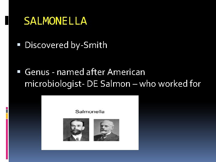
SALMONELLA Discovered by-Smith Genus - named after American microbiologist- DE Salmon – who worked for
SALMONELLA Primarily intestinal parasite of vertebrates which infect man. q colonize intestine of a broad range of animals ( poultry , birds , live stock , reptiles, rodents , domesticated animals and humans)
Diseases caused Causes 1) Enteric fever ( typhoid & paratyphoid ) 2) Food borne Gastroenteritis 3) septicemia - with/ without focal suppuration 4) carrier state
SALMONELLA Source of infection Contaminated food, drink Milk , milk product Eggs of infected fowls Shell fish from contaminated water
GENUS SALMONELLA Comprise >2000 species - all potentially pathogenic ( humans, animals & capable of invading extra intestinal tissues) For practical & clinical purposes , divided into 2 groups 1) Enteric fever group / Typhoid salmonella. Typhoid & paratyphoid bacilli - exclusively or primarily human parasites 2) Food poisoning group/Non Typhoid salmonella. Animal parasites but can also infect human beings , causing gastroenteritis , septicemia or localized infections
DISEASES 1 ) ENTERIC FEVER -- include both Salmonella typhi – typhoid fever Salmonella paratyphi A , B , C – Paratyphoid fever 2) GASTROENTERITIS - ( non typhoidal salmonellae) animal parasites but can also infect man. S. Typhimurium , S. enteritidis , S. newport , S. dublin , S. thompson , S. indiana 3) SEPTICEMIA -- S. cholerae- suis
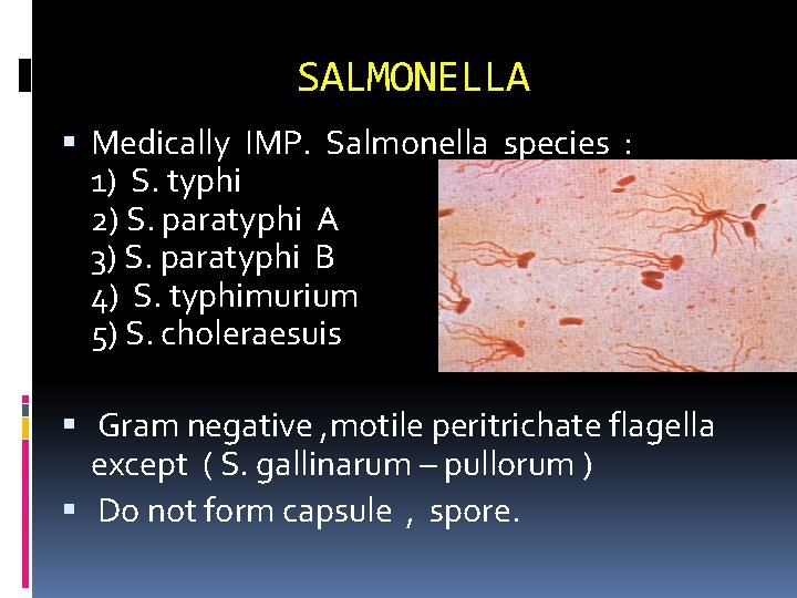
SALMONELLA Medically IMP. Salmonella species : 1) S. typhi 2) S. paratyphi A 3) S. paratyphi B 4) S. typhimurium 5) S. choleraesuis Gram negative , motile peritrichate flagella except ( S. gallinarum – pullorum ) Do not form capsule , spore.
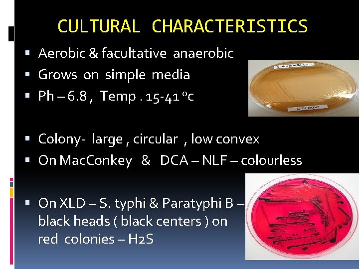
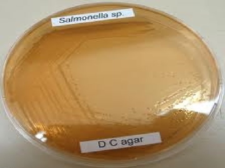
CULTURAL CHARACTERISTICS Aerobic & facultative anaerobic Grows on simple media Ph – 6. 8 , Temp. 15 -41 oc Colony- large , circular , low convex On Mac. Conkey & DCA – NLF – colourless On XLD – S. typhi & Paratyphi B – colonies shows black heads ( black centers ) on red colonies – H 2 S
CULTURAL CHARACTERISTICS On Wilson & Blair bismuth sulphite medium – (selective ) jet black colonies with metallic sheen d/t – H 2 S ( S. paratyphi A – Green coloured – no H 2 S ) Enrichment media – Selenite F , Tetrathionate broth used when isolation is from - faeces Better growth – in presence of Tryptophan
WILSON & BLAIR BISMUTHSULPHITE MEDIUM- Black colored colonies
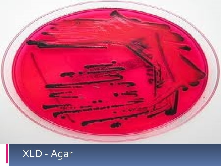
XLD - Agar
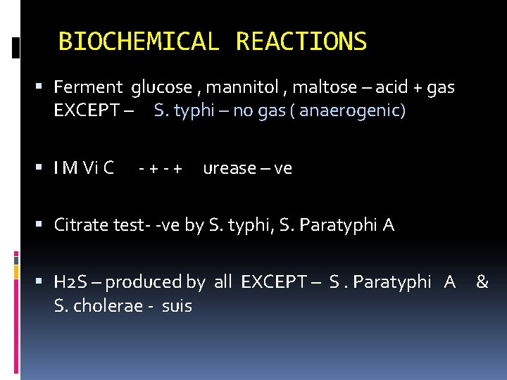
BIOCHEMICAL REACTIONS Ferment glucose , mannitol , maltose – acid + gas EXCEPT – S. typhi – no gas ( anaerogenic) I M Vi C - + urease – ve Citrate test- -ve by S. typhi, S. Paratyphi A H 2 S – produced by all EXCEPT – S. Paratyphi A S. cholerae - suis &
RESISTANCE Killed at 55 o c In 1 hr or 60 o c in 15 min By boiling / chlorination of water & pasteurization of milk Killed within 5 min by mercuric chloride / 5% phenol Survive in polluted water & soil – for weeks in ice – for months
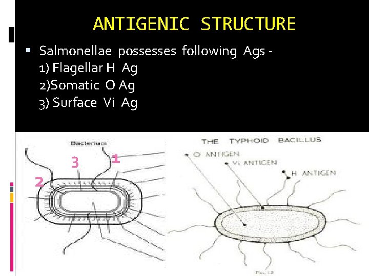
ANTIGENIC STRUCTURE Salmonellae possesses following Ags 1) Flagellar H Ag 2)Somatic O Ag 3) Surface Vi Ag 2 3 1 Ff F Fl a
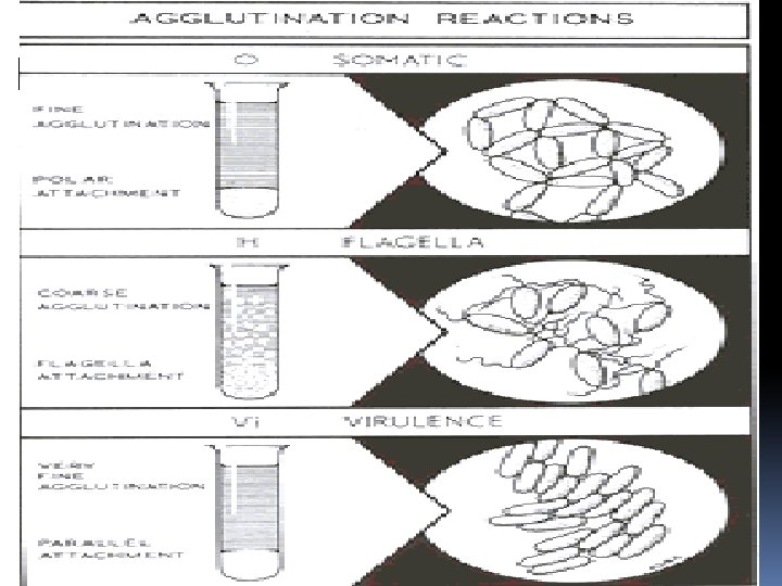
FLAGELLAR H Ag Heat & Alcohol labile protein not destroyed by formaldehyde. Strongly immunogenic. Induces antibody formation in high titres following infection or immunization H Ab Appears late disappears late. Convalescent st Present in 2 forms – phase I & phase II phase I - specific phase- designated as a-z phase II- group phase / non specific – 1, 2
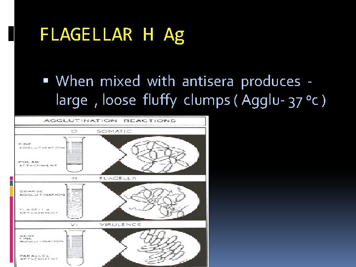
FLAGELLAR H Ag When mixed with antisera produces large , loose fluffy clumps ( Agglu- 37 oc )
SOMATIC O Ag Forms an integral part of cell wall Is a phospholipid-protein-polysaccharide complex. Heat stable / alcohol stable Identical with - endotoxin Less immunogenic than H Ag Titre of O Ab -- after infection or immunization lower than H antibody O Ab appears early , disappears early– recent inf Can be extracted – by t/t – trichloracetic acid – Ist shown by Boivin – called Boivin Ag
SOMATIC O Ag when mixed with antisera - compact , chalky , granular clumps– Agglu- slowly at 50 – 55 o c
Vi ANTIGEN Surface polysaccharide Ag. Envelops O Ag. Many strains S. typhi fail to agglutinate with O antiserum d/t it Become agglutinable after heating 100 O C for 1 hr Destroyed by boiling / heating at 60 oc for 1 hour/ HCL, Na. OH.
Vi antigen Acts as virulence factor by inhibiting phagocytosis by resisting C activation & bacterial lysis Strains possessing Vi Ag – cause clinical disease more consistently than those lacking it
Vi ANTIGEN Poorly immunogenic Low titres of Antibody produced following infection Detection of Vi Ab - not helpful for diagnosis of cases -- not used in Widal test Total absence of Vi Ab in a proven case of typhoid fever --poor prognosis Ab disappears early in convalescence its persistence indicate – carrier state For vaccination
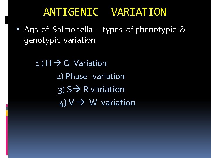
ANTIGENIC VARIATION Ags of Salmonella - types of phenotypic & genotypic variation 1 ) H O Variation 2) Phase variation 3) S R variation 4) V W variation
1) H O Variation Motile non motile Grown on NA + PHENOL ( 1: 800 ) Flagella --inhibited - (phenotypic temporary change ) Flagella reappear -- subcultured on media with out phenol Salmonellae – rarely lose flagella Mutation – form stable non motile mutant of Salmonella Ex- S. typhi 901 -o strain – widely used forpreparation of O – agglutinable bacterial suspensions.
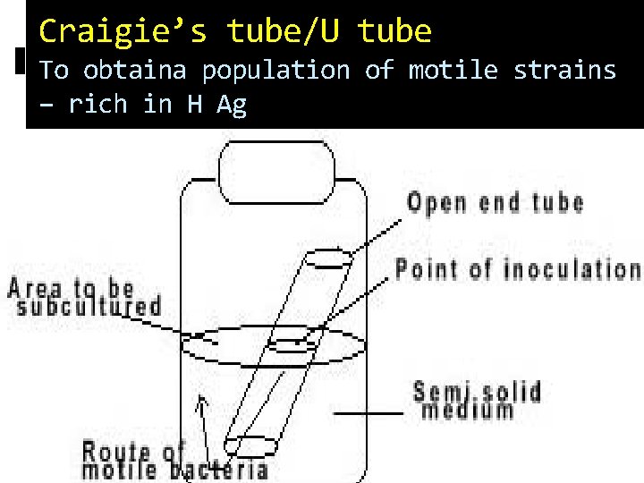
Craigie’s tube/U tube To obtaina population of motile strains – rich in H Ag
Antigenic variation 2) Phase variation – Flagellar Ags phase I – specific for a species / shared by few species –called specific phase – a , b , c , d p hase II – widely shared called – nonspecific/ group – 1, 2, 3 Phase I - --- Phase II by passing them through a Craigie’s tube – specific phase I antiserum
Ag variation 3) S R variation – by mutation Change in colony morphology Loss of O Ag/ of virulence Common in lab strains maintained by- serial subcultures Prevented by- maintaining cultures on Dorset egg media in cold
Ag variation 4) V W variation V form -- Vi Ag - masks O Ag— Agglutinable with Vi antiserum only not with O antiserum W forms -- Complete lose of Vi Ag – Agglutinable by O antisera only & not with Vi antisera Partial loss of vi Ag- Agglutinable with both ( o & vi antisera) – vw form
CLASSIFICATION OF SALMONELLA 1 ) ON THE BASIS OF BIOCHEMICAL REACTIONS – 4 subgenera , subgenus I II IV 2 ) ON THE BASIS OF ANTIGENIC CHARACTERISATION - BY KAUFFMANN – WHITE SCHEME
BY KAUFFMANN – WHITE SCHEME Groups--Based on presence of distinctive O Ag -Salmonellae grouped designated A- Z. Medically Imp Salmonellae belongs to group A-G Gr oup factor--Each group possessing Group factor – This is an O Ag Ex – Factor 2 – belong to group A Within each group , differentiation of serotypes is by identification of phase 1 & phase 2 flagellar Ags
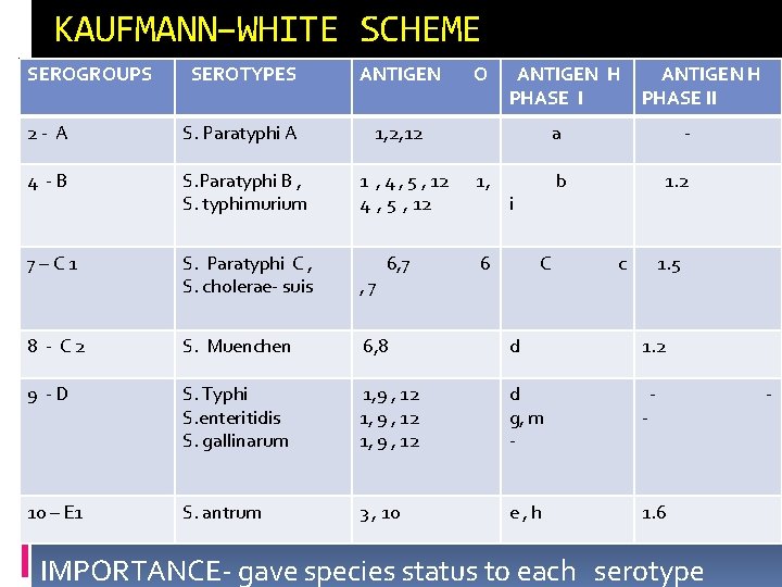
KAUFMANN–WHITE SCHEME SEROGROUPS SEROTYPES ANTIGEN O ANTIGEN H PHASE I 2 - A S. Paratyphi A 1, 2, 12 4 -B S. Paratyphi B , S. typhimurium 1 , 4 , 5 , 12 1, 7–C 1 S. Paratyphi C , S. cholerae- suis , 7 6 8 - C 2 S. Muenchen 6, 8 d 9 -D S. Typhi S. enteritidis S. gallinarum 1, 9 , 12 d g, m - - 10 – E 1 S. antrum 3 , 10 e, h 1. 6 i C ANTIGEN H PHASE II a - b 1. 2 c 1. 5 1. 2 - IMPORTANCE- gave species status to each serotype -
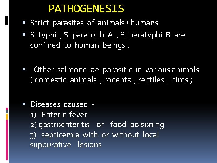
PATHOGENESIS Strict parasites of animals / humans S. typhi , S. paratuphi A , S. paratyphi B are confined to human beings. Other salmonellae parasitic in various animals ( domestic animals , rodents , reptiles , birds ) Diseases caused 1) Enteric fever 2) gastroenteritis or food poisoning 3) septicemia with or without local suppurative lesions
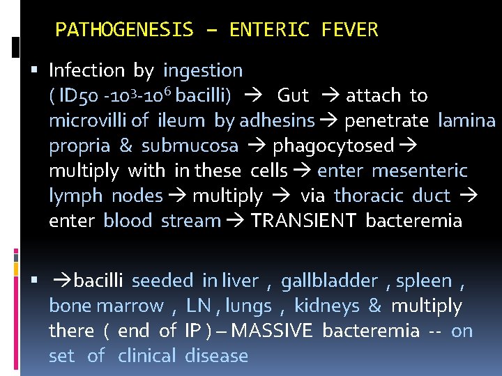
PATHOGENESIS – ENTERIC FEVER Infection by ingestion ( ID 50 -103 -106 bacilli) Gut attach to microvilli of ileum by adhesins penetrate lamina propria & submucosa phagocytosed multiply with in these cells enter mesenteric lymph nodes multiply via thoracic duct enter blood stream TRANSIENT bacteremia bacilli seeded in liver , gallbladder , spleen , bone marrow , LN , lungs , kidneys & multiply there ( end of IP ) – MASSIVE bacteremia -- on set of clinical disease
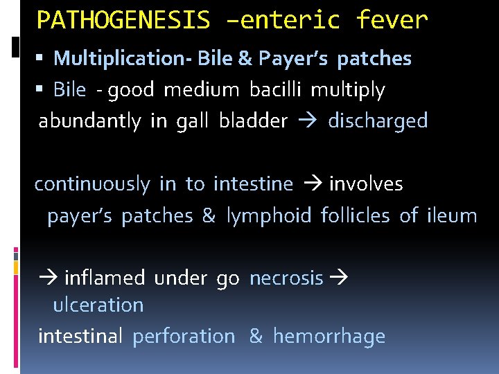
PATHOGENESIS –enteric fever Multiplication- Bile & Payer’s patches Bile - good medium bacilli multiply abundantly in gall bladder discharged continuously in to intestine involves payer’s patches & lymphoid follicles of ileum inflamed under go necrosis ulceration intestinal perforation & hemorrhage
Clinical course I. P. – 7 - 14 days Step ladder pyrexia with bradycardia & toxemia , hepatosplenomegaly with Rose spots - skin during 2 nd & 3 rd week. S. Paratyphi A & B – paratyphoid fever resembles typhoid but milder. S. paratyph. I - C paratyphoid fever with septicemia & suppurative complications S. paratyphi A – prevalent in India , B – rare , C- not seen SOURCE – patient / carrier
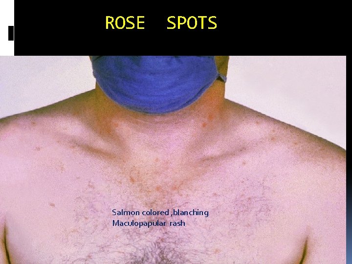
ROSE SPOTS Salmon colored , blanching Maculopapular rash
CARRIERS- ENTERIC FEVER 3 Types of carriers 1) Convalescent - clinical cure – shed bacilli in feces for 3 weeks to 3 months 2) Temporary – for > 3 mts but < a year 3) Chronic- for over a year (2 - 4 % ) pts become chr carriers Bacilli persist in gall bladder / kidney - eliminated faeces or urine Urinary carriage less frequent- some urinary lesions - calculi/ schistosomiasis
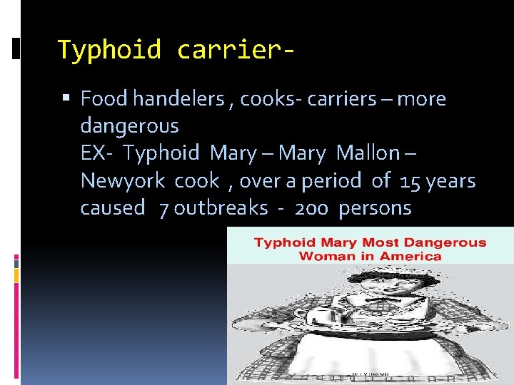
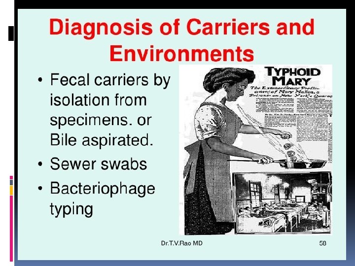
Typhoid carrier Food handelers , cooks- carriers – more dangerous EX- Typhoid Mary – Mary Mallon – Newyork cook , over a period of 15 years caused 7 outbreaks - 200 persons
PATHOGENESIS - Gastroenteritis ( food poisoning ) Zoonotic disease , source – animal products Infection acquired by -- consumption of meat , milk & milk products , egg products , salad , uncooked vegetables , food contaminated with droppings of rats , lizards q Caused by any salmonella except S. typhimurium most common species q I. P. - 24 HOURS q Diarrhoea , vomiting , pain , fever q Lab diagnosis- isolating salmonellae feces , food
PATHOGENESIS – Septicemia S. choleraesuis imp along with focal suppurative lesions - osteomylitis , deep abscesses , arthritis , endocarditis , pneumonia & meningitis Case fatality - 25 % Lab diagnosis- isolation of salmonellae – blood & pus Septicemic salmonella - treated with chloramphenicol
LAB DIAGNOSIS OF ENTERIC FEVER 1) Isolation of bacilli 2) Demonstration of antibodies 3) Demonstration of antigen 4) Other laboratory tests
SPECIMENS COLLECTION Blood , Blood clot , B M, faeces Bone marrow ( valuable - is positive even when blood cultures are negative ) Feces- As valuable as blood culture particularly valuable if pt is on Abs Other specimens urine ( less frequently +ve than culture of blood & faeces ) Rose spots , pus , CSF, sputum Aspirated duodenal fluid ( bile ) --for carriers Selection of relevant sample - duration of illness
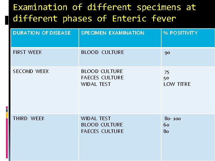
Examination of different specimens at different phases of Enteric fever DURATION OF DISEASE SPECIMEN EXAMINATION % POSITIVITY FIRST WEEK BLOOD CULTURE 90 SECOND WEEK BLOOD CULTURE FAECES CULTURE WIDAL TEST 75 50 LOW TITRE THIRD WEEK WIDAL TEST BLOOD CULTURE FAECES CULTURE 80 - 100 60 80
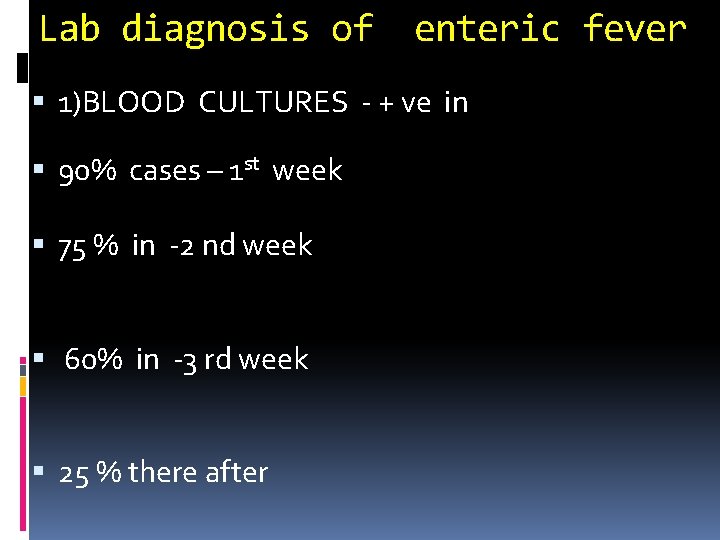
Lab diagnosis of enteric fever 1)BLOOD CULTURES - + ve in 90% cases – 1 st week 75 % in -2 nd week 60% in -3 rd week 25 % there after
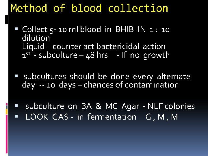
Method of blood collection Collect 5 - 10 ml blood in BHIB IN 1 : 10 dilution Liquid – counter act bactericidal action 1 st - subculture – 48 hrs - If no growth subcultures should be done every alternate day -- 10 days – chances of contamination subculture on BA & MC Agar - NLF colonies LOOK GAS - in fermentation G , M
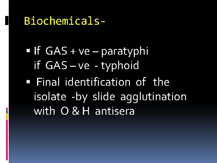
Biochemicals If GAS + ve – paratyphi if GAS – ve - typhoid Final identification of the isolate -by slide agglutination with O & H antisera
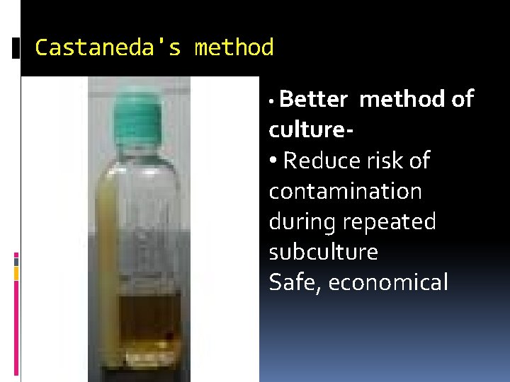
Castaneda's method • Better method of culture • Reduce risk of contamination during repeated subculture Safe, economical
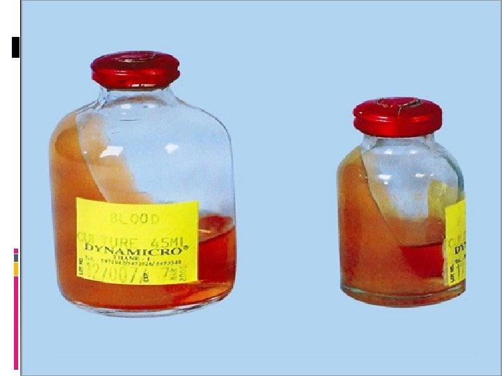
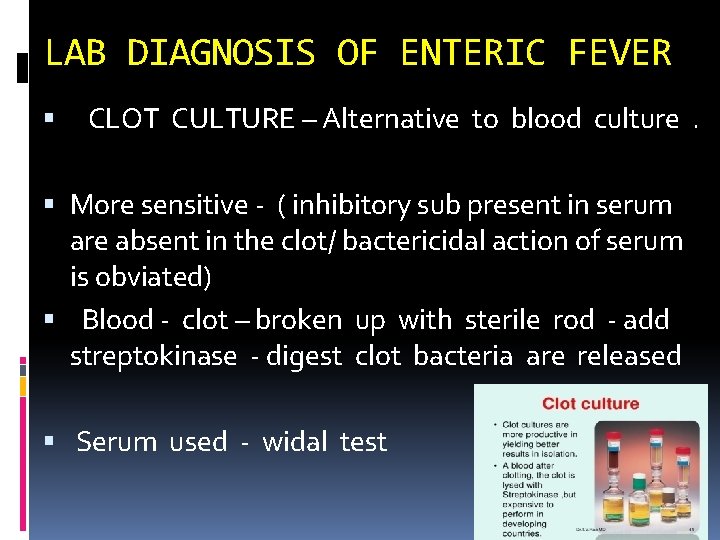
LAB DIAGNOSIS OF ENTERIC FEVER CLOT CULTURE – Alternative to blood culture. More sensitive - ( inhibitory sub present in serum are absent in the clot/ bactericidal action of serum is obviated) Blood - clot – broken up with sterile rod - add streptokinase - digest clot bacteria are released Serum used - widal test
Lab diagnosis of enteric fever Bone marrow – valuable as it positive in most cases even when cultures – ve FAECES CULTURE - shedding through out the disease -- useful in patient / carriers use selective media like – DCA , Wilson Blair media
Lab diagnosis of enteric fever Urine culture- irregular shedding Unusual strain – National salmonella reference centre – CRI – Kasauli For salmonellae of animal origin – Indian veterinary research institute , Izatnagar
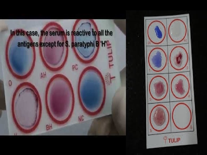
2) Ab detection- WIDAL TEST - Agglutination test Detection & measurement of H & O Abs - for Typhoid & paratyphoid bacilli in patients serum Time---- at end of 1 st wk & rise in Ab titre in paired sera - significant FOR H AGGLUTINATION - Dreyers narrow Tube with conical bottom For O AGGLUTINATION - Felix tube round bottomed tube short
ANTIGENS USED IN WIDALTEST H & O Antigens of S. typhi --TH & TO H Antigens of S. Paratyphi A - AH S. paratyphi B- BH & Patyphoid O antigens not used -- as they cross react with S. typhi O antigens d/t their sharing of factor 12
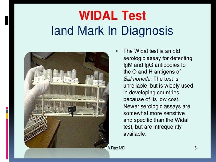
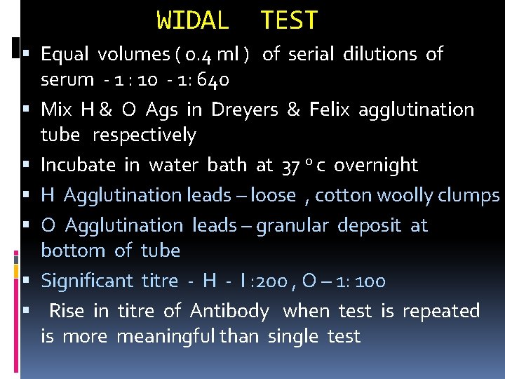
WIDAL TEST Equal volumes ( 0. 4 ml ) of serial dilutions of serum - 1 : 10 - 1: 640 Mix H & O Ags in Dreyers & Felix agglutination tube respectively Incubate in water bath at 37 o c overnight H Agglutination leads – loose , cotton woolly clumps O Agglutination leads – granular deposit at bottom of tube Significant titre - H - I : 200 , O – 1: 100 Rise in titre of Antibody when test is repeated is more meaningful than single test
Preparation of Antigens OR Agglutinable suspension H Ag- 0. 1 % formalin to 24 hr broth culture O Ag- By culturing on Phenol agar-
Things taking in to account 1) Agglu titre – depend on stage of the disease 2) Rise in titre of Ab – more meaningful – at least two tests – performed 3) Prior infection / immunization --Anamnestic response- during unrelated inf 4) Bacterial suspensions- used as Ag- free from fimbria- False + ve 5) Cases treated early – chloramphenicol- show poor Ab response
3)Demonstration of ANTIGEN Present in the blood in early phase of the disease & also in urine Demonstrated by sensitised Staphylococcal coagglutination test ( Cowan I strain ) CIEP ELISA
4) OTHER LABORATORY TESTS 1) TLC - leucopenia with lymphocytosis 2) Diazo test – urine + diazo reagent + 30% ammonium hydroxide -- shake -- red or pink froth ---- + ve ( between 5 th and 14 th day of fever)
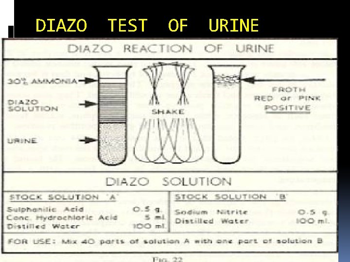
DIAZO TEST OF URINE
DETECTION OF CARRIERS Isolation of bacilli from - bile , faeces Tracing of carriers SEWER SWAB technique – gauze pads left in sewers & drains are cultured By tracing +ve swab – house harbouring a carrier Filtration of sewage through milipore memb , culturing memb selective medium
PROPHYLAXIS Adequate sewage disposal Purification of drinking water supplies Proper hand hygiene Promotion of food hygiene Health education Vaccination Carriers –Detection /T/t, Cholecystectomy
Vaccination 1) TAB vaccine- heat killed whole cell v given SC- 2 doses 2) Live oral (Ty 21 a ) vaccine – typhoral A virulent mutant of S. typhi - Ty 21 a Lacking enz- UDP- galactose – 4 – epimerase Given orally ----enteric coated capsule on days 1, 3, 5 & 7 O n ingestion – initiates infection – self destructs – 4/ 5 cell divisions- no illness 3) Purified Vi polysaccharide vaccine – Typhim – vi Given IM in single dose
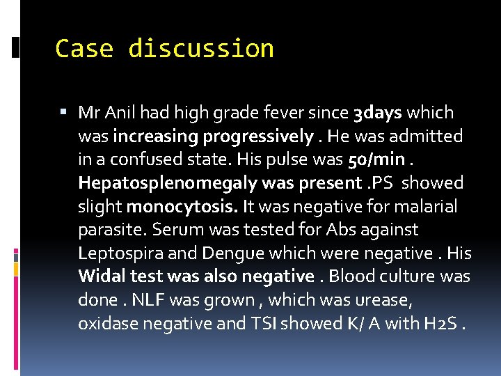
Case discussion Mr Anil had high grade fever since 3 days which was increasing progressively. He was admitted in a confused state. His pulse was 50/min. Hepatosplenomegaly was present. PS showed slight monocytosis. It was negative for malarial parasite. Serum was tested for Abs against Leptospira and Dengue which were negative. His Widal test was also negative. Blood culture was done. NLF was grown , which was urease, oxidase negative and TSI showed K/ A with H 2 S.
What is clinical diagnosis? How diagnosis is justified?

THANK YOU
- Slides: 71