Sahlis Method of Hemoglobin Determination Hemoglobin Determination Apparatus

Sahli’s Method of Hemoglobin Determination

Hemoglobin Determination • Apparatus: • Sahli’s tube which is having red and yellow scales on two sides • Red scale is percentage scale and yellow scale is gram percentage or g/100 ml scale. • Heamometer which is having two standards Sahli’s pipette • Error percentage is 3%.

Procedure: In sahli’s tube, take N/10 hydrochloric acid: HCL which is (1/10 th of the original HCL) up to 10 th level of scale. In sahli’s pipette, take 0. 02 ml (20 microleter) blood. Add blood from pipette into tube. HCL will cause the lysis of the blood cells and hemoglobin is released. Hemoglobin after combining with HCL, forms acid hematin which is of tan color (i. e. to change all forms of hemoglobin to acid hematin to standardize the color index) Put tube in the hemometer and continuously add drops of distilled water and shake with the stirrer until color matches. Then, take the reading. Drabkin’s method cyanide The method of choice for hemoglobin determination is the cyanmethemoglobin method (This is a type of colorimetric method). The principle of this method is that when blood is mixed with a solution containing potassium ferricyanide and potassium cyanide, the potassium ferricyanide oxidizes iron to form methemoglobin. The potassium cyanide then combines with methemoglobin to form cyanmethemoglobin, which is a stable color pigment read photometrically at a wave length of 540 nm. Three advantages of the cyanmethemoglobin method are: 1. measures all forms of hemoglobin except sulfhemoglobin 2. can be easily standardized 3. cyanmethemoglobin reagent (also called Drabkin's solution) is very stable

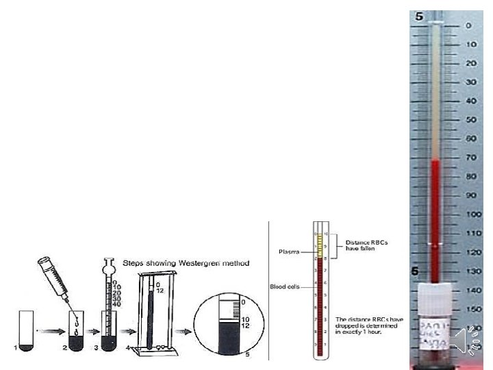
Practical part: Procedure: • • EDTA or any suitable anticoagulant is prepared in a test tube. Obtain 2 ml of blood from a superficial vein. Gently transfer the blood into the test tube; rotate carefully allowing a good mix of the blood with anticoagulant. Do not shake, as this would cause bubbles to form in the blood anticoagulant mixture. Put the Westergren pipette in the tube that contains anti-coagulated blood, the blood is shucked carefully in the Westergren pipette up to zero mark. Fix the Westergren pipette vertically on a sedimentation pipette rack avoiding leakage of blood. Allow the pipette to remain undisturbed for exactly 1 hr. At the end of that time, the upper portion of the blood column will appear clear because erythrocytes will have settled from that region. Read from the graduated scale on the pipette the number of millimeters that the erythrocytes have settled below the zero mark. Record the erythrocyte sedimentation rate in millimeters per hour (mm/hr). Caution: No air bubble should be trapped in the blood placed in Westergren pipette. The Westergren pipette should be absolutely vertical. Slight inclination gives false reading of accelerated ESR and also disturbs the sedimentation forces of cells. Blood should at no stage show any signs of hemolysis or coagulation. Blood should be used within 2 hours of collection.
- Slides: 8