RTCA 218 Fluoroscopy Bushong Chapter 25 Fluoroscopy Abstract

RTCA 218 Fluoroscopy Bushong, Chapter 25

Fluoroscopy Abstract: This section deals with fluoroscopy, its purpose and use. Emphasis will be placed on parts of the fluoroscope, image intensification, fluoroscopic-image monitoring and spot filming. Objectives: At the end of this section, the student will be able to: n discuss the history and development of fluoroscopy. n list and discuss the components of an image intensification tube. n define flux gain and brightness gain. n discuss the fluoroscopic-image monitoring system. n list and discuss the components of the television monitoring system. n discuss spot filming.

Fluoroscopy History of Fluoroscopy n invented by _____________ in 1896 n early fluoroscopy procedures permitted only one person to view the fluoroscopy screen at a time n radiologist had to adapt eyes to the _____ before beginning a fluoroscopy procedure

“Hands Free” Fluoroscope

Fluoroscopy image intensifier invented by William Chamberlain in the 1950 s – increase the ________ of the image n allowed ______ viewers of a procedure n no longer needed to adapt eyes at the beginning of the procedure n

Fluoroscopy and Visual Physiology n Illumination 1. level of brightness 2. measured in lamberts or milli-lamberts 3. radiographs viewed at illumination levels of ___ to ____ m. L

Fluoroscopy n Human Vision 1. rods and cones responsible for vision 2. rods are sensitive to _____ light 3. cones used primarily for ______ vision – rods used at night 4. visual acuity – ability to perceive fine detail – cones are better than rods at visual acuity

Fluoroscopy 5. contrast perception – ability to distinguish between levels in _______ – cones better than rods at contrast perception 6. ____ vision much more preferable than rod vision in fluoroscopy

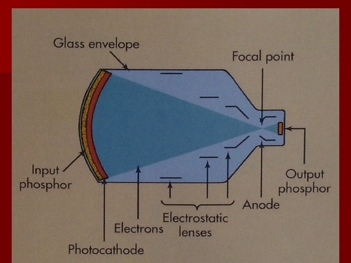
Fluoroscopy Image Intensifier n Image Intensification Tube n Approximately _____ cm long

Fluoroscopy 1. Input Phosphor n ___________ n receives remnant radiation n converts ______ xray energy to visible light

Fluoroscopy 2. Photocathode n bonded directly to the input phosphor n emits _______ when stimulated by light – photoemission n process similar to ______ emission

Fluoroscopy number of electrons emitted is ______ proportional to the intensity of the light energy falling on it n number of light photons is directly proportional to the intensity of the remnant radiation beam n Therefore, the number of electrons emitted by the ________ is directly proportional to the intensity of the remnant radiation beam n

Fluoroscopy 3. Anode n circular plate with a hole in the middle to allow the electrons to pass through to the output phosphor n in order for the image to be precise, the electron path must be _______ n electrostatic focusing ______ maintain a precise electron path from the photocathode to the output phosphor – known as electron optics

Fluoroscopy between the photocathode and the anode on the other side of the Image Intensifier is an electrical potential of about ____ k. V n ______ the electrons to the anode n electrons arrive at the output phosphor with high kinetic energy and contain the image from the input phosphor in minified form n

Fluoroscopy 4. Output Phosphor n made of _________ sulfide n convert electrons to ______ n each photoelectron that strikes the output phosphor results in 50 to 75 times as many light photons that originally created the Photoelectron

Fluoroscopy n Flux Gain 1. ratio of the number of ____ photons at the output phosphor to the number of _____ at the input phosphor 2. # of output light photons / # of input xray photons

Fluoroscopy 1000 x-rays comprise the remnant x-ray beam. If 300, 000 light photons are produced at the output phosphor, what is the flux gain for this image intensifier? Flux gain = # of output light photons / # of input x-ray photons 300, 000 / 1000 = 300

Fluoroscopy n Brightness Gain 1. ability of the image intensifier tube to increase the ________ level of the image 2. equals minification gain flux gain

Fluoroscopy 3. minification gain = the ratio of the ________ of the diameter of the input phosphor to the ________ of the diameter of the output phosphor Minification gain = I. P 2 / O. P 2 4. output phosphor sizes are typically 2. 5 or 5 cm 5. input phosphor sizes vary from 10 to 35 cm 6. brightness gain of most image intensifiers is between _____ and ______

Fluoroscopy If the flux gain of an image intensifier is 300 and the input phosphor has a diameter of 25 cm and the output phosphor has a diameter of 5 cm, what is the brightness gain for this image intensifier? Brightness gain = flux gain x minification gain Minification gain = I. P 2 / O. P. 2 252 / 52 = 625 / 25 = 25

Fluoroscopy If the flux gain of an image intensifier is 300 and the input phosphor has a diameter of 25 cm and the output phosphor has a diameter of 5 cm, what is the brightness gain for this image intensifier? Brightness gain = flux gain x minification gain Flux gain = 300 Minification gain = 25 Brightness gain = 300 x 25 = 7500

Fluoroscopy n Conversion Factor 1. defined as the increased ________ of an image intensifier compared with the standard conventional _______ screen 2. equals the ratio of the output phosphor illumination to the input exposure rate 3. image intensification tubes have conversion factors ranging from 50 to 300 or __/_____ of brightness gain

Fluoroscopy The remnant radiation at the input phosphor has an exposure rate of 100 m. R/sec. If the illumination of the fluoroscopic image is 10, 000, what is the conversion factor for this image intensifier? Conversion factor = output illumination / input exposure rate 10, 000 / 100 = 100

Fluoroscopy Multi-field Image Intensification 1. allow for a variable size of the _____ phosphor 2. examples are 25/17 or 25/17/12 or 23/15/10 3. by decreasing the diameter of the input phosphor, the electron focal point is moved ______ from the output phosphor n
 and magnifying the image")
Fluoroscopy 4. results in reducing the field of view (FOV) and magnifying the image 5. magnification gain is a simple ratio between the ____ diameter to the _____ diameter 6 using the magnified mode results in fewer photoelectrons striking the output phosphor yielding a dimmer image

Fluoroscopy 7. x-ray tube m. A must be increased to maintain the same brightness level – _____________ (ABC) 8. results in better image quality and contrast resolution 9. increase in _____ equals the square of the ratio between the area of input phosphor used

Fluoroscopy If the fluoroscopic image is magnified by moving from a 25 cm input phosphor to a 12 cm input phosphor, how much is the image magnified? Magnification Gain = I. P 1 / I. P. 2 25 / 12 = 2. 08 times increase in image size

Fluoroscopy If the fluoroscopic image is magnified by moving from a 25 cm input phosphor to a 12 cm input phosphor, what is the increase in patient dose? Dose increase = I. P 12 / I. P. 22 252 / 122 = 625 / 144 = 4. 34 times increase in radiation dose

Fluoroscopy Fluoroscopic Image Monitoring n Television Monitoring 1. output phosphor coupled directly to a television camera tube – _____ or _______ 2. converts the light from output phosphor to an electrical signal

Fluoroscopy 3. signal sent to the _______ monitor and reconstructed into an image on the television screen

Fluoroscopy 4. advantages of this system include: – control of ______ and _________ levels – several can view screen at the same time – ________ monitors – storage of procedures on a VCR or DVD

Fluoroscopy Spot Filming 1. spot film cassette placed between the _____ and image intensifier 2. during fluoroscopy the cassette is placed in a lead lined shroud to prevent exposure n

Fluoroscopy 3. when a spot film in desired, the cassette is moved into position, and the x-ray unit changes from ____ m. A fluoroscopic mode to _____ m. A radiographic mode 4. may make multiple exposures per film 5. results in increased patient dose but provide a hard copy image of a possible pathological condition

Fluoroscopy 6. Photo-spot Camera – Similar to a movie camera – Only exposes one frame when activated – No significant interruption of fluoroscopy – Requires ______ patient dose than spot filming – Uses film sizes of 70 to 105 mm – ________ film format results in better image quality but increases patient dose

Fluoroscopy 7. Digital Fluoroscopy
- Slides: 36