ROOT CANAL ANATOMY AND ACCESS CAVITY PREPERATION Dr


 The AC generally")


- Slides: 23
ROOT CANAL ANATOMY AND ACCESS CAVITY PREPERATION Dr. Rakesh kumar yadav Associate professor
ROOT CANAL ANATOMY AND ACCESS CAVITY PREPERATION The hard tissue surrounding the dental pulp can take a variety of configurations and shapes thorough knowledge of tooth morphology Careful interpretation of angled radiographs Adequate access and interior exploration. That are prerequisites for root canal treatment ….
ROOT CANAL ANATOMY
the apical constriction & cementodentinal junction apical foramina(AC, …CDJ, …AF……………. ) The AC generally is considered the part of the root canal with the smallest diameter; it also is the reference point clinicians use most often as the apical termination for shaping, cleaning, and obturation. The CDJ is the point in the canal where cementum meets dentin; it is the point where pulp tissue ends and periodontal tissue begins. The AF is the “circumference or rounded edge, like a funnel or crater, that differentiates the termination of the cemental canal from the exterior surface of the root. .
Types of canals: Vertucci's canal configurations
objectives of access cavity preparation To remove all caries, � To conserve sound tooth structure � To completely unroof the pulp chamber � To remove all coronal pulp tissue. � To locate all root canal orifices � To achieve straight- or direct-line access � To establish restorative margins to minimize marginal leakage of the restored tooth. �
An isthmus is a narrow, ribbon-shaped communication between two root canals that contains pulp or pulpally derived tissue. Type I is an incomplete isthmus; it is a faint communication between two canals. Type II is characterized by two canals with a definite connection between them (complete isthmus). Type III is a very short, complete isthmus between two canals. Type IV is a complete or incomplete isthmus between three or more canals. Type V is marked by two or three canal openings without visible connections
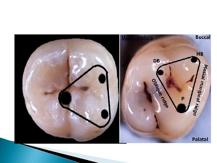
Krasner and Rankow Law of access opening � Law of the CEJ: The distance from the external surface of the clinical crown to the wall of the pulp chamber is the same throughout the circumference of the tooth at the level of the CEJ, making the CEJ is the most consistent repeatable landmark for locating the position of the pulp chamber. � First law of symmetry: Except for the maxillary molars, canal orifices are equidistant from a line drawn in a mesiodistal direction through the center of the pulp chamber floor. � Second law of symmetry: Except for the maxillary molars, canal orifices lie on a line perpendicular to a line drawn in a mesiodistal direction across the center of the pulp chamber floor.
� Law of color change: The pulp chamber floor is always darker in color than the walls. � First law of orifice location: The orifices of the root canals are always located at the junction of the walls and the floor. � Second law of orifice location: The orifices of the root canals are always located at the angles in the floor–wall junction. � Third law of orifice location: The orifices of the root canals are always located at the terminus of the roots’ developmental fusion lines.
Armamentaria of Access Cavity. Preparation � Magnification � Hand � Burs pieces � Endodontic and illumination explorer (DG-16, DE-17) � Endodontic spoon � #17 operative explorer � Ultrasonic unit and tips
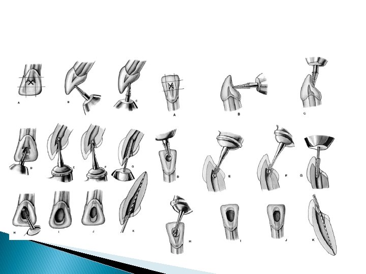
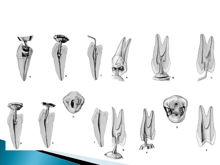
Access Cavity. Preparation Anterior teeth The access cavity is initiated in the middle of the palatal/lingual side of the tooth. Initial preparation should be at 90° to the palatal/lingual aspect of the tooth. Once dentine has been reached the angulation of the bur is changed to follow a long axis of the tooth using a slow handpiece.
Premolar teeth The access cavity is initiated in the middle of the palatal side of the tooth. Initial preparation should be at 90° to the palatal aspect of the tooth. Once dentine has been reached the angulation of the bur is changed to follow a long axis of the tooth using a slow handpiece.
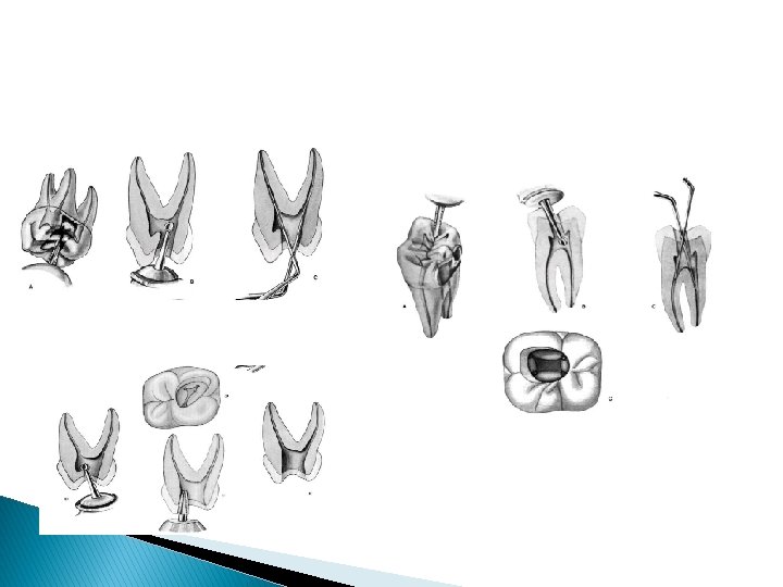
Molars Initial Preparation The cavity is then is done in the mesial pit. extended in the mesial half of the tooth to include all canals. The mesial marginal ridge must not be damaged in upper molars as the cavity should lay mesially to it. However lower molar teeth, have a distal canal, which is located just past the middle of the tooth.
After the roof of the pulp chamber has been penetrated and the access cavity prepared, the entrances to the pulp canals must be probed. � A hooked explorer can be used to determine if enough dentin has been removed � The canal entrances are found by feeling with a thin, stiff explorer. If the explorer sticks in a spot, a size 15 Hedstr 6 m file is used to verify that the spot is indeed the entrance to a root canal and not a perforation. Only �
MCQ 1 Access openings is provide to� a. Facilitate canal medication � b. Provide good access for irrigation � c. Aid in locating canal orifices � d. Provide straight line access to the apex
MCQ 2 � Which instrument is helpful in making access opening- � a. Gates glidden drill � b. K-file � c. Inverted bur � d. Round bur
MCQ 3 Access cavity shape in mandibular 1 st molar with 4 canals a. Trapezoidal b. Round c. Oval d. Triangular
MCQ 4 The fourth root canal if present in a maxillary 1 st molar is usually present in � a. � Mesiolingual root b. Mesiobuccal � c. Palatal root � d. Distal root
MCQ 5 Bifurcations and trifurcations are most commonly observed ina. Maxillary 1 st premolar b. Maxillary 2 nd premolar c. Mandibular 1 st premolar d. Mandibular 2 nd premolar