Role of Vitamin D receptor VDR in HIV
 in HIV induced tubular injury Mohammad Husain Department")

2 D 3 through VDR Functions as a pluripotent")

 A. Schematic of the VDR protein comprised of a DNA")


 is a combined glomerular and tubular injury characterized by:")
 VDR/DAPI Control Tg 26 mouse model")


 VDR Actin C EV HIV C VDA 50")


")


- Slides: 29
Role of Vitamin D receptor (VDR) in HIV induced tubular injury Mohammad Husain Department of Biotechnology, Jamia Millia Islamia New Delhi 07/10/2015
INTRODUCTION • Vitamin D is technically not a Vitamin; rather, a prohormone produced photo chemically in the skin from 7 - dehydrocholestrol. • Expression and nuclear activation of Vitamin D Receptor (VDR) is necessary for the effects of VIT D. • VDR tissue distribution and cellular localization has been reported in almost every organ. • VDR regulates the expression of approximately >500 of the 20488 genes in human genome.
Biological actions of 1α 25 (OH)2 D 3 through VDR Functions as a pluripotent hormone in different arenas are: • Adaptive Immune system • Innate Immune system • Insulin secretion by pancreatic beta cells • Multifactorial heart functioning and Blood pressure regulation • Reduction of Proteinuria • Control of systemic Inflammation. .
Vitamin D synthesis
Vitamin D receptor (VDR) A. Schematic of the VDR protein comprised of a DNA binding domain, a large ligand binding domain and a hinge region that links the two functional domain of the protein together. N, amino terminal end; C, carboxy terminal end; AF 2, activation function 2. Amino acid numbers are shown. B. Crystal structure of the VDR ligand binding domain comprised of 12 α-helices (H 1–H 12). The N-terminal and C-terminal portions of the molecule are shown.
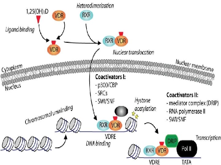
VDR - Member of a family of Nuclear steroid hormone receptors • • Intracellular organization of the VDR. In the absence of 1, 25(OH)2 D, VDR shuttles between cytoplasm & nucleus. The VDR can dimerize with RXR, but the formed heterodimer is not stable and has a low affinity for the VDRE. This results in basal regulation of target gene transcription • • Upon binding 1, 25(OH)2 D, VDR is activated and translocated to the nucleus The VDR/RXR heterodimer is stabilized, giving it a high affinity for the VDRE, which results in an increased transactivation or transrepression of the genes
HIV infection and VITAMIN D deficiency • Observational studies have revealed very high low 25(OH)D levels in HIV-infected patients Could be due to : – Traditional risk factors: Lack of UVB exposure, darker skin pigmentation. – Antiretroviral therapy (ART)–As VIT D and PI( protease inhibitors) and NNRTI ( Non Nucleoside reverse transcriptase inhibitor) are metabolized by Cytochrome P 450 system. • The mechanism of HIV on VDR remains to be investigated?
VDR polymorphism and HIV infection susceptibility and progression • The risk of HIV disease infection and progression has been examined in relation to the presence of a number of polymorphism in the VDR gene. • The study by Nieto et al. and Barber et al. have examined the prevalence of mutations in a region of the VDR gene corresponding to Bsm I restriction enzyme and Fok I in relation to HIV status. • The heterozygous genotype Bb and Ff was significantly associated to progression of AIDS and CD 4 count drop to under 200/mm 3
HIVAN • HIV-associated nephropathy (HIVAN) is a combined glomerular and tubular injury characterized by: – Collapsing glomerulopathy (CG) with collapse of glomerular capillary structures and a striking hyperplasia of podocytes. – Microcystic dilatation of renal tubules. – Interstitial inflammation and fibrosis consequent to the glomerular and tubular injuries. HIVAN GENETIC (Black ancestry and APOL 1 gene) ENVIRONMENTAL HIV Infection HOST FACTORS RAAS
VDR expression in murine model of HIVAN(Tg 26) VDR/DAPI Control Tg 26 mouse model was developed using gag/pol deleted clone of HIV-1 to render the virus non-replicative
VDR localization in Kidney VDR in mouse proximal renal Tubule, Distal tubule, Glomerulus.
HIV induces apoptosis • HIV has been reported to induce apoptotic phenotype in tubular cells. • Oxidative stress is involved in the induction of HIV-induced kidney cell apoptosis. It can contribute to HIV disease pathogenesis by – Enhancing viral replication – Increasing the inflammatory immune response – Accelerating the loss of immune function. • HIV-1 -induced overproduction of ROS, has been proposed to account for the activation of apoptosis in several cell types including tubular cells. • Role of VDR in HIV induced ROS generation in tubular cells?
Morphological effect of HIV-1 expression on tubular cells EV HIV
HIV down regulates VDR in tubular cells VDR Actin C EV HIV m. RNA expression of VDR Actin 0 hr 4 hr 6 hr 12 hr 18 hr 24 hr TIME course of VDR down regulation with HIV Protein expression
Immunofluoroscence study of VDR expression in tubular cells DAPI Control VD-A HIV VDR MERGED
Effect of VDA agonist (EB 1089) VDR Actin C EV HIV C VDA 50 nm 100 nm EV VDA HIV VDA 50 nm 100 nm
HIV activates tubular cell Renin Angiotensin system Renin Actin C p < 0. 0131 EV HIV p< 0. 001
VDR as a negative regulator of RENIN VDR Renin Actin Cont si. RNA VDR si. RNA HIV • Si. RNA- small interfering RNA ANGIOTENSIN II ELISA
ROS generation in Mitochondria Mitotracker green Red. CC 1 Merge EV HIV+VDA HIV+LOS • To determine the site of ROS in HIV milieu and its modulation by VDA and Ang II blockade, Both EV/MC and HIV/MC were treated with either buffer, VDA (50 n. M), or losartan (10 -7 M) and then labeled with Red CC 1 and Mito. Tracker green followed by fluorescence microscopy. HIV stimulated mitochondrial ROS generation; whereas, both VD and losartan partially inhibited HIV-induced ROS generation.
HOW HIV Down regulates VDR? ?
Oxidative stress / Reactive oxygen species VDR Actin EV/HC HIV/HC + CAT VDR Actin 0 25 50 100 H 2 O 2 (µM) 250 500
1. DNA Damage response py. H 2 AX/Actin ratio P-y. H 2 ax KU 80 RAD 51 C/HC EV/HC HIV/HC C EV HIV KU 80 - Non homologous end joining protein In G 2 phase of cell cycle. RAD 51: Homologous recombination protein In S phase of cell cycle KU 80/Actin ratio ACTIN C/HC EV/HC HIV/HC
Cell Cycle arrest genes p 21 Gadd 45 Actin C 1 C 2 EV 1 EV 2 H 1 H 2 p 21/actin Phosp 53/actin phos-p 53 p 21 - Cyclin dependent kinase inhibitor -1 Gadd 45 - Growth arrest and DNA damage inducible 45 alpha Gadd 45/actin p 53 -Tumour Suppressor gene
2. Role of Inflammation • HIV infection induces high TNF-α state (Tumour necrosis factor) • TNF-α is associated with decreased 125(OH)2 D 3 levels. (a) TNF α Actin EV (b) HIV VDR Actin C EV HIV +Anti TNFα Ab Anti TNF α Ab
3. EPIGENETIC Modifications • Two changes are DNA methylation and Histone modification both of which serve to regulate gene expression without altering the underlying DNA sequence. • DNA methylation in vertebrates typically occurs at Cp. G sites (cytosinephosphate-guanine sites, that is, where a cytosine is directly followed by a guanine in the DNA sequence by DNA methyltransferase (DNMT) enzymes.
Methylation Studies Dnmt 3 b Actin EV HIV
Summary HIV • TNFα • Epigenetic modifications ? AGT VDR Neg Regulator of transcription Renin ANGI ACE ROS Generation Endogenous DNA Oxidative Damage Double strand DNA breaks Apoptosis ANGII
THANK YOU!!