
Roger Webb University of Surrey Ion Beam Centre

Roger Webb University of Surrey Ion Beam Centre www. uknibc. co. uk
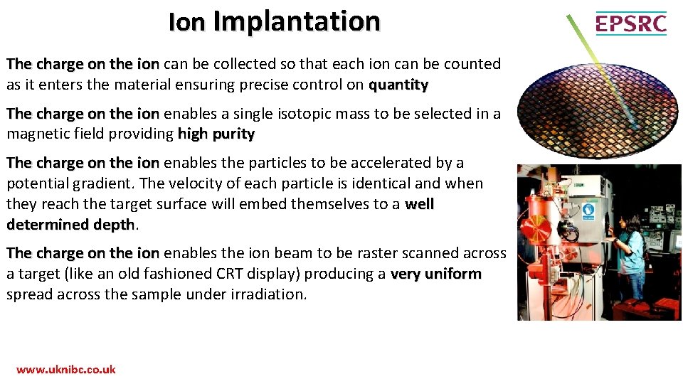
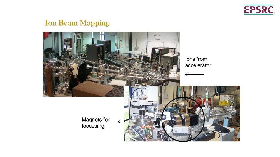
Ion Implantation The charge on the ion can be collected so that each ion can be counted as it enters the material ensuring precise control on quantity The charge on the ion enables a single isotopic mass to be selected in a magnetic field providing high purity The charge on the ion enables the particles to be accelerated by a potential gradient. The velocity of each particle is identical and when they reach the target surface will embed themselves to a well determined depth The charge on the ion enables the ion beam to be raster scanned across a target (like an old fashioned CRT display) producing a very uniform spread across the sample under irradiation. www. uknibc. co. uk
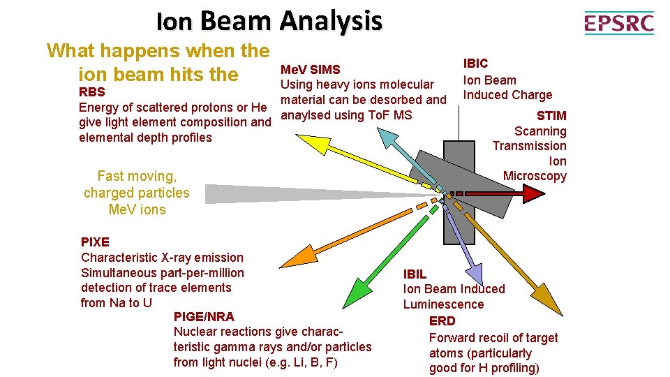
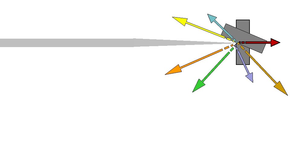
Ion Beam Analysis What happens when the ion beam hits the RBS target? Energy of scattered protons or He give light element composition and PARTICLES elemental depth profiles Me. V SIMS Using heavy ions. Sputtered molecular material can be desorbed Molecules and anaylsed using To. F MS Fast moving, charged particles Me. V ions PIXE Characteristic X-ray emission X RAYS Simultaneous part-per-million detection of trace elements from Na to U PIGE/NRA Nuclear reactions give. GAMMA charac-RAYS teristic gamma rays and/or particles from light nuclei (e. g. Li, B, F) IBIC A Ion Beam Induced Charge STIM Scanning Transmission Transmitted Ion ions Microscopy IBIL Ion Beam Induced Luminescence LIGHT ERD Forward recoil of target RECOILS atoms (particularly good for H profiling)

Bringing together 3 ion beam facilities in the UK www. uknibc. co. uk An EPSRC Mid Range Facility

Huddersfield Miami Facility • Implanter with In situ TEM o Facilities • • • JOEL 2000 FX TEM 2 -100 ke. V Implanter Temperature stage -173 -1000 o. C o Applications • Nuclear Radiation damage • Ion Implantation o Staff • • • Steve Donnelly Johnathan Hinks Graeme Greaves www. uknibc. co. uk
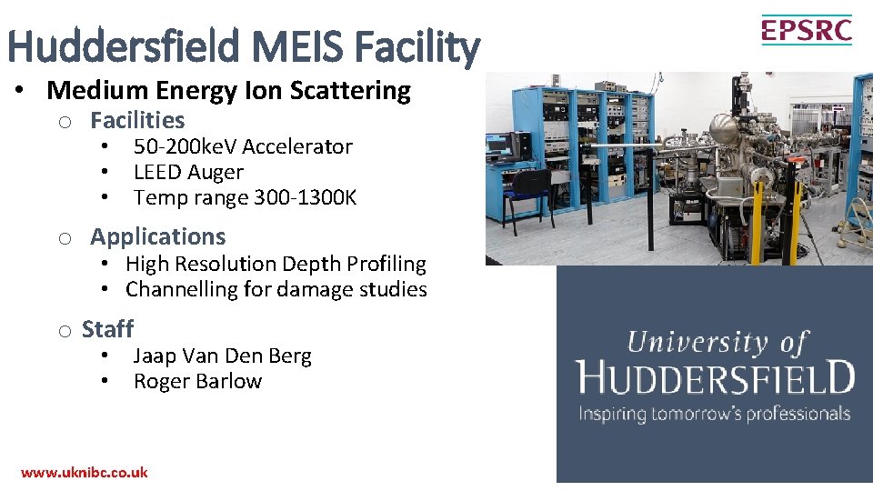
Huddersfield MEIS Facility • Medium Energy Ion Scattering o Facilities • • • 50 -200 ke. V Accelerator LEED Auger Temp range 300 -1300 K o Applications • High Resolution Depth Profiling • Channelling for damage studies o Staff • • Jaap Van Den Berg Roger Barlow www. uknibc. co. uk
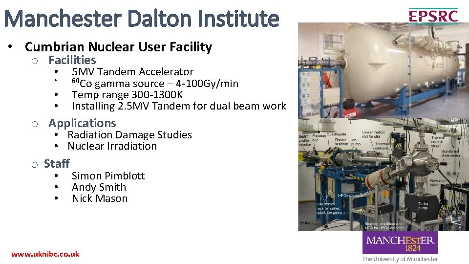
Manchester Dalton Institute • Cumbrian Nuclear User Facility o Facilities • • 5 MV Tandem Accelerator 60 Co gamma source – 4 -100 Gy/min Temp range 300 -1300 K Installing 2. 5 MV Tandem for dual beam work o Applications • Radiation Damage Studies • Nuclear Irradiation o Staff • • • Simon Pimblott Andy Smith Nick Mason www. uknibc. co. uk

Surrey Ion Beam Centre • Investment – Support • £ 5 M investment in machines and beam lines • Approx £ 1. 5 M per annum support • 2 academics • 8 RAs (including 2 senior RAs) • 3 technicians + 1 secretarial – Technology Transfer • 32 University departments around the UK • 53 local and international companies • 15 Ph. D projects • Supporting >50 EPSRC grants worth >£ 50 M www. uknibc. co. uk
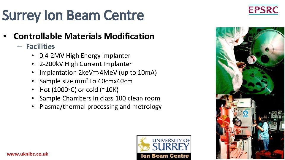
Surrey Ion Beam Centre • Controllable Materials Modification – Facilities • • 0. 4 -2 MV High Energy Implanter 2 -200 k. V High Current Implanter Implantation 2 ke. V 4 Me. V (up to 10 m. A) Sample size mm 2 to 40 cmx 40 cm Hot (1000 o. C) or cold (~10 K) Sample Chambers in class 100 clean room Plasma/thermal processing and metrology www. uknibc. co. uk
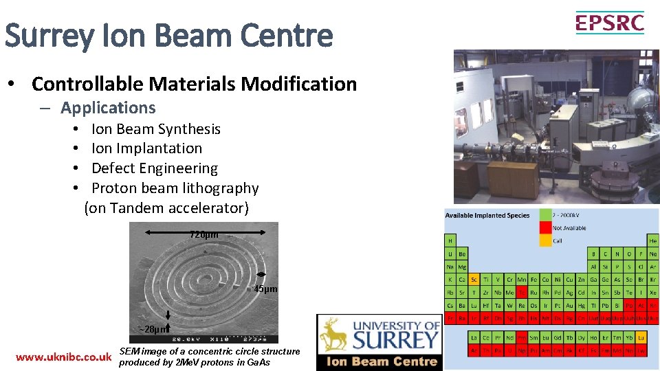
Surrey Ion Beam Centre • Controllable Materials Modification – Applications • • Ion Beam Synthesis Ion Implantation Defect Engineering Proton beam lithography (on Tandem accelerator) 720µm 45µm ~28µm www. uknibc. co. uk SEM image of a concentric circle structure produced by 2 Me. V protons in Ga. As
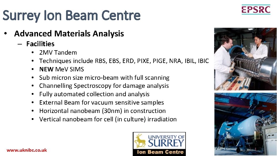
Surrey Ion Beam Centre • Advanced Materials Analysis – Facilities • • • 2 MV Tandem Techniques include RBS, ERD, PIXE, PIGE, NRA, IBIL, IBIC NEW Me. V SIMS Sub micron size micro-beam with full scanning Channelling Spectroscopy for damage analysis Fully automated collection and analysis External Beam for vacuum sensitive samples Horizontal nanobeam (30 nm) in construction Vertical nanobeam for cell (in culture) irradiation www. uknibc. co. uk
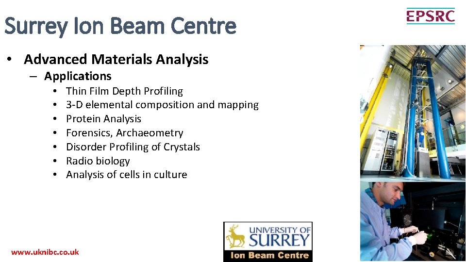
Surrey Ion Beam Centre • Advanced Materials Analysis – Applications • • Thin Film Depth Profiling 3 -D elemental composition and mapping Protein Analysis Forensics, Archaeometry Disorder Profiling of Crystals Radio biology Analysis of cells in culture www. uknibc. co. uk
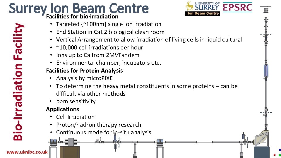
Bio-Irradiation Facility Surrey. Facilities Ion for. Beam Centre bio-irradiation • Targeted (~100 nm) single ion irradiation • End Station in Cat 2 biological clean room • Vertical Arrangement to allow irradiation of living cells in liquid cultural • ~10, 000 cell irradiations per hour • Ions up to Ca from 2 MVTandem • Environmental chamber, incubators etc. Facilities for Protein Analysis • Analysis by micro. PIXE • To determine the heavy metal constituents in some proteins – can be difficult via other methods • ppm sensitivity Applications • Cell Irradiation • Proton/hadron therapy research • Continuous mode for in-situ analysis www. uknibc. co. uk

Surrey Ion Beam Centre Quality Assurance and Standards • Extensive QA program • ISO 9001 certified since 2008 • Working with both NPL and PTB on various fundamental studies • ISO 17025 accreditation for high accuracy RBS www. uknibc. co. uk
Surrey Ion Beam Centre www. uknibc. co. uk
Surrey Ion Beam Centre www. uknibc. co. uk
Surrey Ion Beam Centre www. uknibc. co. uk
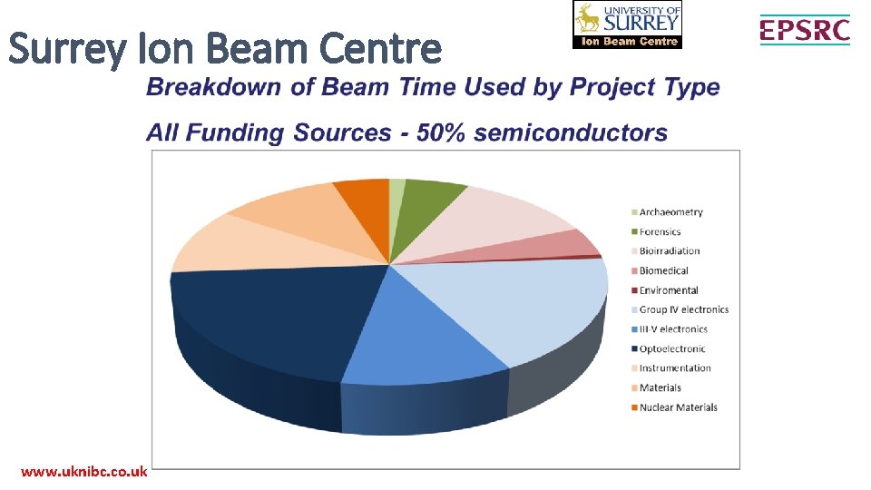
Surrey Ion Beam Centre www. uknibc. co. uk
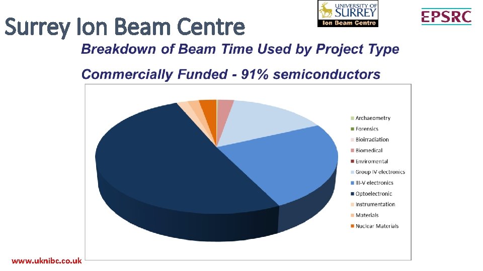
Surrey Ion Beam Centre www. uknibc. co. uk
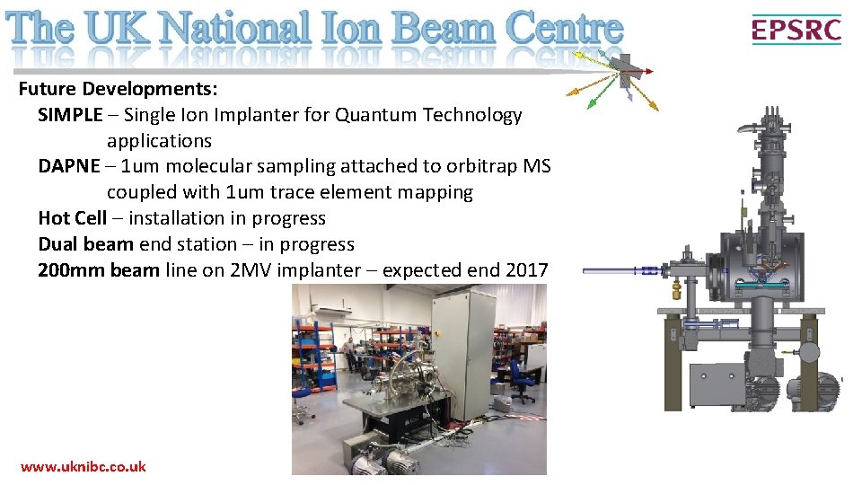
Future Developments: SIMPLE – Single Ion Implanter for Quantum Technology applications DAPNE – 1 um molecular sampling attached to orbitrap MS coupled with 1 um trace element mapping Hot Cell – installation in progress Dual beam end station – in progress 200 mm beam line on 2 MV implanter – expected end 2017 www. uknibc. co. uk
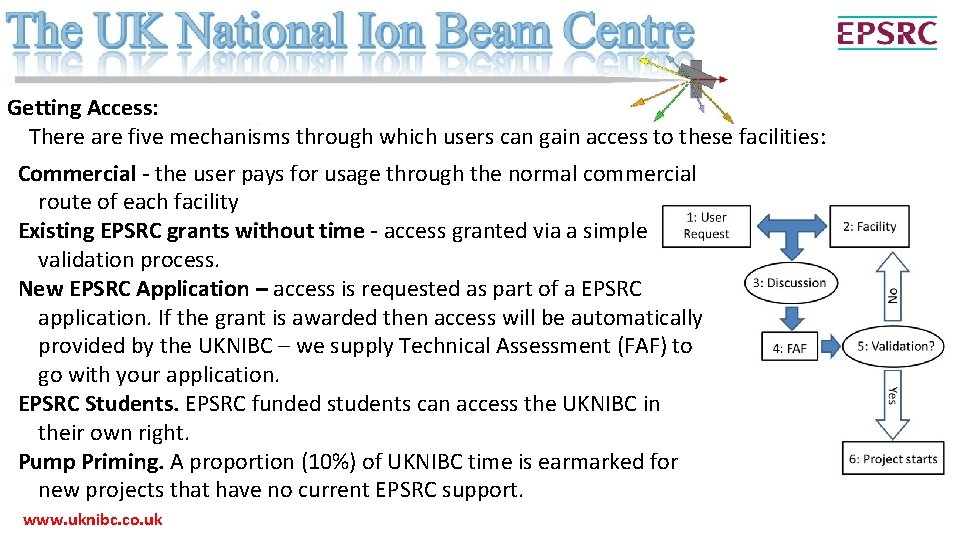
Getting Access: There are five mechanisms through which users can gain access to these facilities: Commercial - the user pays for usage through the normal commercial route of each facility Existing EPSRC grants without time - access granted via a simple validation process. New EPSRC Application – access is requested as part of a EPSRC application. If the grant is awarded then access will be automatically provided by the UKNIBC – we supply Technical Assessment (FAF) to go with your application. EPSRC Students. EPSRC funded students can access the UKNIBC in their own right. Pump Priming. A proportion (10%) of UKNIBC time is earmarked for new projects that have no current EPSRC support. www. uknibc. co. uk
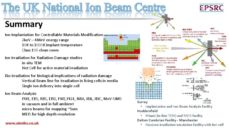
Summary Ion Implantation for Controllable Materials Modification 2 ke. V – 4 Me. V energy range 10 K to 1000 K implant temperature Class 100 clean room Ion Irradiation for Radiation Damage studies in-situ TEM Hot Cell for active material irradiation Bio-irradiation for biological implications of radiation damage Vertical Beam line for irradiation in living cells in media Single ion delivery into single cell Ion Beam Analysis PIXE, EBS, RBS, ERD, PIXE, PIGE, NRA, IBIL, IBIC, Me. V-SIMS in vacuum and in full ambient micro beams for mapping ~1 um MEIS for high depth resolution www. uknibc. co. uk Surrey • Implantation and Ion Beam Analysis facility Huddersfield • Miami (in-line TEM) and MEIS facility Dalton Cumbrian Facility - Manchester • Neutron irradiation emulation facility with hot cell

• Additional slides…
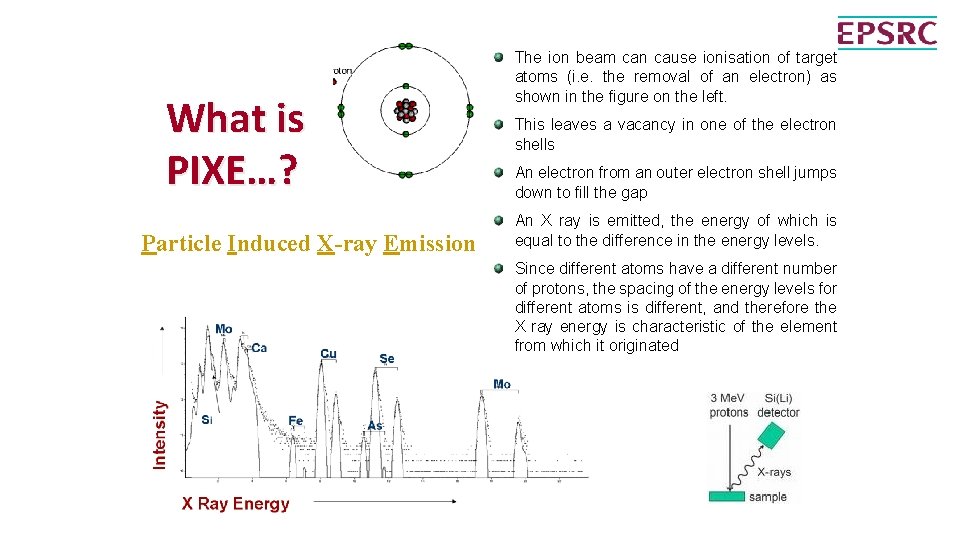
What is PIXE…? Particle Induced X-ray Emission The ion beam can cause ionisation of target atoms (i. e. the removal of an electron) as shown in the figure on the left. This leaves a vacancy in one of the electron shells An electron from an outer electron shell jumps down to fill the gap An X ray is emitted, the energy of which is equal to the difference in the energy levels. Since different atoms have a different number of protons, the spacing of the energy levels for different atoms is different, and therefore the X ray energy is characteristic of the element from which it originated
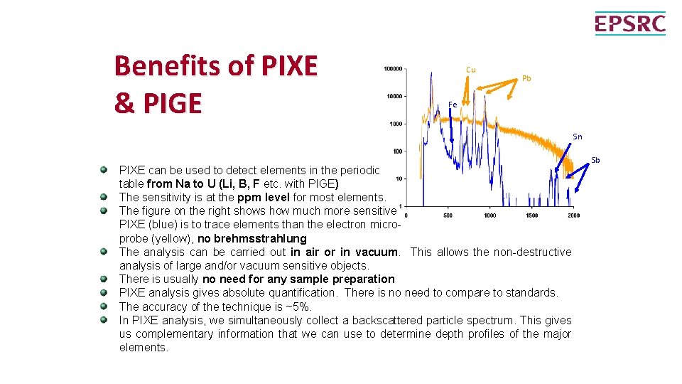
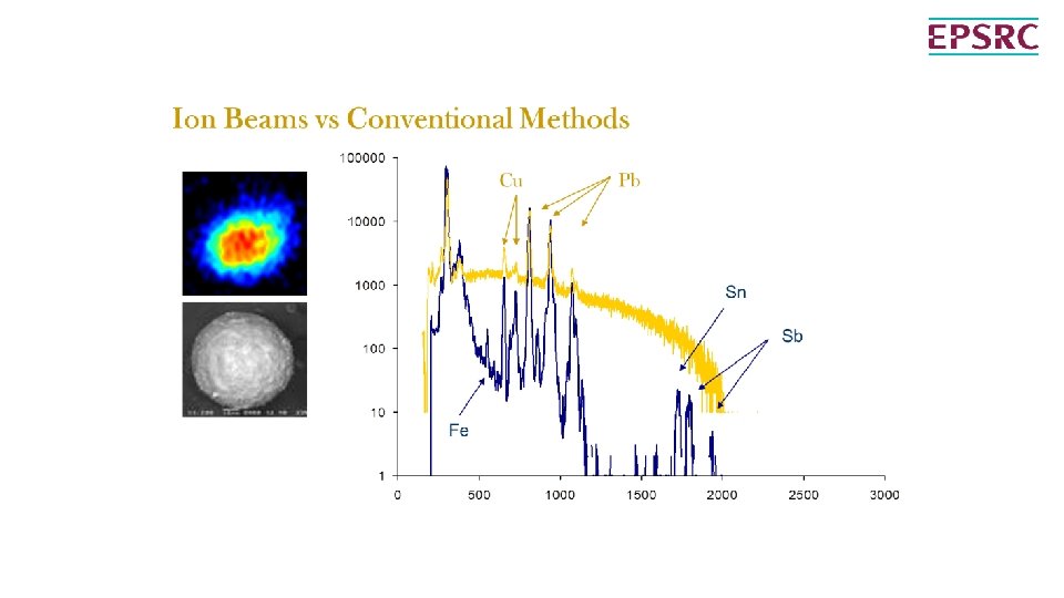
Benefits of PIXE & PIGE Cu Pb Fe Sn PIXE can be used to detect elements in the periodic table from Na to U (Li, B, F etc. with PIGE) The sensitivity is at the ppm level for most elements. The figure on the right shows how much more sensitive PIXE (blue) is to trace elements than the electron microprobe (yellow), no brehmsstrahlung The analysis can be carried out in air or in vacuum. This allows the non-destructive analysis of large and/or vacuum sensitive objects. There is usually no need for any sample preparation PIXE analysis gives absolute quantification. There is no need to compare to standards. The accuracy of the technique is ~5%. In PIXE analysis, we simultaneously collect a backscattered particle spectrum. This gives us complementary information that we can use to determine depth profiles of the major elements. Sb
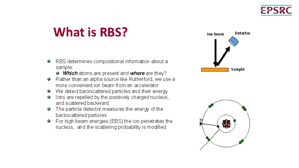
What is RBS? RBS determines compositional information about a sample: Which atoms are present and where are they? Rather than an alpha source like Rutherford, we use a more convenient ion beam from an accelerator We detect backscattered particles and their energy Ions are repelled by the positively charged nucleus, and scattered backward. The particle detector measures the energy of the backscattered particles. For high beam energies (EBS) the ion penetrates the nucleus, and the scattering probability is modified. Ion beam Detector Sample
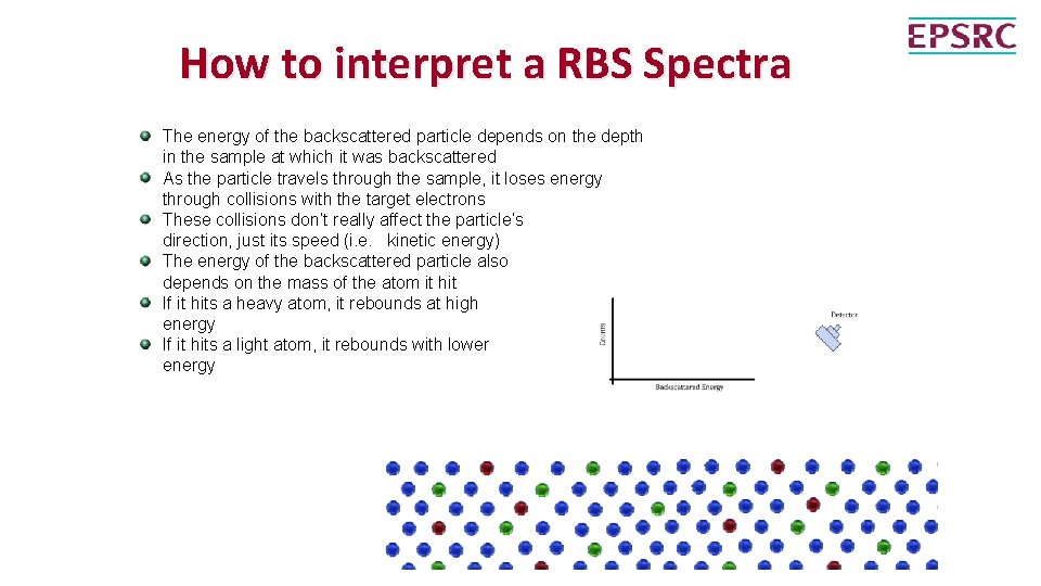
How to interpret a RBS Spectra The energy of the backscattered particle depends on the depth in the sample at which it was backscattered As the particle travels through the sample, it loses energy through collisions with the target electrons These collisions don’t really affect the particle’s direction, just its speed (i. e. kinetic energy) The energy of the backscattered particle also depends on the mass of the atom it hit If it hits a heavy atom, it rebounds at high energy If it hits a light atom, it rebounds with lower energy
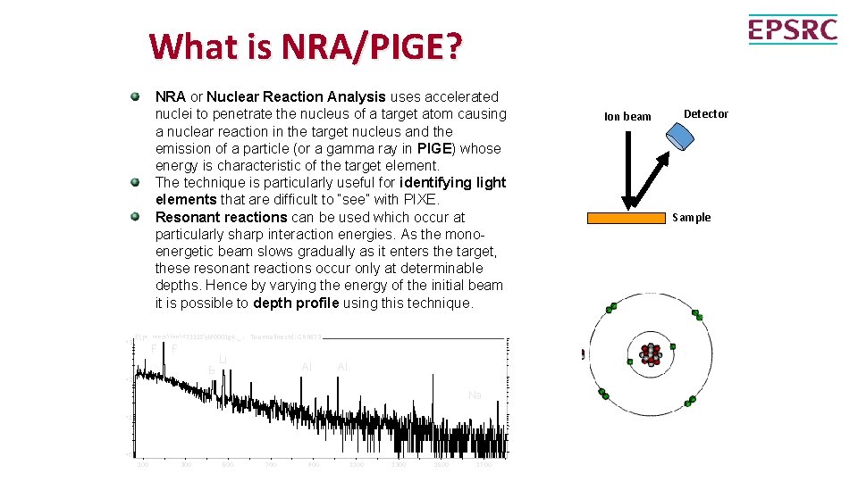
What is NRA/PIGE? NRA or Nuclear Reaction Analysis uses accelerated nuclei to penetrate the nucleus of a target atom causing a nuclear reaction in the target nucleus and the emission of a particle (or a gamma ray in PIGE) whose energy is characteristic of the target element. The technique is particularly useful for identifying light elements that are difficult to “see” with PIXE. Resonant reactions can be used which occur at particularly sharp interaction energies. As the monoenergetic beam slows gradually as it enters the target, these resonant reactions occur only at determinable depths. Hence by varying the energy of the initial beam it is possible to depth profile using this technique. ^3 f: pc_usersjpn971118490001 g 4. _ : Tourmaline std: GRR 573 F F B ^2 Li Al Al Na ^1 ^0 100 300 500 700 900 1100 1300 1500 1700 Ion beam Detector Sample
- Slides: 31