
Rheumatologic disorders The rheumatic diseases of childhood are


- Slides: 79
Rheumatologic disorders
• The rheumatic diseases of childhood are characterized by autoimmunity and inflammation, which may be localized or generalized. • The classic rheumatic diseases of children include juvenile idiopathic arthritis (JIA) formerly called juvenile rheumatoid Arthritis and systemic lupus erythematosus (SLE) • Vasculitis is a component of many rheumatic diseases.
• Musculoskeletal pain syndromes are a set of overlapping conditions characterized by poorly localized pain involving the extremities. • Scleroderma, Behçet syndrome, and Sjögren syndrome are rare in childhood. • The differential diagnosis of rheumatologic disorders typically includes infections, postinfectious processes, and malignancies
Henoch–Schönlein Purpura (HSP) • Definition. A systemic Ig. A-mediated vasculitis that primarily involves the skin, joints, gastrointestinal (GI) tract, and kidneys. • Epidemiology • HSP is primarily a disease of children (ages 3– 15 years); the peak incidence is at 4– 6 years. • HSP is the most common form of vasculitis of childhood. • Males are more likely to be affected. • HSP occurs primarily in the fall, winter, and spring.
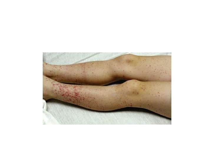
Clinical features • A viral syndrome or upper respiratory infection precedes HSP in about 50% of patients. • Distinctive skin, GI, and joint manifestations follow the prodrome. • Skin manifestations : petechiae and palpable purpuric lesions, concentrated on the buttocks and lower extremities. • Children may also present with edema of hands, feet, scrotum, and scalp.
Clinical features • Joint manifestations occur in 80% of patients as arthralgia or arthritis. • The knees and ankles are most commonly involved. • Primarily involves large joints in the lower extremities and can be very painful. • Swelling is periarticular but lacks effusions or increased warmth and does not cause chronic damage.
Clinical features • GI manifestations occur in about 50% of patients. • Colicky abdominal pain that may be severe • GI bleeding (either occult blood or grossly bloody stool) • Increased risk of intussusception (most common GI complication of HSP)
Clinical features • Renal manifestations: • Mild hematuria and trace proteinuria • Gross hematuria • Nephrotic syndrome • Chronic renal insufficiency • End-stage renal disease
Diagnosis • History and characteristic physical examination establish a clinical diagnosis. • Routine laboratory tests are neither specific nor diagnostic. • Skin biopsies show leukocytoclastic vasculitis with Ig. A deposition. • Increased serum Ig. A levels are present in 50% of patients. • Platelet counts are normal ( the skin rash is a nonthrombocytopenic purpura).
Management. Treatment is based on relief of symptoms, including use of analgesics and hydration, and monitoring for complications. • Steroids may be effective for relief of abdominal pain and arthritis but do not impact the clinical course. • Patients should be followed up with urinalysis and blood pressure monitoring every 2 weeks for 1– 2 months after presentation, then monthly for at least 6 months. • Hematuria, proteinuria, or hypertension should prompt referral to a nephrologist.
Prognosis • Most patients recover within 4 weeks. • HSP recurs at least once in one-third of patients, usually within 3– 4 months of presentation. • Long-term morbidity is dependent on the severity of renal disease
Kawasaki Disease • Definition. An acute febrile vasculitis of childhood; this disease involves multiple organ systems, including the heart, skin, mucous membranes, GI tract, central nervous system (CNS), and joints • KD is the most common cause of acquired heart disease in children in the United States. • Males are slightly more commonly affected than females.
• Most common in children of Asian ethnicity but can occur in any ethnicity. • The mean age at presentation is 18– 24 months • 80– 90% of cases occur in children younger than 5 years). • It is uncommon in children younger than 6 months
Diagnostic criteria • Temperature > 38. 9°C (102°F) lasting ≥ 5 days most consistent manifestation of KD • The patient must also have four of the following five clinical manifestations: • Bilateral conjunctivitis: bulbar injection with limbic sparing and without exudate • Mucous membrane changes: pharyngitis; strawberry tongue; or most commonly, red, cracked, swollen lips
Diagnostic criteria • Cervical adenopathy: a unilateral nonsuppurative cervical lymph node ≥ 1. 5 cm in diameter. • Rash: primarily on the trunk. The rash may assume many forms (“polymorphous • It often begins as perineal erythema and desquamation.
• Changes in distal extremities • Early (first 7– 10 days of illness): swelling and induration of the hands and feet with erythematous palms and soles • Later (7– 10 days after the start of fever): peeling around the nail beds of the distal extremities
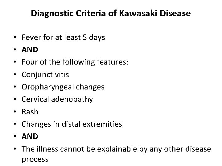
Diagnostic Criteria of Kawasaki Disease • • • Fever for at least 5 days AND Four of the following features: Conjunctivitis Oropharyngeal changes Cervical adenopathy Rash Changes in distal extremities AND The illness cannot be explainable by any other disease process
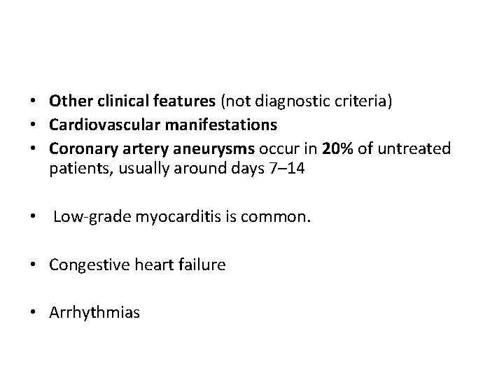
• Other clinical features (not diagnostic criteria) • Cardiovascular manifestations • Coronary artery aneurysms occur in 20% of untreated patients, usually around days 7– 14 • Low-grade myocarditis is common. • Congestive heart failure • Arrhythmias
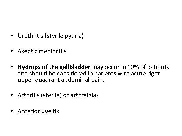
• Urethritis (sterile pyuria) • Aseptic meningitis • Hydrops of the gallbladder may occur in 10% of patients and should be considered in patients with acute right upper quadrant abdominal pain. • Arthritis (sterile) or arthralgias • Anterior uveitis
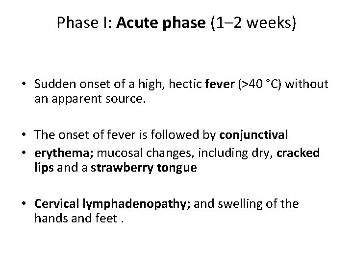
Phase I: Acute phase (1– 2 weeks) • Sudden onset of a high, hectic fever (>40 °C) without an apparent source. • The onset of fever is followed by conjunctival • erythema; mucosal changes, including dry, cracked lips and a strawberry tongue • Cervical lymphadenopathy; and swelling of the hands and feet.
Phase I: Acute phase (1– 2 weeks) • Extreme irritability is prominent, especially in infants. • Carditis in the acute phase may be manifested by tachycardia, shortness of breath, or overt congestive heart failure. • Giant coronary artery aneurysms, which are rare but occur most commonly in very young children, can appear during this phase.
Phase II: Subacute phase (weeks to months) • Characterized by gradual resolution of fever (if untreated) and other symptoms. • Desquamation of the skin, particularly of the fingers and toes, appears at this point. • The platelet count, previously normal or slightly depressed, increases to a significant degree (often >1 million/mm 3).
Phase II: Subacute phase (weeks to months) • Coronary artery aneurysms, which may also appear in the convalescent phase and pose the highest risk of morbidity and mortality. • Risk factors for development of coronary artery aneurysms include: • Prolonged fever • Prolonged elevation of inflammatory parameters such as (ESR) • Age younger than 1 year • Older than 6 years, and male gender
Phase III: Convalescent phase (weeks to years) • The convalescent phase begins with the disappearance of clinical symptoms and continues until the ESR returns to normal • usually 6 to 8 weeks after the onset of illness.
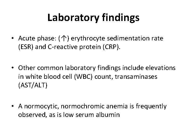
Laboratory findings • Acute phase: (↑) erythrocyte sedimentation rate (ESR) and C-reactive protein (CRP). • Other common laboratory findings include elevations in white blood cell (WBC) count, transaminases (AST/ALT) • A normocytic, normochromic anemia is frequently observed, as is low serum albumin
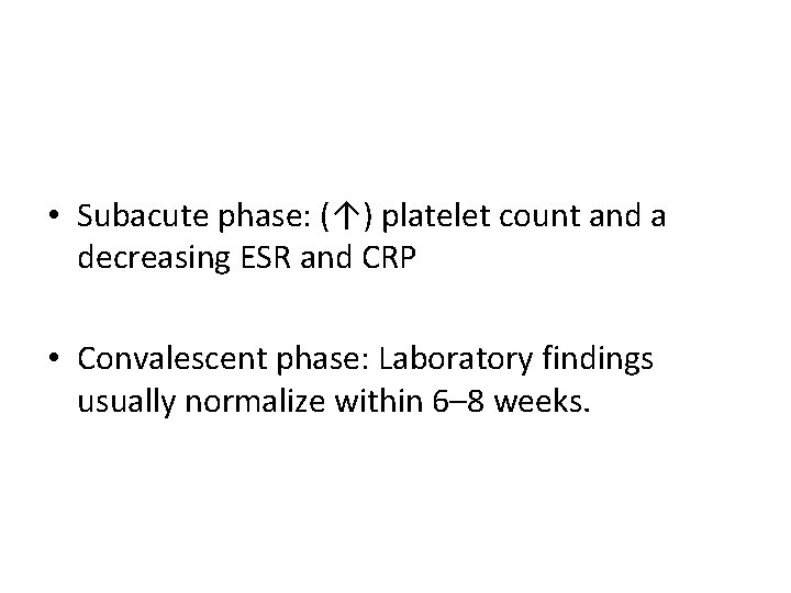
• Subacute phase: (↑) platelet count and a decreasing ESR and CRP • Convalescent phase: Laboratory findings usually normalize within 6– 8 weeks.
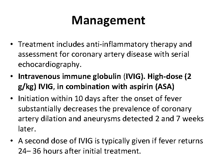
Management • Treatment includes anti-inflammatory therapy and assessment for coronary artery disease with serial echocardiography. • Intravenous immune globulin (IVIG). High-dose (2 g/kg) IVIG, in combination with aspirin (ASA) • Initiation within 10 days after the onset of fever substantially decreases the prevalence of coronary artery dilation and aneurysms detected 2 and 7 weeks later. • A second dose of IVIG is typically given if fever returns 24– 36 hours after initial treatment.
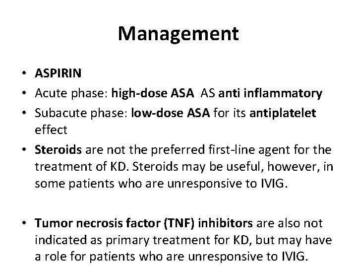
Management • ASPIRIN • Acute phase: high-dose ASA AS anti inflammatory • Subacute phase: low-dose ASA for its antiplatelet effect • Steroids are not the preferred first-line agent for the treatment of KD. Steroids may be useful, however, in some patients who are unresponsive to IVIG. • Tumor necrosis factor (TNF) inhibitors are also not indicated as primary treatment for KD, but may have a role for patients who are unresponsive to IVIG.
Incomplete or atypical KD • A subset of patients suspected of having KD may not fulfill all of the clinical diagnostic criteria. These patients have “incomplete” or “atypical” KD. • Patients must still have at least 5 days of fever but otherwise have only have 2– 3 clinical features and no alternative explanation for symptoms. • Laboratory and echocardiographic studies are used to make the diagnosis in these children. • Treatment is the same as for typical KD.
Prognosis • If coronary artery disease is absent, mortality is rare. Recurrence is also rare. • Even if coronary artery disease is present, the mortality is <1%, and aneurysms, especially those <8 mm, commonly regress over time. • Long-term prognosis is unclear. • It’s possible that there may be an increased risk of atherosclerotic heart disease in adulthood.
Juvenile Idiopathic Arthritis (JIA) • Definition. JIA is a disorder characterized by chronic joint inflammation in children, with or without extraarticular involvement. • JIA is the most common pediatric rheumatic disease with arthritis as the distinguishing manifestation. • The mean age of onset is 1– 3 years; presentation before 6 months of age is unusual. • JIA most commonly occurs in females • Males are equally likely to have systemic-onset JIA
• Diagnosis of JIA is based on history and clinical features. • Classification. The classification of JIA is determined on the basis of the number and types of joints involved, along with extraarticular manifestations. • Regardless of type, all patients have arthritis during their clinical course.
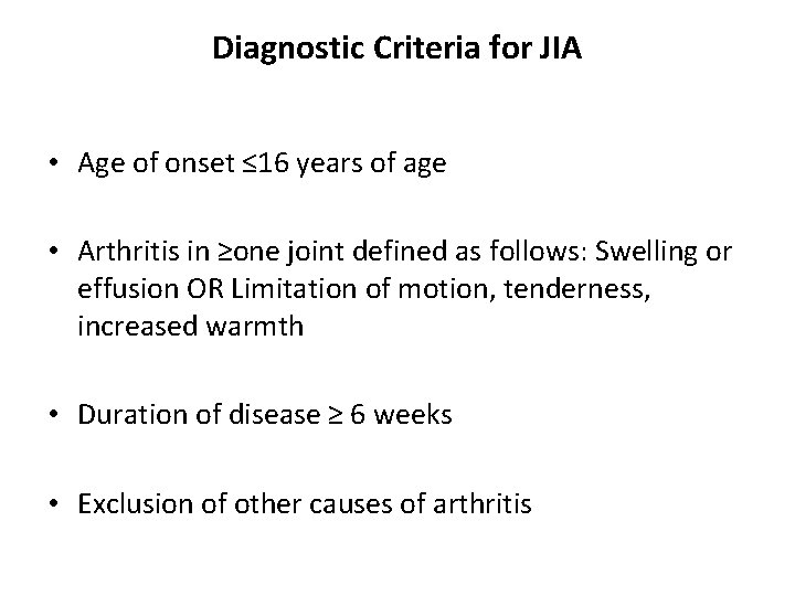
Diagnostic Criteria for JIA • Age of onset ≤ 16 years of age • Arthritis in ≥one joint defined as follows: Swelling or effusion OR Limitation of motion, tenderness, increased warmth • Duration of disease ≥ 6 weeks • Exclusion of other causes of arthritis
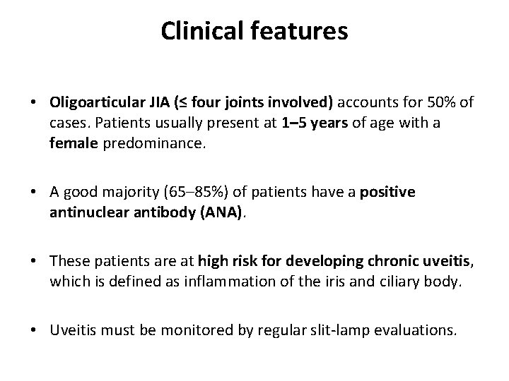
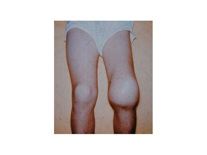
Clinical features • Oligoarticular JIA (≤ four joints involved) accounts for 50% of cases. Patients usually present at 1– 5 years of age with a female predominance. • A good majority (65– 85%) of patients have a positive antinuclear antibody (ANA). • These patients are at high risk for developing chronic uveitis, which is defined as inflammation of the iris and ciliary body. • Uveitis must be monitored by regular slit-lamp evaluations.
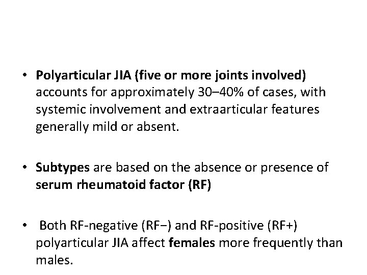
• Polyarticular JIA (five or more joints involved) accounts for approximately 30– 40% of cases, with systemic involvement and extraarticular features generally mild or absent. • Subtypes are based on the absence or presence of serum rheumatoid factor (RF) • Both RF-negative (RF−) and RF-positive (RF+) polyarticular JIA affect females more frequently than males.
• RF− disease presents both early and late in childhood. • RF+ disease presents in children older than 8 years and is generally more severe than RF− disease • Articular involvement is characterized by a symmetric polyarthritis that typically involves both small and large joints
• Systemic-onset JIA accounts for 10– 15% of cases. • High spiking fevers (temperature > 39°C [102. 2°F]) occur most commonly in the late afternoon or evening and subsequently return quickly to baseline or subnormal levels • Systemic JIA is typically included in the differential diagnosis of “fever of unknown origin. ”
• Transient salmon-pink macular rash is most commonly found on the trunk and proximal extremities, especially during febrile episodes. • The rash is evanescent (occurs with fever spikes and then fades) and nonpruritic. • Hepatosplenomegaly • Lymphadenopathy
• Fatigue, anorexia, weight loss, and failure to thrive are common. • Serositis, including pericarditis and pleuritis, is also common. • CNS involvement, including meningitis and encephalopathy, may occasionally be present. • Myositis and tenosynovitis can also be seen in addition to the arthritis.
Laboratory findings • Laboratory results are nonspecific; if present, they reflect the existence or extent of inflammation and are useful to evaluate for other disease processes. • Anemia is usually microcytic and hypochromic, consistent with anemia of chronic disease. • Acute-phase reactants are elevated, including ESR, CRP, Ferritin, and platelet count.
Laboratory findings • Rheumatoid markers • RF is negative in most patients with JIA. RF positivity is not diagnostic but can help determine prognosis for patients with polyarticular disease. • ANA is present in 65– 85% of patients with oligoarticular JIA and in 50% of patients with polyarticular JIA. • An ANA is not a diagnostic test for JIA but can help determine the risk of uveitis. • ANA is rare in children with systemic-onset JIA.
Management • The goal of treatment is to decrease joint inflammation and preserve function. • Control of inflammation • NSAIDs reduce pain and inflammation. • Immunomodulatory medications glucocorticoids, methotrexate, sulfasalazine, TNF inhibitors
Management • Mechanical and physical measures include physical and occupational therapy. • Surgery (joint replacement, synovectomy) is generally reserved for patients who have joint contractures or destruction. • Owing to advances in pharmacotherapy, surgery is becoming much less common. • Psychosocial support
Systemic Lupus Erythematosus (SLE) • Definition. This multisystem autoimmune disorder is characterized by widespread chronic inflammation and immune complex–mediated vasculitis. • Epidemiology • Female-to-male ratio is 8: 1 • Age of onset is rarely younger than 10 years and peaks in adolescence.
• Etiology. The cause of SLE is unknown, but the disease may be triggered by: • drug reactions • excessive sun exposure • infections • or hormonal changes (e. g. , menarche menopause, pregnancy).
Clinical features • Constitutional symptoms such as fever, weight loss, and fatigue are common. • CNS involvement includes headache, encephalopathy, seizures, psychosis • Skin findings may include a malar rash (“butterfly distribution” covering the nasal bridge and photosensitivity, both common at presentation or during an illness flare. • Other common skin findings include alopecia and Raynaud phenomenon.
Clinical features • Arthralgias and arthritis are typically migratory and transient. • SLE, unlike JIA, rarely causes joint deformity or erosion. Myositis may also occur. • GI involvement may manifest as hepatosplenomegaly, splenic infarction, mesenteric thrombosis (secondary to vasculitis), and sterile peritonitis.
Clinical features • Cardiovascular involvement is variable, with pericarditis as the most frequent manifestation. Congestive heart failure, arrhythmias. • Neonates born to mothers with SLE may have congenital heart block secondary to transplacental passage of maternal antibodies. • Pulmonary involvement may include pleuritis, pulmonary hemorrhage, and interstitial fibrosis.
Clinical features • Renal involvement is nearly universal, although lupus nephritis may be subclinical. Glomerulonephritis, nephrotic syndrome, hypertension, and renal failure are common. • Hematologic manifestations typically include Leukopenia, Anemia of chronic disease, ITP, and Coombs-positive hemolytic anemia.
Diagnostic Criteria for SLE • • • • Mnemonic “SOAP BRAIN MD” Serositis (pleuritis or pericardial inflammation) Oral or nasal mucocutaneous ulcerations Arthritis, nonerosive Photosensitivity Blood cytopenias (leukopenia, hemolytic anemia, or thrombocytopenia) Renal disease (hematuria, proteinuria, hypertension) ANA-positive Immunoserology abnormalities (antibodies to double-stranded DNA, Smith antigen, false-positive RPR or VDRL assays, or antiphospholipid antibodies) Neurologic symptoms (encephalopathy, seizures, or psychosis) Malar rash (butterfly rash) Discoid lupus (red raised patches with scaling, can cause scarring)
Laboratory findings • Elevated ESR • Anemia of chronic disease or hemolytic anemia • Leukopenia • Thrombocytopenia • Urinalysis may show proteinuria depending on the extent of renal disease.
Laboratory findings • ANA is almost universally elevated (>99%) in SLE and is an excellent screening test. but is nonspecific • RF is often elevated in SLE, but is nonspecific. • Anti–double-stranded DNA (anti-ds. DNA) antibodies are much more specific (found only in SLE), and their levels can be used as markers for active disease, especially nephritis • Anti-Smith (anti-Sm) antibodies are less prevalent in patients with SLE but, when present, are very specific.
Laboratory findings • Antiphospholipid antibodies (e. g. , positive lupus anticoagulant or anticardiolipin antibodies) present in patients with SLE reflect an increased risk of thrombotic events. • Decreased complement (C 3 and C 4) is seen especially with active disease and represents immune complex–mediated complement activation. • Ro (SSA) and La (SSB) antibodies are less strongly associated with SLE (more specific for Sjögren syndrome). Babies born to mothers with these antibodies are at risk for neonatal lupus.
Management • Therapy is based on a multidisciplinary team approach that attempts: • To prevent or minimize inflammation and end-organ dysfunction • To treat complications • To provide psychosocial and family support, as follows: • Control of inflammation • NSAIDs are useful in the treatment of minor inflammatory symptoms.
Management • Immunosuppressive medications • Glucocorticoids are the mainstay of therapy for children with SLE. • Treatment ranges from low-dose oral to high-dose intravenously pulsed steroids. • Cyclophosphamide, is useful for children with severe clinical manifestations, including renal and CNS disease. • Adverse effects of this cytotoxic agent include infertility and gonadal failure, secondary malignancies, and hemorrhagic cystitis.
Management • Hydroxychloroquine is useful for patients with mild disease and is often used as maintenance therapy. • Numerous studies have shown a beneficial effect on long-term disease activity and survival. • Patients require ophthalmologic screening owing to the side-effect of potential vision damage with its use.
Management • • Other agents include Mycophenolate MMF Rituximab Azathioprine Methotrexate Cyclosporine Mycophenolate, in particular, is being used more often as an alternative to cyclophosphamide to treat lupus nephritis.
Treatment of complications • Patients with thrombosis and antiphospholipid antibodies should be anticoagulated with low molecular weight heparin or warfarin. • Patients with renal failure may require dialysis, fluid and electrolyte management, and ultimately renal transplant. • Psychosocial and family support
Prognosis • Before the advent of effective therapy, the outlook for patients with SLE was historically poor and uniformly fatal • However, with current treatment, the survival rate is now 85– 100% at 5– 10 years after initial diagnosis • The major causes of mortality are infection (because of immunosuppression), renal failure, and CNS complications.
Rheumatic Fever • Rheumatic fever is a delayed, nonsuppurative, autoimmune complication occurring 2– 4 weeks after an URTI with group A β-hemolytic streptococcus (GABHS) (Streptococcus pyogenes). • It is characterized by inflammation of the heart, blood vessels, joints, CNS, and skin.
Epidemiology • Rheumatic fever is now rare in the United States and Western Europe but was a worldwide problem during the 1960 s. • It remains a primary cause of cardiac death in the developing world in patients younger than 50 years. • It is most common in children 5– 15 years of ages. • There is no gender predilection. • The streptococcal strains that cause streptococcal skin infection (i. e. , impetigo) do not cause rheumatic fever.
Etiology • The cause of rheumatic fever is unknown, but the disease appears to be autoimmune in nature and may represent “molecular mimicry, ” in which streptococcal antigens resemble autoantigens and trigger an autoimmune response.
Clinical features • Cardiac involvement is the hallmark and most important complication of rheumatic fever. • The inflammatory process may involve all layers of the heart (pancarditis) • Valvulitis is frequent and typically causes insufficiency of the left-sided valves (mitral and aortic). • Mitral regurgitation is the most common early valvular manifestation
Clinical features • It rarely affects the pulmonic or tricuspid valves. • Myocarditis is usually manifested by tachycardia out of proportion to the extent of the fever. • Other severe manifestations include cardiac dilatation and heart failure. • Pericarditis and pericardial effusions are less common
Clinical features • Arthritis is classically migratory, asymmetric, and exquisitely painful and often the earliest manifestation of acute rheumatic fever. • It occurs in 70% of patients • Most commonly involves the elbows, knees, ankles, and wrists. • It does not result in chronic joint disease and responds quickly to NSAIDs and aspirin.
Clinical features • Sydenham chorea occurs later than the other rheumatic fever manifestations, often beginning subtly, months after GABHS pharyngitis. • It reflects involvement of the basal ganglia and caudate nuclei • Chorea may start as hand clumsiness and progress to choreoathetoid movements with emotional lability
Clinical features • Skin involvement • Erythema marginatum is an evanescent, nonpruritic rash that starts as pink to red macules • Subcutaneous nodules, although rarely seen, are associated with severe cardiac involvement. • These small, mobile, and painless nodules occur on the bony prominences of the extensor surfaces of the extremities.
Clinical features • Minor features include: • Fever • Arthralgias • Leukocytosis • Increased ESR • Prolonged PR interval on electrocardiogram.
Diagnosis • Diagnosis requires evidence of recent streptococcal infection and either two major criteria or one major plus two minor criteria
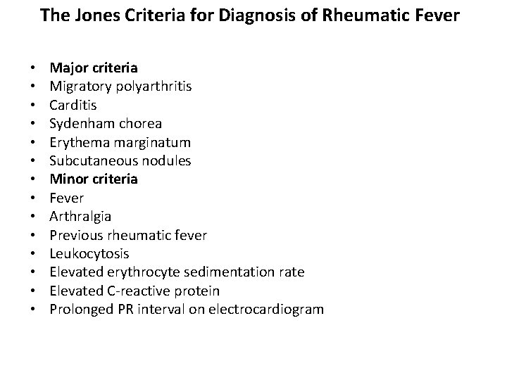
The Jones Criteria for Diagnosis of Rheumatic Fever • • • • Major criteria Migratory polyarthritis Carditis Sydenham chorea Erythema marginatum Subcutaneous nodules Minor criteria Fever Arthralgia Previous rheumatic fever Leukocytosis Elevated erythrocyte sedimentation rate Elevated C-reactive protein Prolonged PR interval on electrocardiogram
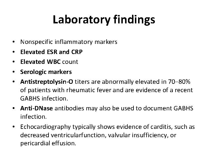
Laboratory findings Nonspecific inflammatory markers Elevated ESR and CRP Elevated WBC count Serologic markers Antistreptolysin-O titers are abnormally elevated in 70– 80% of patients with rheumatic fever and are evidence of a recent GABHS infection. • Anti-DNase antibodies may also be used to document GABHS infection. • Echocardiography typically shows evidence of carditis, such as decreased ventricularfunction, valvular insufficiency, or pericardial effusion. • • •
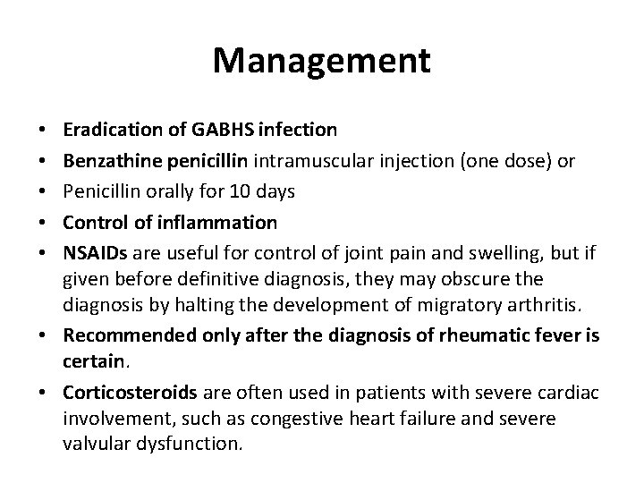
Management Eradication of GABHS infection Benzathine penicillin intramuscular injection (one dose) or Penicillin orally for 10 days Control of inflammation NSAIDs are useful for control of joint pain and swelling, but if given before definitive diagnosis, they may obscure the diagnosis by halting the development of migratory arthritis. • Recommended only after the diagnosis of rheumatic fever is certain. • Corticosteroids are often used in patients with severe cardiac involvement, such as congestive heart failure and severe valvular dysfunction. • • •
• Supportive therapy • Congestive heart failure is treated with diuretics, dietary salt restriction, digoxin, and bed rest. • Sydenham chorea, if severe, may be treated with haloperidol. • Long-term management includes continuous antimicrobial prophylaxis to prevent recurrent episodes of rheumatic fever.
Prognosis • There are no chronic sequelae of the joint, skin, and CNS manifestations of rheumatic fever. • Cardiac inflammation often leads to severe valvular dysfunction, which may require intervention immediately or many years after the event. • Valvular insufficiency or stenosis is usually delayed (usually more than 3 years after rheumatic fever) and, if severe, may require valve replacement or valvuloplasty.
MUSCULOSKELETAL PAIN SYNDROMES • Growing pains, or benign musculoskeletal pain syndrome, occur in 10% to 20% of school-age children. • The peak age range is 3 to 7 years; the syndrome seems to be more common in boys than girls. • There is no known etiology, although there seems to be a familial predisposition. • Children with growing pains complain of deep, crampy pain in the calves and thighs.
• It most typically occurs in the evening or as nocturnal pain that occasionally can waken the child from sleep. • Growing pains tend to be more common in children who are extremely active • The physical examination is unremarkable • Laboratory studies or x-rays, if performed, are normal.
• The diagnosis of growing pains is based on a typical history and a normal physical examination. • It is important to consider leukemia as a cause of nocturnal leg pain in children of this age group, so it is prudent to document a normal complete blood count. • The treatment of growing pains consists of reassurance and a regular bedtime ritual of stretching and relaxation. • The pain can be relieved by massage. Some patients may benefit from acetaminophen or NSAIDS.