Rhematoid arthritis Dr Saremi BUMS Associated professor of

Rhematoid arthritis Dr Saremi. BUMS, Associated professor of rheumatology @drzsaremi www. drzeinabsaremi. ir

Clinical manifestation
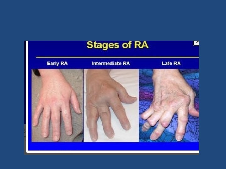

Articular manifestation Pain in affected joint aggravated by movemnt is the most common symptom. Morning stiffness ≥ 1 hr Joints involved -

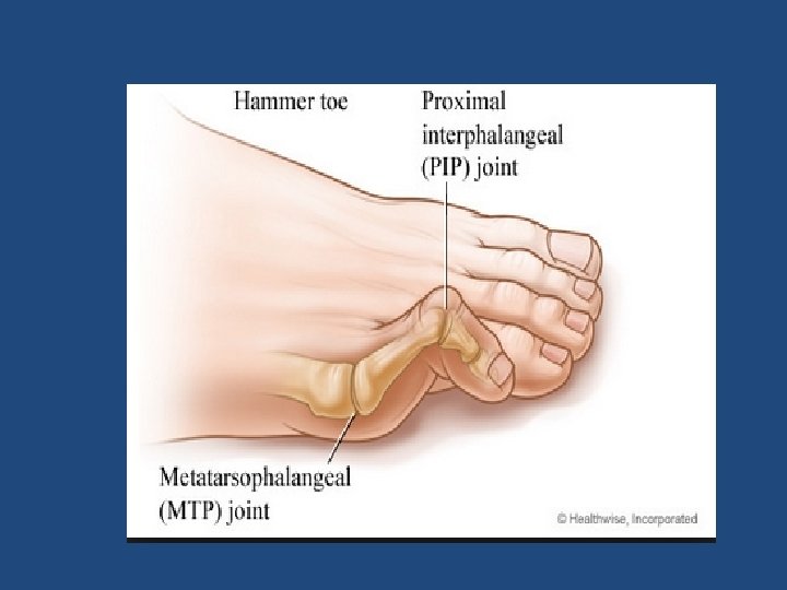
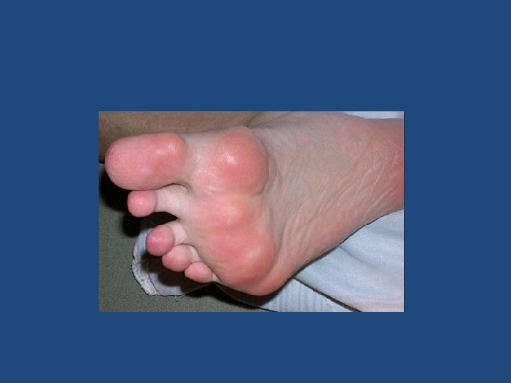
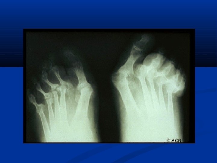
Relative incidence of joint involvement in RA MCP and PIP joints of hands & MTP of feet Knees, ankles & wrists. Shoulders. Elbows. TM, Acromio - clavicular & SC joints- 90% 80% 60% 50% 30%

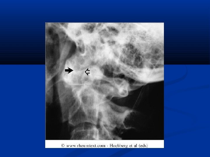
Joints involved in RA Don’t forget the cervical spine!! Instability at cervical spine can lead to impingement of the spinal cord. Thoracolumbar, sacroiliac, and distal interphalangeal joints (DIP)of the hand are NOT involved.

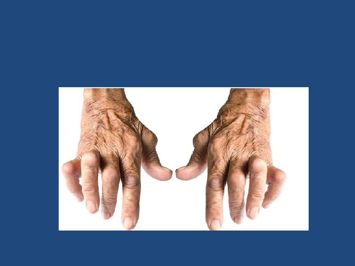
PIP Swelling

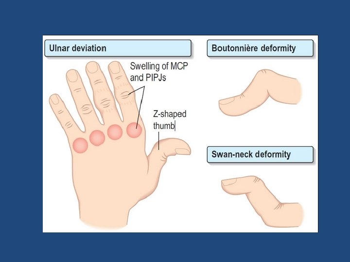
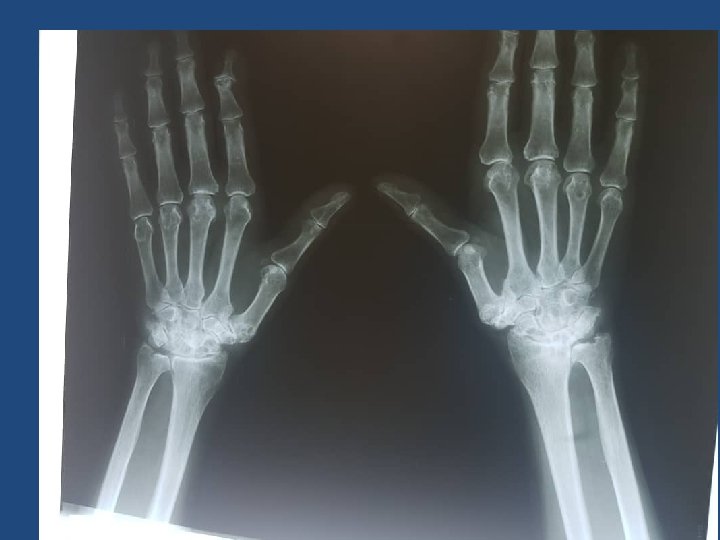
Ulnar Deviation, MCP Swelling, Left Wrist Swelling
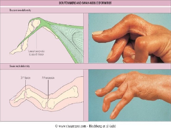

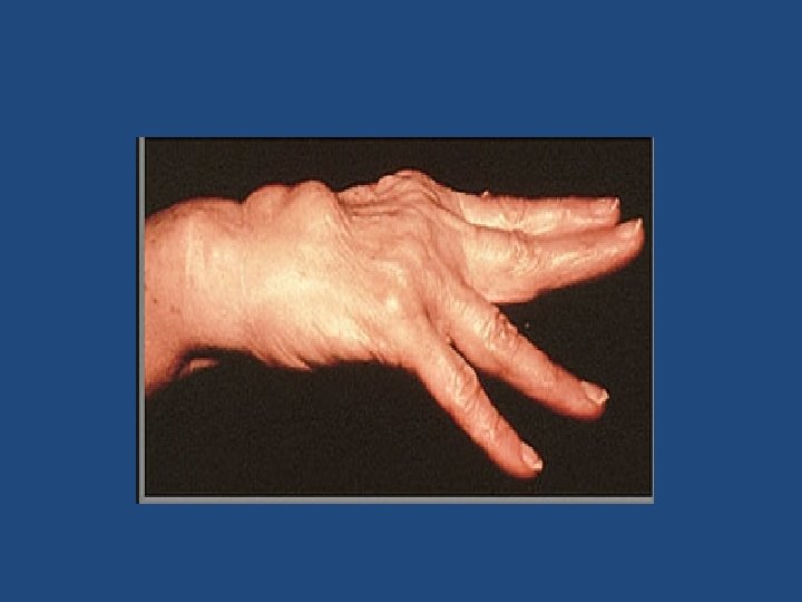
Z deformity

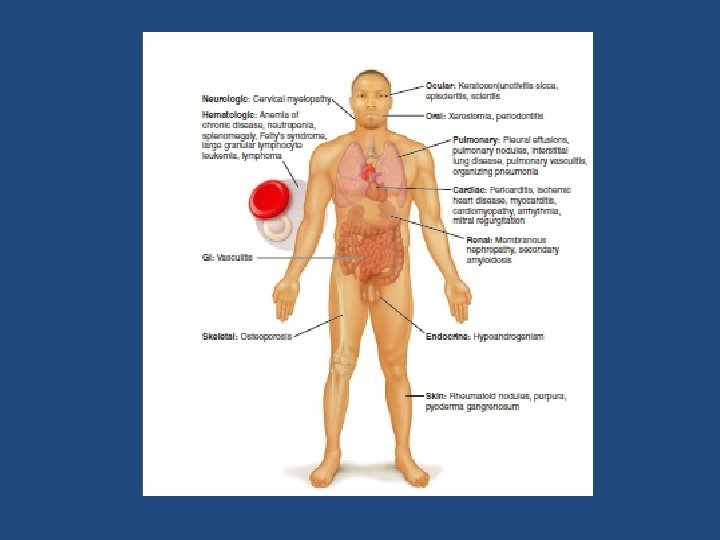
Extra-articular manifestation Present in 30 -40% May occur prior to arthritis Patients that are more likely to get are: High titres of RF/ anti-CCP HLA DR 4+ Male Early onset disability History of smoking
 Rheumatoid nodules(30%) Hematological normocytic normochromic anemia leucocytosis")
Extraarticular Involvement Constitutional symptoms ( most common) Rheumatoid nodules(30%) Hematological normocytic normochromic anemia leucocytosis /leucopenia thrombocytosis Felty’s syndrome- Chronic nodular Rheumatoid Arthritis Spleenomegaly Neutropenia

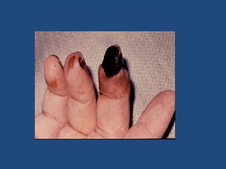
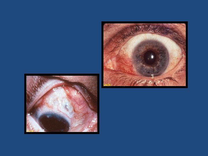
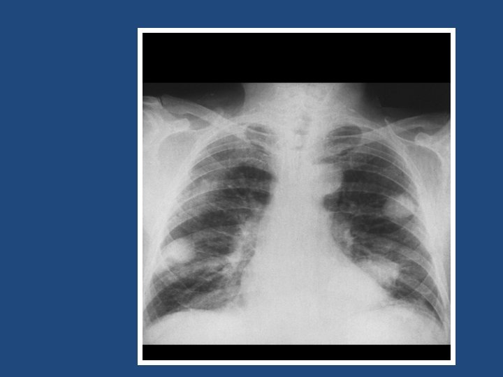
Respiratory- pleural effusion, pneumonitis , pleuro-pulmonary nodules, ILD CVS-asymptomatic pericarditis , pericardial effusion, cardiomyopathy Rheumatoid vasculitis- mononeuritis multiplex, cutaneous ulceration, digital gangrene, visceral infarction CNS- peripheral neuropathy, cord-compression from atlantoaxial/midcervical spine subluxation, entrapment neuropathies EYE- kerato cunjunctivitis sicca, episcleritis, scleritis

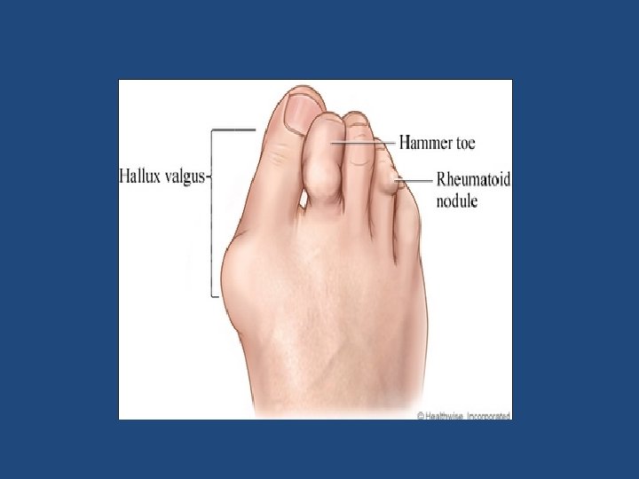
Rheumatoid nodule
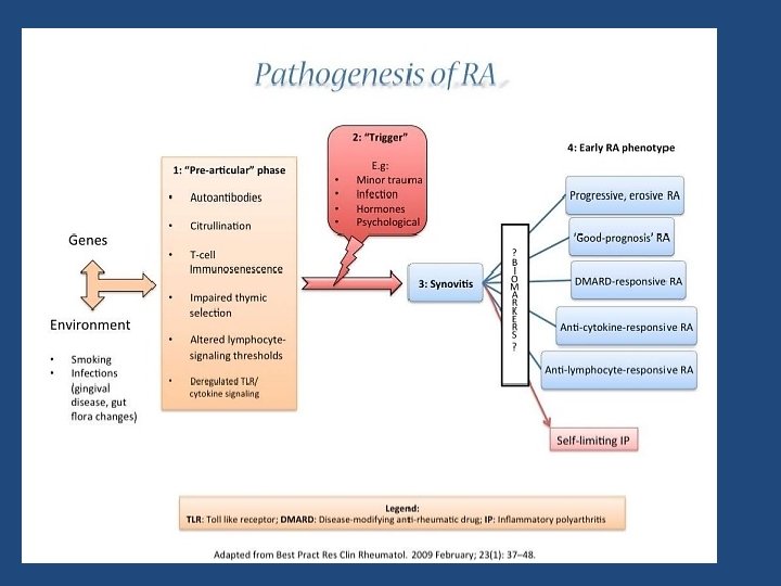

Arthritogenic Antigen Endogenous Bacterial viral Exogenous EBV Mycoplasma Hepatitis Myco. Bacterium Paro Virus B 19 Yarsenia Streptococcus Citrullinated Peptide

genetic • HLA class II is strongly linked to RA. • HLA DR 4 is the major halo-type in ethnic group, HLA DR 1 in Indians and HLA DW 15 in Japanese.

Laboratory investigations in RA CBC- TLC, DLC, Hb, ESR & GBP Acute phase reactants Rheumatoid Factor (RF) Anti- CCP antibodies
 Antibodies that recognize Fc portion of Ig. G Can be Ig.")
Rheumatoid Factor (RF) Antibodies that recognize Fc portion of Ig. G Can be Ig. M , Ig. G , Ig. A 85% of patients with RA over the first 2 years become RF+ • A negative RF may be repeated 4 -6 monthly for the first two year of disease, since some patients may take 18 -24 months to become seropositive. • PROGNISTIC VALUE- Patients with high titres of RF, in general, tend to have POOR PROGNOSIS, MORE EXTRA ARTICULAR MANIFESTATION.

Causes of positive test for RF Rheumatoid arthritis Sjogrens syndrome Vasculitis such as polyarteritis nodosa Sarcoidosis Systemic lupus erythematosus Cryoglobulinemia Chronic liver disease Infections- tuberculosis , bacterial endocarditis, infectious mononucleosis, leprosy, syphilis, leishmaniasis. Malignancies Old age(5% women aged above 60)
 & Specificity")
Anti-CCP Ig. G against synovial membrane peptides damaged via inflammation Sensitivity (65%) & Specificity (95%) Both diagnostic & prognostic value Predictive of Erosive Disease severity Radiologic progression Poor functional outcomes
 Thrombocytosis Leukocytosis ANA 30 -40% Inflammatory synovial fluid")
Elevated APRs( ESR, CRP ) Thrombocytosis Leukocytosis ANA 30 -40% Inflammatory synovial fluid Hypoalbuminemia

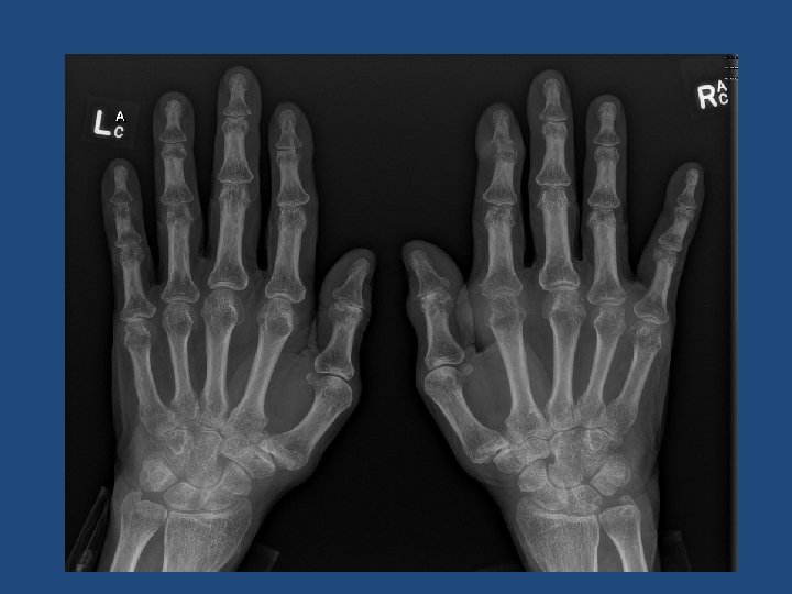
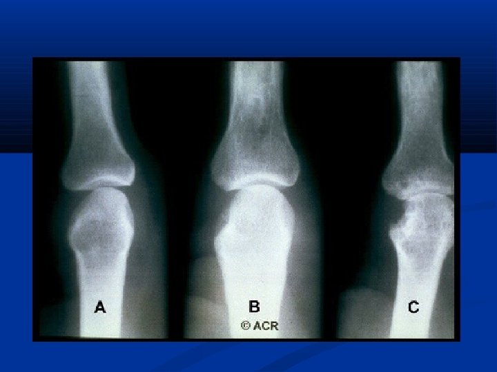
Radiographic Features Peri-articular osteopenia Uniform symmetric joint space narrowing Marginal subchondral erosions Joint Subluxations Joint destruction Collapse Ultrasound detects early soft tissue lesions. MRI has greatest sensitivity to detect synovitis and marrow changes.

2010 ACR/EULAR Classification Criteria • • a score of ≥ 6/10 is needed for classification of a patient as having definite RA A. Joint involvement SCORE 1 large joint 0 2− 10 large joints 1 1− 3 small joints (with or without involvement of large joints) 2 4− 10 small joints (with or without involvement of large joints) 3 >10 joints (at least 1 small joint)†† 5 • • B. Serology (at least 1 test result is needed for classification) Negative RF and negative ACPA Low-positive RF or low-positive ACPA High-positive RF or high-positive ACP • • • C. Acute-phase reactants (at least 1 test result is needed for classification) Normal CRP and normal ESR Abnormal CRP or normal ESR 0 1 • • • D. Duration of symptoms <6 weeks ≥ 6 weeks 0 1 • • 0 2 3

treatment

Goals of management Focused on relieving pain Preventing damage/disability Patient education about the disease Physical Therapy for stretching and range of motion exercises Occupational Therapy for splints and adaptive devices Treatment should be started early and should be individualised. EARLY AGGRESSIVE TREATEMNT

Treatment modalities for RA NSAIDS Steroids DMARDs Immunosuppressive therapy Biological therapies Surgery
 / Coxibs for symptom control 1) Reduce pain and swelling")
NSAIDS Non-Steroidal anti-inflammatories (NSAIDS) / Coxibs for symptom control 1) Reduce pain and swelling by inhibiting COX 2) Do not alter course of the disease. 3) Chronic use should be minimised. 4) Most common side effect related to GI tract.

Corticosteroids in RA Corticosteroids , both systemic and intra-articular are important adjuncts in management of RA. Indications for systemic steroids are: 1. 2. 3. 4. 5. For treatment of rheumatoid flares. For extra-articular RA like rheumatoid vasculitis and interstitial lung disease. As bridge therapy for 6 -8 weeks before the action of DMARDs begin. Maintainence dose of 10 mg or less of predinisolone daily in patients with active RA. Sometimes in pregnancy when other DMARDs cannot be used.

Disease Modifying Anti-rheumatic Agents Drugs that actually alter the disease course. Should be used as soon as diagnosis is made. Appearance of benefit delayed for weeks to months. NSAIDS must be continued with them until true remission is achieved. Induction of true remission is unusual.
 Sulphasalazine Cyclosporine. A")
DMARDs Commonly used Less commonly used Methotrexate Chloroquine Hydroxychloroquine Gold(parenteral &oral) Sulphasalazine Cyclosporine. A Leflunomide D-penicillamine/bucillamine Minocycline/Doxycycline Levamisole Azathioprine, cyclophosphamide, chlorambucil

Immunosuppresive therapy Agent Usual dose/route Side effects Azathioprine 50 -150 mg orally GI side effects , myelosuppression, infection, Cyclosporin A Cyclophosphamide . 3 -5 mg/kg/day. 50 -150 mg orally Nephrotoxic , hypertension , hyperkalemia Myelosuppression , gonadal toxicity , hemorrhagic cystitis , bladder cancer
 • Anti CD 20( rituximab) • Anti")
Biologic drugs • TNF inhibitors( infliximab- adalimumabetanercept) • Anti CD 20( rituximab) • Anti IL 6
- Slides: 50