Review and Revised July 20 2015 Radiographic Projections

 – referred as degrees right or left anterior oblique")


 RAO caudal projection • Usually large (5 -10")
 • Lies in anterior interventricular groove •")
 • Lies in left atrioventricular groove • Obtuse")
 • Lies in right atrioventricular groove •")


 Nodal Artery • RCA (55 – 65%)")


- Slides: 32
Review and Revised - July 20, 2015
Radiographic Projections Transverse Plane (Axial) – referred as degrees right or left anterior oblique Sagital Plane – referred to as degrees cranial or caudal Defined by the position of the imaging detector not the x-ray tube
Angiographic Projections Cranial Caudal Image detector X-ray tube
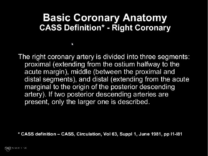
Coronary Anatomy Circulation Vol 63 Supp I, June 1981, I-1 to I-81
Coronary Angiographic Anatomy
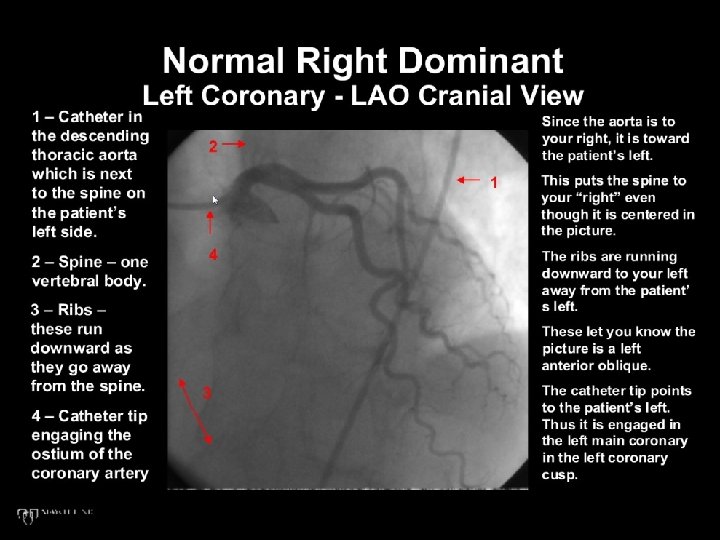
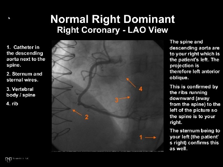
Coronary Angiographic Anatomy Normal Coronary Ostia Positions • Sinotubular junction • LAO View LMCA – to your right, and superior RCA – to your left, and inferior
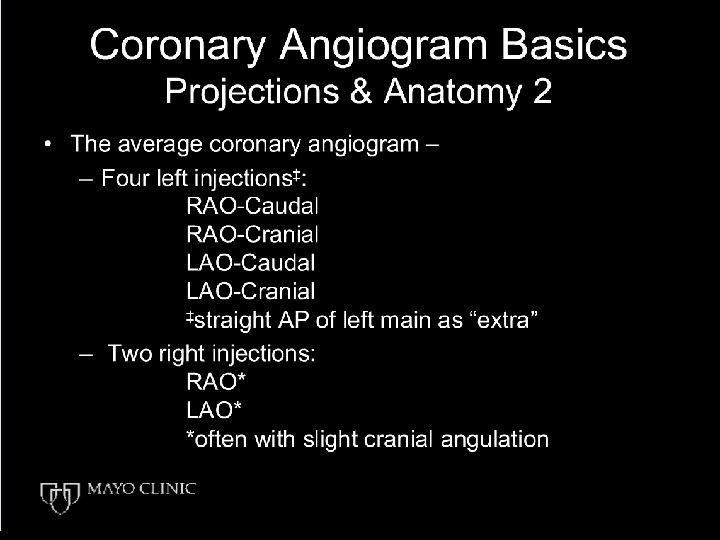
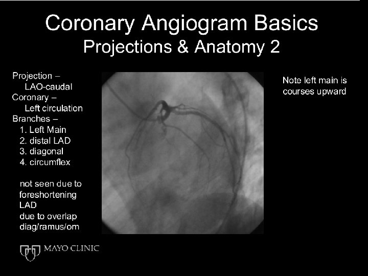
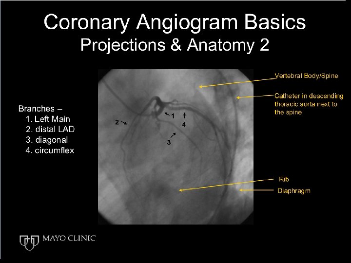
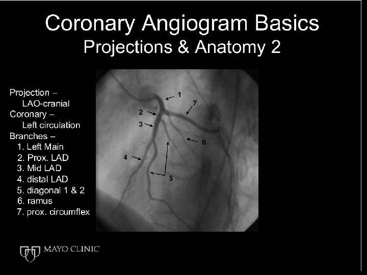
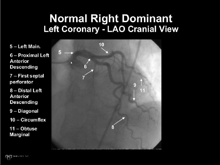
Coronary Angiographic Anatomy Left Main (LM) RAO caudal projection • Usually large (5 -10 mm) • Variable length • Bifurcates into LAD and LCx (most common) • Trifurcates into ramus intermedius, LAD and LCx in 10 -15% of cases • Straight PA – best for ostial disease • LAO caudal best for distal disease (spider view)
Coronary Angiographic Anatomy Left Anterior Descending (LAD) • Lies in anterior interventricular groove • Usually “wraps around” the apex and ascends a variable distance up the posterior interventricular groove • Septal perforators supply anterior two thirds of the interventricular septum • Diagonal branches supply anterolateral wall
Coronary Angiographic Anatomy Left Circumflex (LCx) • Lies in left atrioventricular groove • Obtuse marginal (OM) arteries supply the lateral wall • Coronary dominance = artery supplying the posterior descending artery (PDA) (10% LCx, 90% RCA)
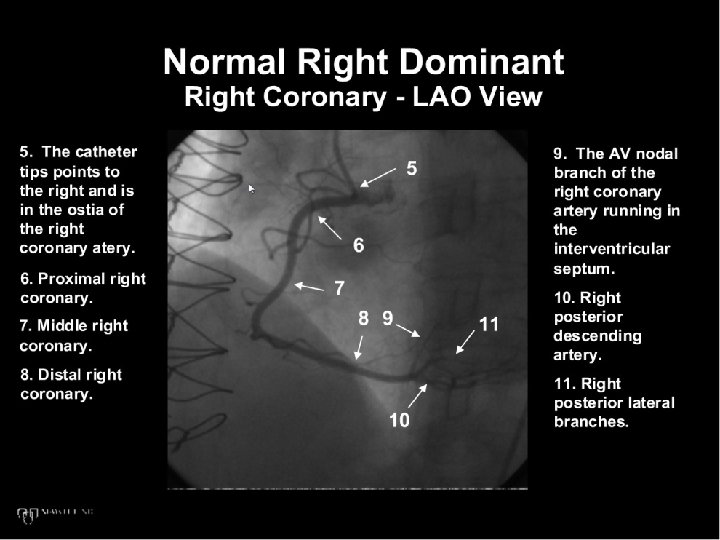
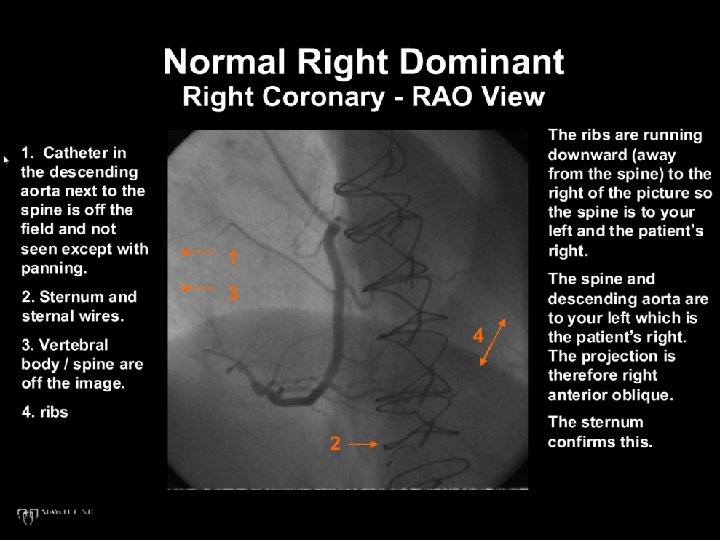
Coronary Angiographic Anatomy Right coronary artery (RCA) • Lies in right atrioventricular groove • Major Branches Conus artery (50% separate ostium) SA nodal artery (45% from LCx) Acute marginal (right ventricular branches) • Bifurcates into PDA and posterolateral branch at crux
Coronary Angiographic Anatomy Dominance By definition – the vessel that gives the PDA
Coronary Venous Anatomy • Anterior cardiac veins drain RV and empty into RA • Coronary sinus and tributaries (Anterior interventricular vein, Great cardiac vein) drain LV and empty via coronary sinus into RA • Thebesian veins – drain directly into underlying chanber (LV or RV) If drain into LV, right to left shunt, usually tiny < 1% of cardiac output
Conducting System Blood Supply • Sinoatrial (SA) Nodal Artery • RCA (55 – 65%) • LCx (35 -45%) • Atrioventricular Nodal Artery • RCA (90%) • His Bundle • RCA and LAD septals • Left Anterior Fascicle • LAD • Left Posterior Fascicle • PDA and LAD
16
- End of this slide set - Andre Lapeyre, MD © Mayo Clinic