Retinopathy of Prematurity Dr Soujanya K Assistant Professor


 is a proliferative retinopathy affecting premature")


- Slides: 74
Retinopathy of Prematurity Dr Soujanya K Assistant Professor, Dept. of Ophthalmology, YMCH
Specific learning objectives • To describing pathogenesis of ROP • To describe prevention and screening protocol of ROP • To understand management of ROP • To describe clinical features of Retinitis pigmentosa • To describe clinical features of pathological myopia
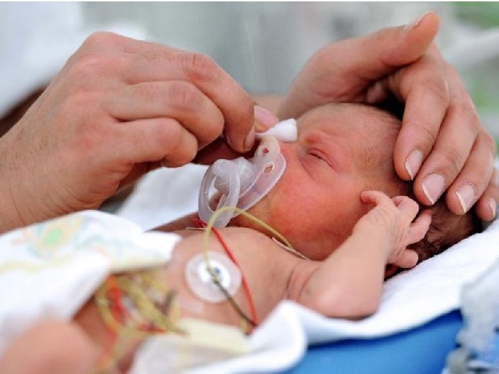
What is prematurity?
What is ROP? • Retinopathy of prematurity (ROP) is a proliferative retinopathy affecting premature infants of very low birth weight, who have often been exposed to high ambient oxygen concentrations
Risk factors • Birth weight < 1500 gm • </= 32 wks GA • Exposure to high concentration of oxygen
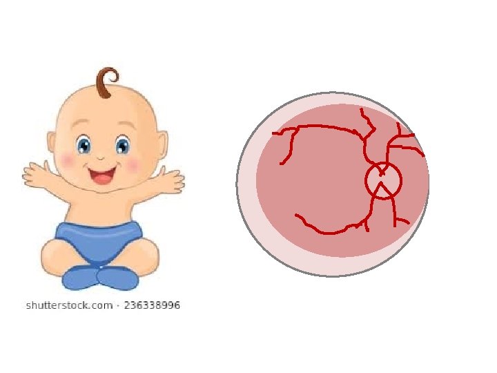
Normal angiogenesis
4 months Temporal Retina Nasal Retina
8 months Temporal Retina Nasal Retina
10 months Temporal Retina Nasal Retina
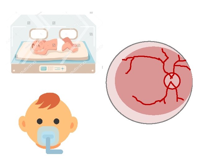
PATHOGENESIS
Increased oxygen saturation
Obliteration of retinal vessels
Release of VEGF
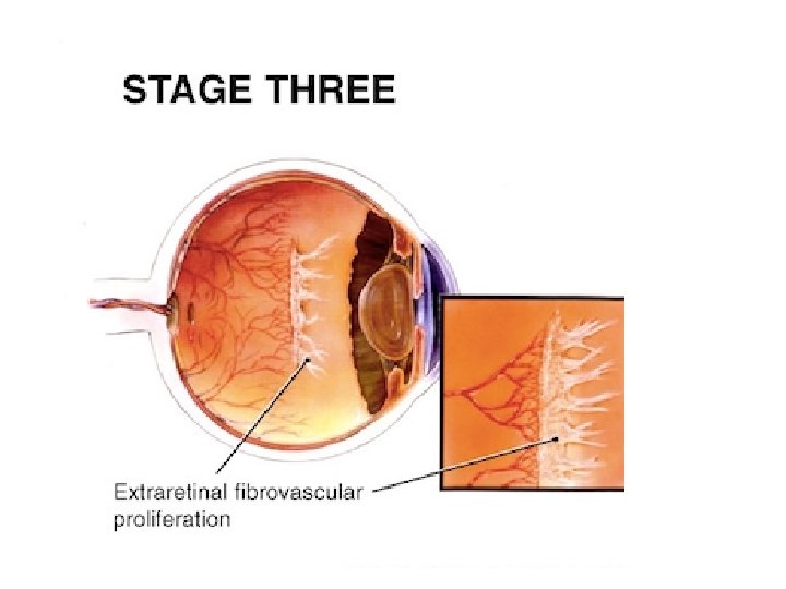
Neovascularisation
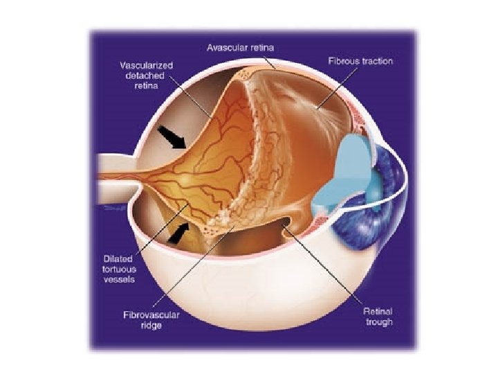
Fibrous tissue proliferatio
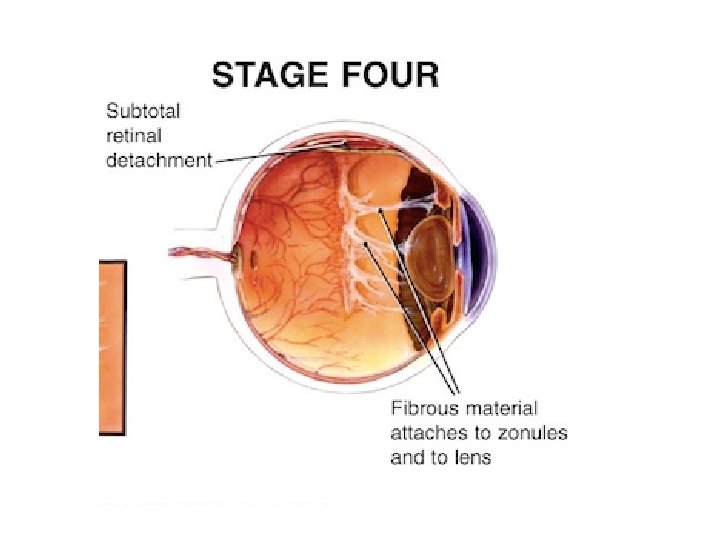
Retinal detachment
Retinal detachment
Sub-Total Retinal detachment
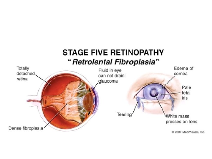
Total Retinal detachment
Pseudoglioma
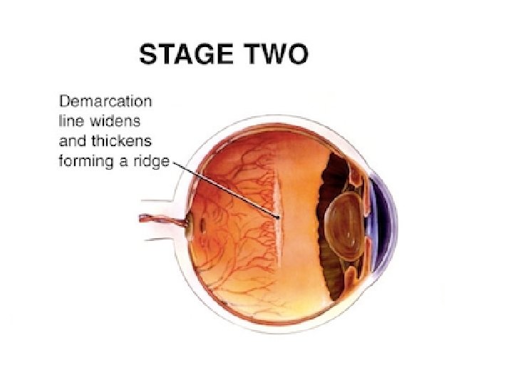
STAGES OF ROP
A discrete line of demarcation separates avascular retina from vascular retina
ZONES IN ROP
Zone 1: Radius- twice the distance from the disc & fovea
Zone 2: Radius- centre of the disc to the nasal ora serrata
Zone 3: Temporal crescent
Screening • Prophylaxis: • Newborn – incubator O 2 saturation <30%
Who need screening? • All premature babies born at – less than or equal to 32 weeks of gestational age – Weighing 1500 g or less
When to screen? • First examination: – Between 6 and 7 weeks post-natal age or – 34 weeks post-conceptual age (whichever is earlier). Eg: Baby born at 32 wks on 1/1/2018 After 6 wks : 15/02/2018 or When baby is 34 wks: 15/01/2018
How to screen? • Dilated fundus examination with Indirect ophthalmoscopy with 28 D lens
Tele-medicine
Treatment • Indirect laser photocoagulation
Treatment
Treatment
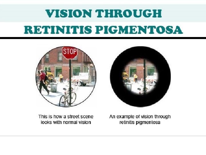
Retinitis pigmentosa
• Retinitis pigmentosa is a hereditary disorder predominantly affecting the rods more than the cones.
What is the function of rods?
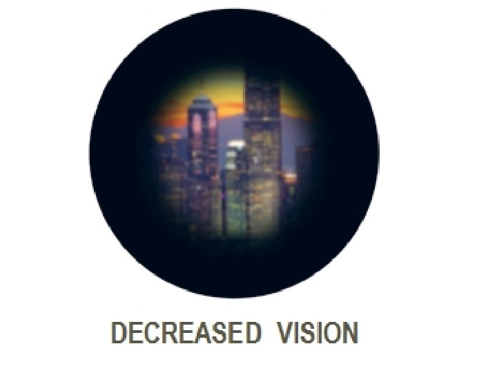
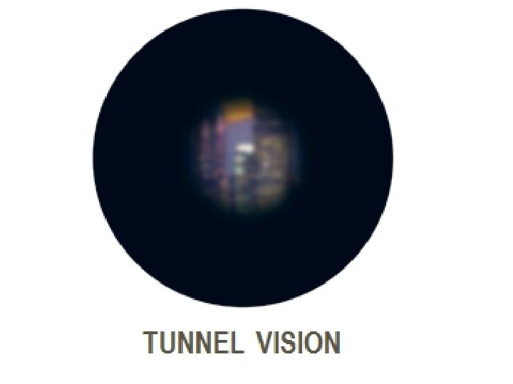
Symptoms • Night blindness. • Tubular vision occurs in advanced cases.
Arteriolar Attenuation Waxy Disc Pallor. Retinal Bone-spicule Pigmentation
Advanced Changes
End-stage Disease
FIELD OF VISION
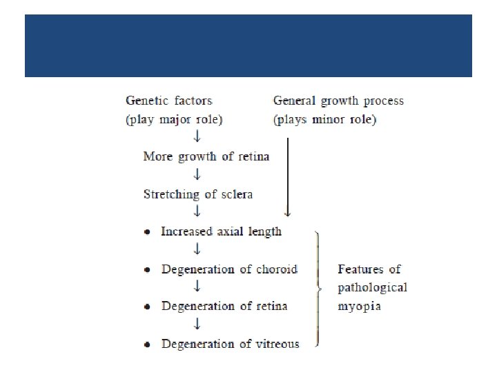
Pathological myopia
• Includes a rapid axial growth of the eyeball beyond normal variation.
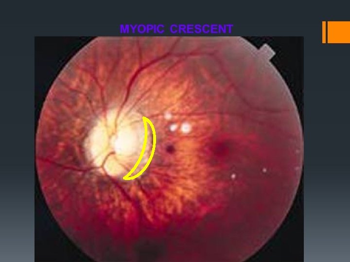
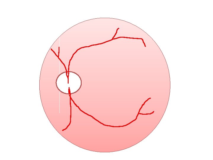
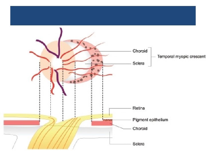
Myopic crescent
Myopic crescent & Super-traction crescent
• Foster-Fuchs' spot: dark red circular patch due to sub-retinal neovas-cularization and choroidal haemorrhage
Foster-Fuchs' spot
Foster Fuchs’ spot
Posterior staphyloma
Posterior vitreous detachment
Vitreous liquefaction
LACQUER CRACKS • spontaneous ruptures of the Bruch’s membrane that appear yellowish-white and are usually located in the posterior pole.
Peripheral retinal degeneration
Questions • When should a premature baby be screened to prevent retinopathy of prematurity? • What are the differential diagnosis of pseudoglioma? • What are the clinical features of retinitis pigmentosa? • Draw a neat labeled diagram depicting features of pathological myopia.